(New page: {{BioPsy}} {{Infobox Anatomy | Name = Vestibule of the ear | Latin = vestibulum auris | GraySubject = 232 | GrayPage = 1047 | Image = Gray919.png| ...) |
No edit summary |
||
| Line 22: | Line 22: | ||
DorlandsSuf = 12856151 | |
DorlandsSuf = 12856151 | |
||
}} |
}} |
||
| + | |||
| − | ''This is a page about the part of the [[ear]]. For other uses see [[Vestibule (disambiguation)]].'' |
||
The '''vestibule''' is the central part of the [[osseous labyrinth]], and is situated medial to the tympanic cavity, behind the cochlea, and in front of the semicircular canals. |
The '''vestibule''' is the central part of the [[osseous labyrinth]], and is situated medial to the tympanic cavity, behind the cochlea, and in front of the semicircular canals. |
||
Latest revision as of 23:46, 14 May 2007
Assessment |
Biopsychology |
Comparative |
Cognitive |
Developmental |
Language |
Individual differences |
Personality |
Philosophy |
Social |
Methods |
Statistics |
Clinical |
Educational |
Industrial |
Professional items |
World psychology |
Biological: Behavioural genetics · Evolutionary psychology · Neuroanatomy · Neurochemistry · Neuroendocrinology · Neuroscience · Psychoneuroimmunology · Physiological Psychology · Psychopharmacology (Index, Outline)
| Vestibule of the ear | ||
|---|---|---|
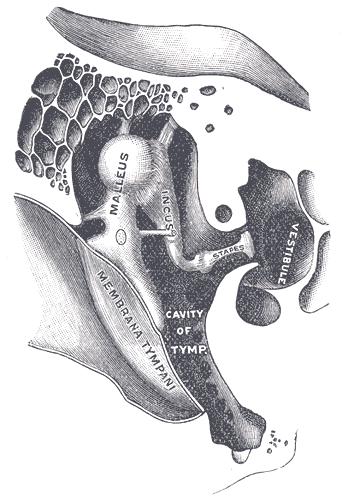 | ||
| Chain of ossicles and their ligaments, seen from the front in a vertical, transverse section of the tympanum. (Vestibule visible at center right.) | ||
| Latin | vestibulum auris | |
| Gray's | subject #232 1047 | |
| System | ||
| MeSH | [1] | |
| [[Image:|190px|center|]] | ||
The vestibule is the central part of the osseous labyrinth, and is situated medial to the tympanic cavity, behind the cochlea, and in front of the semicircular canals.
It is somewhat ovoid in shape, but flattened transversely; it measures about 5 mm. from before backward, the same from above downward, and about 3 mm. across.
In its lateral or tympanic wall is the fenestra vestibuli, closed, in the fresh state, by the base of the stapes and annular ligament.
On its medial wall, at the forepart, is a small circular depression, the recessus sphæricus, which is perforated, at its anterior and inferior part, by several minute holes (macula cribrosa media) for the passage of filaments of the acoustic nerve to the saccule; and behind this depression is an oblique ridge, the crista vestibuli, the anterior end of which is named the pyramid of the vestibule.
This ridge bifurcates below to enclose a small depression, the fossa cochlearis, which is perforated by a number of holes for the passage of filaments of the acoustic nerve which supply the vestibular end of the ductus cochlearis.
As the hinder part of the medial wall is the orifice of the aquæductus vestibuli, which extends to the posterior surface of the petrous portion of the temporal bone.
It transmits a small vein, and contains a tubular prolongation of the membranous labyrinth, the ductus endolymphaticus, which ends in a cul-de-sac between the layers of the dura mater within the cranial cavity.
On the upper wall or roof is a transversely oval depression, the recessus ellipticus, separated from the recessus sphæricus by the crista vestibuli already mentioned.
The pyramid and adjoining part of the recessus ellipticus are perforated by a number of holes (macula cribrosa superior).
The apertures in the pyramid transmit the nerves to the utricle; those in the recessus ellipticus the nerves to the ampullæ of the superior and lateral semicircular ducts.
Behind are the five orifices of the semicircular canals.
In front is an elliptical opening, which communicates with the scala vestibuli of the cochlea.
Additional images
")
")
External links
This article was originally based on an entry from a public domain edition of Gray's Anatomy. As such, some of the information contained herein may be outdated. Please edit the article if this is the case, and feel free to remove this notice when it is no longer relevant.
Sensory system: Auditory and Vestibular systems (TA A15.3, GA 10.1029) | |||||||||||||||
|---|---|---|---|---|---|---|---|---|---|---|---|---|---|---|---|
| Outer ear |
Pinna (Helix, Antihelix, Tragus, Antitragus, Incisura anterior auris, Earlobe) • Ear canal • Auricular muscles | ||||||||||||||
| Middle ear |
| ||||||||||||||
| Inner ear/ (membranous labyrinth, bony labyrinth) |
| ||||||||||||||
| {| class="navbox collapsible nowraplinks" style="margin:auto; " | |||||||||||||||
| |||||||||||||||
|}
| This page uses Creative Commons Licensed content from Wikipedia (view authors). |