m (Vein moved to Veins (anatomy): ALign thesaurus) |
No edit summary |
||
| Line 1: | Line 1: | ||
| + | {{BioPsy}} |
||
| − | {{dablink|For the geological use, see [[vein (geology)]].}} |
||
| + | {{PsyPerspective}} |
||
| ⚫ | |||
| − | In [[ |
+ | In the [[circulatory system]], '''veins''' (from the [[Latin language|Latin]] ''vena'') are [[blood vessel]]s that carry [[blood]] '''toward the [[heart]]'''. Most veins carry deoxygenated blood from the tissues back to the heart; exceptions are the [[pulmonary vein|pulmonary]] and [[umbilical vein]]s, both of which carry oxygenated blood. They differ from [[artery|arteries]] in structure and function; for example, arteries are more muscular than veins and they carry blood away from the heart. |
| ⚫ | |||
| ⚫ | |||
| + | Veins generally function to return deoxygenated blood to the [[heart]], and are essentially tubes that collapse when their [[Lumen (anatomy)|lumens]] are not filled with blood. The thick outermost layer of a vein is made of [[connective tissue]], called tunica adventitia or tunica externa. Deeper are bands of [[smooth muscle]] called tunica media, which are generally thin as veins do not function primarily in a contractile manner. The interior is lined with [[Endothelium|endothelial cells]] called tunica intima. Most veins have one-way flaps called venous valves that prevent blood from flowing back and pooling in the lower extremities due to the effects of [[gravity]]. These are infoldings of the tunica intima. The precise location of veins is much more variable from person to person than that of [[Artery|arteries]].<ref>{{cite book | last = Maton | first = Anthea | authorlink = | coauthors = Jean Hopkins, Charles William McLaughlin, Susan Johnson, Maryanna Quon Warner, David LaHart, Jill D. Wright | title = Human Biology and Health | publisher = Prentice Hall | date = 1993 | location = Englewood Cliffs, New Jersey | pages = | url = | doi = | id = | isbn = 0-13-981176-1}}</ref> |
||
| ⚫ | |||
| ⚫ | |||
| + | The greater saphenous vein is the most important superficial vein of the lower limb. First described by the Persian physician Avicenna, Saphenous derives its name from ''Safina'', meaning hidden. This vein is "hidden" in its own fascial compartment in the thigh and only exits the fascia near the knee. Incompetence of this vein is an important cause of [[varicose vein]]s of lower limbs. |
||
| − | In [[systemic circulation]] de-[[oxygen]]ated blood from the [[capillary]] blood vessels is taken by veins to the right part of the |
||
| − | heart. As an exception, in the [[pulmonary circulation]] oxygenated blood from the [[lung]]s is taken to the left part of the heart by [[pulmonary vein]]s. |
||
| + | The [[pulmonary vein]]s carry relatively oxygenated blood from the lungs to the heart. The [[superior vena cava|superior]] and [[inferior vena cava|inferior venae cavae]] carry relatively deoxygenated blood from the upper and lower systemic circulations, respectively. |
||
| − | Another special case is [[portal circulation]] where the [[portal vein]] transports blood rich in products of digestion from the [[intestine]]s to the [[liver]]. |
||
| + | A [[portal venous system]] is a series of veins or venules that directly connect two [[capillary bed]]s. Examples of such systems include the [[hepatic portal vein]] and [[hypophyseal portal system]]. |
||
| ⚫ | |||
| − | Veins have one-way valves called [[venous valve]] to prevent backflow caused by gravity. They also have a thick [[collagen]] outer layer, which helps maintain [[blood pressure]] and stop blood pooling. The hollow internal cavity in which the blood flows is called the [[lumen (anatomy)|lumen]]. |
||
| + | ===Color=== |
||
| ⚫ | |||
| + | Vein color is determined in large part by the color of [[venous blood]], which is usually dark red as a result of its low oxygen content. Veins appear blue because the [[subcutis|subcutaneous fat]] absorbs low frequency light, permitting only the highly energetic blue wavelengths to penetrate through to the dark vein and reflect off. This results from a phenomenon called [[Rayleigh scattering]]. |
||
| − | Names of important veins: |
||
| ⚫ | |||
| − | * [[Portal vein]] |
||
| − | * [[Superior vena cava]] |
||
| − | * [[Inferior vena cava]] |
||
| − | * [[Femoral vein]] |
||
| − | * [[Great saphenous vein]] |
||
| ⚫ | |||
| − | Names of important venule systems: |
||
| + | Veins serve to return blood from organs to the heart. In [[systemic circulation]] oxygenated blood is pumped by the [[left ventricle]] through the [[artery|arteries]] to the muscles and organs of the body, where its nutrients and gases are exchanged at [[capillary|capillaries]], entering the veins filled with cellular waste and [[carbon dioxide]]. The de-[[oxygen]]ated blood is taken by veins to the [[right atrium]] of the heart, which transfers the blood to the [[right ventricle]], where it is then pumped through the pulmonary arteries to the [[lung]]s. In [[pulmonary circulation]] the [[pulmonary vein]]s return oxygenated blood from the lungs to the [[left atrium]], which empties into the left ventricle, completing the cycle of blood circulation. |
||
| − | *[[Portal venous system]] |
||
| − | *[[Pulmonary venous system]] |
||
| − | *[[Systemic venous system]] |
||
| + | The return of blood to the heart is assisted by the action of the [[skeletal-muscle pump]] which helps maintain the extremely low [[blood pressure]] of the venous system. [[Fainting]] can be caused by failure of the skeletal-muscular pump. Long periods of standing can result in blood pooling in the legs, with blood pressure too low to return blood to the heart. [[Neurogenic shock|Neurogenic]] and [[shock (circulatory)#Hypovolaemic shock|hypovolaemic shock]] can also cause fainting. In these cases the smooth muscles surrounding the veins become slack and the veins fill with the majority of the blood in the body, keeping blood away from the brain and causing unconsciousness. |
||
| − | ==Medical interest== |
||
| ⚫ | |||
| − | Veins are used medically as points of access to the blood stream, permitting the withdrawal of blood specimens ([[venipuncture]]) for testing purposes, and enabling the infusion of fluid, [[electrolyte]]s, nutrition, and medications. The latter is called '''intravenous''' delivery. It can be done by an injection with a [[syringe]], or by inserting a [[catheter]] (a flexible tube). |
||
| + | The arteries are perceived as carrying oxygenated blood to the tissues, while veins carry deoxygenated blood back to the heart. This is true of the systemic circulation, by far the larger of the two circuits of blood in the body, which transports oxygen from the heart to the tissues of the body. However, in pulmonary circulation the arteries carry deoxygenated blood from the heart to the lungs and veins return blood from the lungs to the heart. The difference between veins and arteries is their direction of flow (out of the heart by arteries, returning to the heart for veins), not their oxygen content. In addition, deoxygenated blood that is carried from the tissues back to the heart for reoxygenation in systemic circulation still carries some oxygen, though it is considerably less than that carried by the systemic arteries or pulmonary veins. |
||
| ⚫ | If an intravenous catheter has to be inserted, for most purposes this is done into a peripheral |
||
| + | ==Classification== |
||
| − | The precise location of veins is much more variable from person to person than that of arteries. |
||
| + | Veins are classified in a number of ways, including superficial vs. deep, pulmonary vs. systemic, and large vs. small. |
||
| − | + | ;Superficial veins |
|
| + | :[[Superficial vein]]s are those whose course is close to the surface of the body, and who have no corresponding artery. |
||
| − | In the light, blood appears red because most colors are absorbed except for red, which bounces back from the blood. Every colour but red is absorbed by the oxygen-carrying pigment [[hemoglobin]] (Hb). If a filter that blocks the reflected color is positioned between the blood and the eyes of the person watching, the perceived color changes. In the case of humans, the skin serves as a filter for the color red, and the remaining color ends up being green. The exact [[color spectra]] is determined by the relative levels of oxygenated iron (HbO) and [[carbon dioxide|CO<sub>2</sub>]] in the blood. High oxygen reflects red and high CO<sub>2</sub> reflects blue, which mixed with the yellowish color of the fat and/or skin ends up showing as green. |
||
| + | |||
| + | ;Deep veins |
||
| + | :[[Deep vein]]s are deeper in the body and have corresponding arteries. |
||
| + | |||
| + | ;Pulmonary veins |
||
| + | :The [[pulmonary vein]]s are a set of veins that deliver oxygenated blood from the [[lung]]s to the heart. |
||
| + | |||
| + | ;Systemic veins |
||
| + | :[[Systemic vein]]s drain the tissues of the body and deliver deoxygenated blood to the heart. |
||
| + | |||
| + | ==Clinical significance== |
||
| ⚫ | |||
| + | |||
| + | ===Intravenous access=== |
||
| + | Veins are used medically as points of access to the blood stream, permitting the withdrawal of blood specimens ([[venipuncture]]) for testing purposes, and [[Intravenous therapy|intravenous delivery]] of fluid, [[electrolyte]]s, nutrition, and medications through injection with a [[syringe]], or by inserting a [[catheter]]. In contrast to arterial blood which is uniform throughout the body, the blood removed from veins for testing can vary in its contents depending on the part of the body the vein drains; blood drained from a working muscle will contain significantly less oxygen and [[glucose]] than blood drained from the [[liver]]. However the more blood from different veins mixes as it returns to the heart, the more homogeneous it becomes. |
||
| + | |||
| ⚫ | If an intravenous catheter has to be inserted, for most purposes this is done into a peripheral vein near the surface of the skin in the [[hand]] or [[arm]], or less desirably, the [[Human leg|leg]]. Some highly concentrated fluids or irritating medications must flow into the large central veins, which are sometimes used when peripheral access cannot be obtained. Catheters can be threaded into the [[superior vena cava]] for these uses: if long term use is thought to be needed, a more permanent access point can be inserted surgically. |
||
| + | |||
| + | ===Phlebology=== |
||
| + | Phlebology is the medical discipline that involves the diagnosis and treatment of disorders of venous origin. Diagnostic techniques used include the history and physical examination, venous imaging techniques and laboratory evaluation related to venous [[Thrombosis|thromboembolism]]. The American Medical Association has added phlebology to their list of self-designated practice specialties. |
||
| + | |||
| + | The [[American College of Phlebology]] (ACP) is a professional organization of physicians and health care professionals from a variety of backgrounds. ACP meetings are conducted to facilitate learning and sharing of knowledge regarding venous disease. The equivalent body for countries in the Pacific is the [[Australasian College of Phlebology]], active in Australia and New Zealand. |
||
| + | |||
| + | ===Venous diseases=== |
||
| + | ====Venous insufficiency==== |
||
| + | {{main|Venous insufficiency}} |
||
| + | |||
| + | Venous insufficiency is the most common disorder of the venous system, and is usually manifested as [[Telangiectasia|spider veins]] or [[varicose veins]]. A variety of treatments are used depending on the patient's particular type and pattern of veins and on the physician's preferences. Treatment can include [[Somnoplasty|radiofrequency ablation]], [[vein stripping]], [[ambulatory phlebectomy]], foam [[sclerotherapy]], [[laser]]s, or compression. |
||
| + | |||
| + | [[Postphlebitic syndrome]] is venous insufficiency that develops following [[deep vein thrombosis]].<ref name="pmid16822286">{{cite journal |author=Kahn SR |title=The post-thrombotic syndrome: progress and pitfalls |journal=Br. J. Haematol. |volume=134 |issue=4 |pages=357–65 |year=2006 |month=August |pmid=16822286 |doi=10.1111/j.1365-2141.2006.06200.x |url=}}</ref> |
||
| + | |||
| + | ====Deep vein thrombosis==== |
||
| + | {{main|Deep vein thrombosis}} |
||
| + | |||
| + | Deep vein thrombosis is a condition where a [[Thrombus|blood clot]] forms in a deep vein, which can lead to [[pulmonary embolism]] and chronic venous insufficiency. |
||
| + | |||
| + | ====Thrombophlebitis==== |
||
| + | {{main|Thrombophlebitis}} |
||
| + | |||
| + | Thrombophlebitis is an inflammatory condition of the veins related to [[thrombus|blood clots]]. |
||
==See also== |
==See also== |
||
| + | *[[Artery]] |
||
*[[Deep vein]] |
*[[Deep vein]] |
||
*[[Deep vein thrombosis]] |
*[[Deep vein thrombosis]] |
||
| − | *[[ |
+ | *[[Peripheral vein]] |
| ⚫ | |||
*[[Superficial vein]] |
*[[Superficial vein]] |
||
| − | *[[Varicose |
+ | *[[Varicose veins]] |
| + | |||
| + | ==References== |
||
| + | {{Refimprove|date=January 2008}} |
||
| + | {{reflist}} |
||
| + | |||
| + | ==External links== |
||
| + | {{wiktionary}} |
||
| + | *[http://www.merck.com/mmhe/sec03/ch036/ch036a.html Merck Manual article on veins.] |
||
| + | *[http://www.phlebology.org/ American College of Phlebology] |
||
| + | *[http://www.americanboardofphlebology.org/ American Board of Phlebology] |
||
| + | *[http://www.phlebologyfoundation.org/ American College of Phlebology Foundation] |
||
| + | *[http://www.phlebology.com.au/forms/selmenu.aspx?selmenu=5 Australasian College of Phlebology] Information from the Australasian College of Phlebology Website |
||
| + | *[http://ocw.dmc.keio.ac.jp/economics/02A-003_e/lecture_contents/ln03e.pdf In economics: Arterial and venous industries] |
||
| + | *[http://www.simmed.com/Demo/Default.aspx Animated Venous Access tutorials] |
||
| + | |||
| + | ===Scientific publications=== |
||
| + | *[http://phleb.rsmjournals.com/ Phlebology: The Journal of Venous Disease] |
||
| + | *[http://www.medi-data.co.uk/phlebology/index.htm Phlebology: 1986-1999] |
||
| + | {{VeinsHeadNeck}} |
||
| + | {{Veins of the upper extremity}} |
||
{{Veins}} |
{{Veins}} |
||
| + | {{Veins of the lower extremity}} |
||
{{cardiovascular_system}} |
{{cardiovascular_system}} |
||
| − | [[Category:Anatomy]] |
||
[[Category:Cardiovascular system]] |
[[Category:Cardiovascular system]] |
||
[[Category:Veins]] |
[[Category:Veins]] |
||
| + | <!-- |
||
[[af:Aar]] |
[[af:Aar]] |
||
| + | [[ar:وريد]] |
||
| + | [[zh-min-nan:Chēng-me̍h]] |
||
[[bs:Vena]] |
[[bs:Vena]] |
||
| + | [[bg:Вена]] |
||
| + | [[ca:Vena]] |
||
| + | [[cy:Gwythïen]] |
||
[[da:Vene]] |
[[da:Vene]] |
||
[[de:Vene]] |
[[de:Vene]] |
||
| + | [[el:Φλέβα]] |
||
[[es:Vena]] |
[[es:Vena]] |
||
| + | [[eo:Vejno]] |
||
| + | [[eu:Zain]] |
||
| + | [[fa:سیاهرگ]] |
||
[[fr:Veine]] |
[[fr:Veine]] |
||
| + | [[ko:정맥]] |
||
| + | [[hr:Vena]] |
||
| + | [[id:Pembuluh balik]] |
||
| + | [[is:Bláæð]] |
||
[[it:Vena]] |
[[it:Vena]] |
||
[[he:וריד]] |
[[he:וריד]] |
||
| + | [[jv:Pambuluh balik]] |
||
| + | [[ku:Xwînwerîn]] |
||
| + | [[la:Vena]] |
||
| + | [[lv:Vēnas]] |
||
[[lt:Vena]] |
[[lt:Vena]] |
||
| + | [[hu:Véna]] |
||
[[mk:Вена]] |
[[mk:Вена]] |
||
| − | [[ |
+ | [[ms:Vena]] |
| + | [[mn:Вена (анатоми)]] |
||
| + | [[nl:Ader (anatomie)]] |
||
[[ja:静脈]] |
[[ja:静脈]] |
||
[[no:Vene]] |
[[no:Vene]] |
||
[[nn:Vene]] |
[[nn:Vene]] |
||
| + | [[pag:Ulat]] |
||
| ⚫ | |||
| + | [[nds:Veen]] |
||
| ⚫ | |||
[[pt:Veia]] |
[[pt:Veia]] |
||
[[ru:Вена (анатомия)]] |
[[ru:Вена (анатомия)]] |
||
[[sq:Vena]] |
[[sq:Vena]] |
||
| + | [[scn:Vina (anatumìa)]] |
||
[[simple:Vein]] |
[[simple:Vein]] |
||
[[sk:Žila]] |
[[sk:Žila]] |
||
| + | [[sl:Vena]] |
||
| + | [[sh:Vena]] |
||
[[fi:Laskimo]] |
[[fi:Laskimo]] |
||
[[sv:Ven (blodkärl)]] |
[[sv:Ven (blodkärl)]] |
||
| + | [[ta:சிரை]] |
||
| + | [[te:సిర]] |
||
| + | [[th:หลอดเลือดดำ]] |
||
| + | [[tr:Toplardamar]] |
||
| + | [[uk:Вена]] |
||
| + | [[vi:Tĩnh mạch]] |
||
| + | [[fiu-vro:Verisuun]] |
||
| + | [[diq:Damare]] |
||
[[zh:静脉]] |
[[zh:静脉]] |
||
| + | --> |
||
{{enWP|Vein}} |
{{enWP|Vein}} |
||
Revision as of 12:55, 27 March 2009
Assessment |
Biopsychology |
Comparative |
Cognitive |
Developmental |
Language |
Individual differences |
Personality |
Philosophy |
Social |
Methods |
Statistics |
Clinical |
Educational |
Industrial |
Professional items |
World psychology |
Biological: Behavioural genetics · Evolutionary psychology · Neuroanatomy · Neurochemistry · Neuroendocrinology · Neuroscience · Psychoneuroimmunology · Physiological Psychology · Psychopharmacology (Index, Outline)
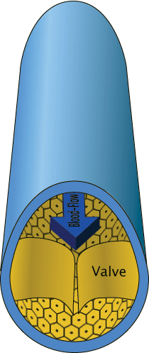
Cross section of a vein showing a valve which prevents backflow
In the circulatory system, veins (from the Latin vena) are blood vessels that carry blood toward the heart. Most veins carry deoxygenated blood from the tissues back to the heart; exceptions are the pulmonary and umbilical veins, both of which carry oxygenated blood. They differ from arteries in structure and function; for example, arteries are more muscular than veins and they carry blood away from the heart.
Anatomy
Veins generally function to return deoxygenated blood to the heart, and are essentially tubes that collapse when their lumens are not filled with blood. The thick outermost layer of a vein is made of connective tissue, called tunica adventitia or tunica externa. Deeper are bands of smooth muscle called tunica media, which are generally thin as veins do not function primarily in a contractile manner. The interior is lined with endothelial cells called tunica intima. Most veins have one-way flaps called venous valves that prevent blood from flowing back and pooling in the lower extremities due to the effects of gravity. These are infoldings of the tunica intima. The precise location of veins is much more variable from person to person than that of arteries.[1]
Notable veins and vein systems
The greater saphenous vein is the most important superficial vein of the lower limb. First described by the Persian physician Avicenna, Saphenous derives its name from Safina, meaning hidden. This vein is "hidden" in its own fascial compartment in the thigh and only exits the fascia near the knee. Incompetence of this vein is an important cause of varicose veins of lower limbs.
The pulmonary veins carry relatively oxygenated blood from the lungs to the heart. The superior and inferior venae cavae carry relatively deoxygenated blood from the upper and lower systemic circulations, respectively.
A portal venous system is a series of veins or venules that directly connect two capillary beds. Examples of such systems include the hepatic portal vein and hypophyseal portal system.
Color
Vein color is determined in large part by the color of venous blood, which is usually dark red as a result of its low oxygen content. Veins appear blue because the subcutaneous fat absorbs low frequency light, permitting only the highly energetic blue wavelengths to penetrate through to the dark vein and reflect off. This results from a phenomenon called Rayleigh scattering.
Function
Veins serve to return blood from organs to the heart. In systemic circulation oxygenated blood is pumped by the left ventricle through the arteries to the muscles and organs of the body, where its nutrients and gases are exchanged at capillaries, entering the veins filled with cellular waste and carbon dioxide. The de-oxygenated blood is taken by veins to the right atrium of the heart, which transfers the blood to the right ventricle, where it is then pumped through the pulmonary arteries to the lungs. In pulmonary circulation the pulmonary veins return oxygenated blood from the lungs to the left atrium, which empties into the left ventricle, completing the cycle of blood circulation.
The return of blood to the heart is assisted by the action of the skeletal-muscle pump which helps maintain the extremely low blood pressure of the venous system. Fainting can be caused by failure of the skeletal-muscular pump. Long periods of standing can result in blood pooling in the legs, with blood pressure too low to return blood to the heart. Neurogenic and hypovolaemic shock can also cause fainting. In these cases the smooth muscles surrounding the veins become slack and the veins fill with the majority of the blood in the body, keeping blood away from the brain and causing unconsciousness.
The arteries are perceived as carrying oxygenated blood to the tissues, while veins carry deoxygenated blood back to the heart. This is true of the systemic circulation, by far the larger of the two circuits of blood in the body, which transports oxygen from the heart to the tissues of the body. However, in pulmonary circulation the arteries carry deoxygenated blood from the heart to the lungs and veins return blood from the lungs to the heart. The difference between veins and arteries is their direction of flow (out of the heart by arteries, returning to the heart for veins), not their oxygen content. In addition, deoxygenated blood that is carried from the tissues back to the heart for reoxygenation in systemic circulation still carries some oxygen, though it is considerably less than that carried by the systemic arteries or pulmonary veins.
Classification
Veins are classified in a number of ways, including superficial vs. deep, pulmonary vs. systemic, and large vs. small.
- Superficial veins
- Superficial veins are those whose course is close to the surface of the body, and who have no corresponding artery.
- Deep veins
- Deep veins are deeper in the body and have corresponding arteries.
- Pulmonary veins
- The pulmonary veins are a set of veins that deliver oxygenated blood from the lungs to the heart.
- Systemic veins
- Systemic veins drain the tissues of the body and deliver deoxygenated blood to the heart.
Clinical significance

Venous valves prevent reverse blood flow.
Intravenous access
Veins are used medically as points of access to the blood stream, permitting the withdrawal of blood specimens (venipuncture) for testing purposes, and intravenous delivery of fluid, electrolytes, nutrition, and medications through injection with a syringe, or by inserting a catheter. In contrast to arterial blood which is uniform throughout the body, the blood removed from veins for testing can vary in its contents depending on the part of the body the vein drains; blood drained from a working muscle will contain significantly less oxygen and glucose than blood drained from the liver. However the more blood from different veins mixes as it returns to the heart, the more homogeneous it becomes.
If an intravenous catheter has to be inserted, for most purposes this is done into a peripheral vein near the surface of the skin in the hand or arm, or less desirably, the leg. Some highly concentrated fluids or irritating medications must flow into the large central veins, which are sometimes used when peripheral access cannot be obtained. Catheters can be threaded into the superior vena cava for these uses: if long term use is thought to be needed, a more permanent access point can be inserted surgically.
Phlebology
Phlebology is the medical discipline that involves the diagnosis and treatment of disorders of venous origin. Diagnostic techniques used include the history and physical examination, venous imaging techniques and laboratory evaluation related to venous thromboembolism. The American Medical Association has added phlebology to their list of self-designated practice specialties.
The American College of Phlebology (ACP) is a professional organization of physicians and health care professionals from a variety of backgrounds. ACP meetings are conducted to facilitate learning and sharing of knowledge regarding venous disease. The equivalent body for countries in the Pacific is the Australasian College of Phlebology, active in Australia and New Zealand.
Venous diseases
Venous insufficiency
- Main article: Venous insufficiency
Venous insufficiency is the most common disorder of the venous system, and is usually manifested as spider veins or varicose veins. A variety of treatments are used depending on the patient's particular type and pattern of veins and on the physician's preferences. Treatment can include radiofrequency ablation, vein stripping, ambulatory phlebectomy, foam sclerotherapy, lasers, or compression.
Postphlebitic syndrome is venous insufficiency that develops following deep vein thrombosis.[2]
Deep vein thrombosis
- Main article: Deep vein thrombosis
Deep vein thrombosis is a condition where a blood clot forms in a deep vein, which can lead to pulmonary embolism and chronic venous insufficiency.
Thrombophlebitis
- Main article: Thrombophlebitis
Thrombophlebitis is an inflammatory condition of the veins related to blood clots.
See also
- Artery
- Deep vein
- Deep vein thrombosis
- Peripheral vein
- Pulmonary circulation
- Superficial vein
- Varicose veins
References
| This article needs additional citations for verification. Please help improve this article by adding reliable references. Unsourced material may be challenged and removed. (January 2008) |
- ↑ Maton, Anthea; Jean Hopkins, Charles William McLaughlin, Susan Johnson, Maryanna Quon Warner, David LaHart, Jill D. Wright (1993). Human Biology and Health, Englewood Cliffs, New Jersey: Prentice Hall.
- ↑ Kahn SR (August 2006). The post-thrombotic syndrome: progress and pitfalls. Br. J. Haematol. 134 (4): 357–65.
External links
Look up this page on
Wiktionary:
Veins (anatomy)
- Merck Manual article on veins.
- American College of Phlebology
- American Board of Phlebology
- American College of Phlebology Foundation
- Australasian College of Phlebology Information from the Australasian College of Phlebology Website
- In economics: Arterial and venous industries
- Animated Venous Access tutorials
Scientific publications
Veins (emissary, jugular and others) of head and neck (drainage patterns can vary) (TA A12.3.04–06, GA 7.644) | |||||||||||||||||||||||||
|---|---|---|---|---|---|---|---|---|---|---|---|---|---|---|---|---|---|---|---|---|---|---|---|---|---|
| External jugular |
| ||||||||||||||||||||||||
| Internal jugular |
| ||||||||||||||||||||||||
| Brachiocephalic |
| ||||||||||||||||||||||||
| Template:Vascular navs | |||||||||||||||||||||||||
Template:Veins of the upper extremity
- REDIRECT Template:Veins of the torso
Template:Veins of the lower extremity
Cardiovascular system |
|---|
|
Blood | Heart → Aorta → Arteries → Arterioles → Capillaries → Venules → Veins → Vena cava → Heart → Pulmonary arteries → Lungs → Pulmonary vein |
| This page uses Creative Commons Licensed content from Wikipedia (view authors). |