Assessment |
Biopsychology |
Comparative |
Cognitive |
Developmental |
Language |
Individual differences |
Personality |
Philosophy |
Social |
Methods |
Statistics |
Clinical |
Educational |
Industrial |
Professional items |
World psychology |
Biological: Behavioural genetics · Evolutionary psychology · Neuroanatomy · Neurochemistry · Neuroendocrinology · Neuroscience · Psychoneuroimmunology · Physiological Psychology · Psychopharmacology (Index, Outline)
| Spermatozoon | ||
|---|---|---|
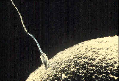 | ||
| A sperm cell attempts to penetrate an ovum coat to fertilize it. | ||
| Latin | spermiusmatro | |
| Gray's | subject #258 1243 | |
| System | ||
| MeSH | A05.360.490.890 | |
| Diagram of a human spermatozoon | ||
A spermatozoon (alternate spellings spermatozoan, spermatozoön; plural spermatozoa) is a motile sperm cell, or moving form of the haploid cell that is the male gamete. (A non-motile sperm cell is called a spermatium.) A spermatozoon joins an ovum to form a zygote. (A zygote is a single cell, with a complete set of chromosomes, that normally develops into an embryo.) The term spermatozoon comes from the ancient Greek word σπέρμα (seed) and ζῷον (living being).
Sperm cells contribute approximately half of the nuclear genetic information to the diploid offspring. In mammals, the sex of the offspring is determined by the sperm cell: a spermatozoon bearing a Y-chromosome will lead to a male (XY) offspring, while one bearing an X-chromosome will lead to a female (XX) offspring (the ovum always provides an X-chromosome). Sperm cells were first observed by Anton van Leeuwenhoek in 1677.[1]
Mammalian spermatozoan structure, function, and size
Electron micrograph of human spermatozoa magnified 3140 times.
Humans
The human sperm cell is the reproductive cell in males and will only survive in warm environments, once leaving the body the sperm's survival is reduced and may cause the cell to die, decreasing the sperm quality. Sperm cells come in two types, "female" and "male". Sperm cells that give rise to female (XX) offspring after fertilization differ in that they carry an X-chromosome, while sperm cells that give rise to male (XY) offspring carry a Y-chromosome.
In male humans, sperm cells consists of a head 5 µm by 3 µm and a tail 41 µm long. The tail flagellates, which propels the sperm cell (at about 1–3 mm/minute in humans) by whipping in an elliptical cone.[2] Semen has an alkaline nature, and they do not reach full motility (hypermotility) until they reach the vagina where the alkaline pH is neutralized by acidic vaginal fluids. This gradual process takes 20–30 minutes. In this time, fibrinogen from the seminal vesicles forms a clot, securing and protecting the sperm. Just as they become hypermotile, fibrinolysin from the prostate dissolves the clot, allowing the sperm to progress optimally.
The spermatozoon is characterized by a minimum of cytoplasm and the most densely packed DNA known in eukaryotes. Compared to mitotic chromosomes in somatic cells, sperm DNA is at least sixfold more highly condensed.[3]
The specimen contributes with DNA/chromatin, a centriole and perhaps also an oocyte-activating factor (OAF).[4] It may also contribute with paternal Messenger RNA (mRNA), also contributing to embryonic development.[4]
Avoidance of immune system response
Glycoprotein molecules on the surface of sperm cells are recognized by all human immune systems, and interpreted as a signal that the cell should not be rejected. The male immune system might otherwise attack sperm whilst in the testes, and the female immune system would attack sperm in the reproductive tract. The specific glycoproteins coating sperm cells are also utilized by some cancerous and bacterial cells, some parasitic worms, and HIV-infected white blood cells, thereby avoiding an immune response from the host organism.[5]
The blood-testis barrier, maintained by the tight junctions between the Sertoli cells of the seminiferous tubules, prevents communication between the forming spermatozoa and the blood vessels within the interstitial space. This gives the forming spermatozoa an immunological advantage and thus prevents them from eliciting an immune response. The blood-testis barrier is also important in preventing toxic substances from disrupting spermatogenesis.
Spermatozoa in other organisms
Motile sperm cells of algae and seedless plants.
- See also: Sperm
- See also: Female Sperm Storage
Animals
Fertilization relies on spermatozoa for most sexually reproductive animals.
The fruit fly has the largest known spermatozoon relative to its size.[6] Drosophila melanogaster produces sperm that can be up to 1.8 cm[7] in size. The incredibly long tail is thought to block other sperm from entering the egg. The entire sperm, tail included, gets incorporated into the oocyte cytoplasm.[8]
The wood mouse Apodemus slvaticus possesses spermatozoa with falciform morphology. What makes these gametocytes even more unique is the presence of an apical hook on the sperm head. This hook is used to attach to the hooks or to the flagella of other spermatozoa. Aggregation is caused by these attachments and mobile trains result. These trains provide improved motility in the female reproductive tract and are a means by which fertilization is promoted.[9]
Sea urchins such as Arbacia punctulata—are ideal organisms to use in sperm research, they spawn large numbers of sperm into the sea, making them well-suited as model organisms for experiments.
Spermatozoa production in mammals
- Main article: Spermatogenesis
Spermatozoa are produced in the seminiferous tubules of the testes in a process called spermatogenesis. Round cells called spermatogonia divide and differentiate eventually to become spermatozoa. During copulation the cloaca or vagina gets inseminated, and then the spermatozoa move through chemotaxis to the ovum inside a Fallopian tube or the uterus.
Spermatozoa activation
- Main article: Acrosome reaction
Acrosome reaction on a Sea Urchin cell
Approaching the egg cell is a rather complex, multistep process of chemotaxis guided by different chemical substances/stimuli on individual levels of phylogeny. One of the most significant, common signaling character of the event is that a prototype of professional chemotaxis receptors, formyl peptide receptor (60.000 receptor/cell) as well as the activator ability of its ligand formyl Met-Leu-Phe have been demonstrated in the surface membrane even in the case of human sperms.[10] Mammalian sperm cells become even more active when they approach an egg cell in a process called sperm activation. Sperm activation has been shown to be caused by calcium ionophores in vitro, progesterone released by nearby cumulus cells and binding to ZP3 of the zona pellucida. The cumulus cells are embedded in a gel-like substance made primarily of hyaluronic acid, and developed in the ovary with the egg and support it as it grows.
The initial change is called "hyperactivation", which causes a change in spermatozoa motility. They swim faster and their tail movements become more forceful and erratic.
A recent discovery links hyperactivation to a sudden influx of calcium ion into the tails. The whip-like tail (flagellum) of the sperm is studded with ion channels formed by proteins called CatSper. These channels are selective, allowing only calcium ion to pass. The opening of CatSper channels is responsible for the influx of calcium. The sudden rise in calcium levels causes the flagellum to form deeper bends, propelling the sperm more forcefully through the viscous environment. Sperm hyperactivity is necessary for breaking through two physical barriers that protect the egg from fertilization.
The second process in sperm activation is the acrosome reaction. This involves releasing the contents of the acrosome, which disperse, and the exposure of enzymes attached to the inner acrosomal membrane of the sperm. This occurs after the sperm first meets the egg. This lock-and-key type mechanism is species-specific and prevents the sperm and egg of different species from fusing. There is some evidence that this binding is what triggers the acrosome to release the enzymes that allow the sperm to fuse with the egg.
ZP3, one of the proteins that make up the zona pellucida, then binds to a partner molecule on the sperm. Enzymes on the inner acrosomal membrane digests the zona pellucida. After the sperm penetrates the zona pellucida, part of the sperm's cell membrane then fuses with the egg cell's membrane, and the contents of the head diffuse into the egg.
Upon penetration, the oocyte is said to have become activated. It undergoes its secondary meiotic division, and the two haploid nuclei (paternal and maternal) fuse to form a zygote. In order to prevent polyspermy and minimise the possibility of producing a triploid zygote, several changes to the egg's zona pellucida renders them impenetrable shortly after the first sperm enters the egg.
See also
References
- ↑ includeonly>"Timeline: Assisted reproduction and birth control", CBC News. Retrieved on 2006-04-06.
- ↑ Sumio Ishijima, Shigeru Oshio, Hideo Mohri, "Flagellar movement of human spermatozoa", Gamete research, 1986, vol. 13, no3, pp. 185–197 (27 ref.) [1]
- ↑ Ward WS, Coffey DS (1991). DNA packaging and organization in mammalian spermatozoa: comparison with somatic cells. Biol. Reprod. 44 (4): 569–74.
- ↑ 4.0 4.1 Developmental sperm contributions: fertilization and beyond Gerardo Barroso, M.D., M.Sc.a, Carlos Valdespin, M.D.a, Eva Vega, M.Sc.a, Ruben Kershenovich, M.D.a, Rosaura Avila, B.Sc.a, Conrado Avendaño, M.D.b, Sergio Oehninger, M.D., Ph.D.b. FertStert, Volume 92, Issue 3, Pages 835-848 (September 2009)
- ↑ includeonly>"Sperm clue to 'disease immunity'", BBC News, 2007-12-17.
- ↑ http://www.livescience.com/animalworld/060616_big_sperm.html
- ↑ Pitnick, S., Spicer, G. S., & Markow, T. A. (1995). How long is a giant sperm Nature, 375(6527), 109. doi:10.1038/375109a0
- ↑ Gilbert, Scott F., Developmental Biology, Eighth Edition. 2006. Sinauer Associates, pp. 254
- ↑ Moore, Harry et al., Exceptional sperm cooperation in Wood Mouse.Nature 418, 174-177 (2002)
- ↑ Gnessi L, Fabbri A, Silvestroni L, Moretti C, Fraioli F, Pert CB, Isidori A. (1986). Evidence for the presence of specific receptors for N-formyl chemotactic peptides on human spermatozoa.. J Clin Endocrinol Metab 63 (4): 841–6.
External links
Template:Commons cat
- The Handbook of Andrology
- Sperm hyperactivity
- Slower conception 'leads to boys'
- Human Sperm Under A Microscope
Male reproductive system
| |
|---|---|
| Scrotum | layers (skin, Dartos, External spermatic fascia, Cremaster, Internal spermatic fascia) • Perineal raphe • Spermatic cord |
| Testes | layers (Tunica vaginalis, Tunica albuginea) • Appendix • Mediastinum • Lobules • Septa • Leydig cell • Sertoli cell • Blood-testis barrier |
| Spermatogenesis | Spermatogonium • Spermatocytogenesis • Spermatocyte • Spermatidogenesis • Spermatid • Spermiogenesis • Spermatozoon |
| seminal tract | Seminiferous tubules (Tubuli seminiferi recti, Rete testis, Efferent ducts) • Epididymis (Appendix) • Vas deferens • Ejaculatory duct Seminal colliculus |
| urinary tract | Internal urethral orifice • Urethra (Prostatic, Intermediate, Spongy) • Urethral crest • Urethral gland • External urethral orifice |
| Penis | Corpus cavernosum • Corpus spongiosum • Navicular fossa of male urethra • Glans penis • Fundiform ligament • Suspensory ligament • Foreskin • Frenulum |
| accessory glands | Seminal vesicles (Excretory duct of seminal gland) • Prostate (Prostatic utricle, Prostatic sinus) • Bulbourethral glands |
| This page uses Creative Commons Licensed content from Wikipedia (view authors). |