No edit summary |
m (Reverted edits by 182.186.65.133 (talk | block) to last version by Dr Joe Kiff) |
||
| (12 intermediate revisions by 4 users not shown) | |||
| Line 1: | Line 1: | ||
{{BioPsy}} |
{{BioPsy}} |
||
| + | {{PsyPerspective}} |
||
| − | '''Skin''' is an [[organ (anatomy)|organ]] of the [[integumentary system]] made up of multiple layers of [[epithelial]] [[biological tissue|tissues]] that guard underlying [[muscle]]s and [[organ (anatomy)|organ]]s. As the interface with the surroundings, it plays the most important role in protecting against [[pathogen]]s. Its other main functions are [[Thermal insulation|insulation]] and [[temperature]] regulation, and [[vitamin D]] and [[Vitamin B|B]] synthesis. Skin is considered one of the most important parts of the body. |
||
| + | [[Image:Skin.jpg|thumb |300px| '''Skin''' layers: [[Epidermis (skin)|epidermis]], [[dermis]], and [[subcutis]], showing a [[hair follicle]], [[sweat gland]] & [[sebaceous gland]].]] |
||
| + | '''Skin''' or '''Epithelium''' is one of the four basic types of [[animal]] [[Tissue (biology)|tissue]], along with [[connective tissue]], [[muscle|muscle tissue]] and [[nervous tissue]]. Epithelial tissues line the [[body cavities|cavities]] and surfaces of structures throughout the body, and also form many [[glands]]. Functions of epithelial cells include secretion, selective absorption, protection, transcellular transport and detection of sensation. In Greek "Epi" means, "on, upon," and "Theli" meaning "tissue." |
||
| − | Skin has [[pigment]]ation, or [[melanin]], provided by [[melanocyte]]s, which absorb some of the potentially dangerous [[ultraviolet radiation]] in [[sunlight]]. It also contains [[DNA]] repair [[enzyme]]s which help to reverse UV damage, and people who lack the [[gene]]s for these enzymes suffer high rates of [[skin cancer]]. One form predominantly produced by UV light, [[malignant]] [[melanoma]], is particularly invasive, causing it to [[metastasize|spread]] quickly, and can often be deadly. Human skin pigmentation varies among populations in a striking manner. This has sometimes led to the classification of people(s) on the basis of [[human skin color|skin color]]. |
||
| + | Epithelial layers are [[avascular]], so they must receive nourishment via diffusion of substances from the underlying connective tissue, through the basement membrane. Epithelia can also be organized into clusters of cells that function as exocrine and endocrine glands. Exocrine and endocrine epithelial cells are highly vascular. |
||
| + | ==General structure== |
||
| − | [[Mammal]]ian skin often contains hairs, which in sufficient density is called [[fur]]. The [[hair]] mainly serves to augment the insulation the skin provides, but can also serve as a [[Secondary sex characteristic|secondary sexual characteristic]] or as [[camouflage]]. On some animals the skin is very hard and thick, and can be processed to create [[leather]]. [[Reptile]]s and [[fish]] have hard protective scales on their skin for protection, and [[bird]]s have hard feathers, all made of tough β-[[keratin]]s. [[Amphibian]] skin is not a strong barrier to passage of chemicals and is often subject to [[osmosis]]. A [[frog]] sitting in an [[anesthetic]] solution will quickly go to sleep. |
||
| + | Cells in epithelium are very densely packed together like bricks in a wall, leaving very little intercellular space. The cells form continuous sheets which are attached to each other at many locations by [[tight junctions]] and [[desmosomes]].<ref name="marieb-p103-104" /> The epithelial tissues cover the interior and exterior part of our skin. |
||
| + | ===Basement membrane=== |
||
| − | Damaged skin will try to heal by forming [[scar|scar tissue]], often giving rise to discoloration and depigmentation of the skin. |
||
| + | All epithelial cells rest on a [[basement membrane]], which acts as a [[scaffolding]] on which epithelium can grow and regenerate after injuries.<ref>{{Cite book|author=csxcasdfrg4y24q5qdwsedrMcConnell, Thomas H.|title=The nature of disease: pathology for the health professions|publisher=Lippincott Williams & Wilkins|year=2006|isbn=9780781753173|page=55|url=http://books.google.com/books?id=chs_lilPFLwC&pg=PA55}}</ref> Epithelial tissue is [[innervated]], but [[avascular]]. Thus epithelial tissue must be nourished by substances diffusing from the blood vessels in the underlying tissue. The basement membrane acts as a selectively permeable membrane that determines which substances will be able to enter the epithelium.<ref name="Eurell-2006-p18">{{cite book|editors=Eurell, Jo Ann C. et al.|title=Dellmann's textbook of veterinary histology|publisher=Wiley-Blackwell|year=2006|isbn=9780781741484|page=18|url=http://books.google.com/books?id=FnS4uiOlRT0C&pyg=PA18}}</ref><ref>Freshney, 2002: [http://books.google.com/books?id=KqKNxeWlU6MC&pg=PA3 p. 3]</ref> |
||
| + | ===Cell junctions=== |
||
| − | The skin is often known as "the largest organ of the human body". This applies to exterior surface, as it covers the body, ''appearing'' to have the largest surface area of all the organs. Moreover, it applies to weight, as it weighs more than any single internal organ, accounting for about 15 percent of body weight. For the average adult human, the skin has a surface area of between 1.5-2.0 square meters, most of it is between 2-3 mm thick. The average square inch of skin holds 650 sweat glands, 20 blood vessels, 60,000 melanocytes, and more than a thousand nerve endings. |
||
| + | [[Cell junctions]] are especially abundant in epithelial tissues. They consist of protein complexes and provide contact between neighbouring cells, between a cell and the extracellular matrix, or they build up the paracellular barrier of epithelia and control the [[paracellular transport]].{{citation needed|date=January 2011}} |
||
| + | Cell junctions are the contact points between plasma membrane and tissue cells. There are mainly 5 different types of cell junctions. They are tight junctions, adherens junctions, desmosomes, hemidesmosomes, and gap junctions. |
||
| − | The use of natural or synthetic [[cosmetics]] to treat the appearance of the face and condition of the skin (such as [[pore control]] and [[black head]] cleansing) is common among many cultures. |
||
| + | Tight junctions are a pair of trans-membranar protein fused on outer plasma membrane. |
||
| + | Adherens junctions are a plaque (protein layer on the inside plasma membrane) which attaches both protein and microfilaments. |
||
| + | Desmosomes attach to the microfilaments of cytoskeleton made up of keratin protein. |
||
| + | Hemidesmosomes resemble desmsomes on a section. They are made up of the integrin (a transmembraner protein) instead of cadherin. They attach the epithelial cell to the basement membrane. |
||
| + | Gap junctions connect the cytoplasm of two cells and are made up of proteins called connexins (six of which come together to make a connexon). |
||
| + | ==Classification of epithelial tissue== |
||
| ⚫ | |||
| − | [[ |
+ | [[File:Illu epithelium.jpg|thumb|350px|Types of epithelium]] |
| + | Tissues are generally classified by the morphology of their cells, and the number of layers they are composed of.<ref name="marieb-p103-104" /><ref name="Eurell-2006-p18" /><ref>{{cite book|author=Platzer, Werner|title=Color atlas of human anatomy: Locomotor system|publisher=Thieme|year=2008|isbn=9783135333069|page=8|url=http://books.google.com/books?id=T9bb4T422j8C&pg=PA8}}</ref> Epithelial tissue that is only one cell thick is known as [[simple epithelium]].<ref>van Lommel, 2002: [http://books.google.com/books?id=EvYjLNKLu9sC&pg=PA94 p. 94]</ref> If it is two or more cells thick, it is known as [[stratified epithelium]].<ref name="vanLommel-2002-p97">van Lommel, 2002: [http://books.google.com/books?id=EvYjLNKLu9sC&pg=PA97 p. 97]</ref> However, when taller simple epithelial cells (see [[#Simple columnar epithelium|columnar]], below) are viewed in cross section with several nuclei appearing at different heights, they can be confused with stratified epithelia. This kind of epithelium is therefore described as [[pseudostratified epithelium|"pseudostratified"]] epithelium.<ref>{{cite book|editors=Melfi, Rudy C. & Alley, Keith E.|title=Permar's oral embryology and microscopic anatomy: a textbook for students in dental hygiene|publisher=Lippincott Williams & Wilkins|year=2000|isbn=9780683306446|page=9|url=http://books.google.com/books?id=fGonan0UwhQC&pg=PA9}}</ref> |
||
| ⚫ | '''Skin''' is composed of three primary layers: the ''[[epidermis (skin)|epidermis]]'', which provides waterproofing and serves as a barrier to infection; the ''[[dermis]]'', which serves as a location for the [[appendages]] of skin; and the ''[[hypodermis]] (subcutaneous adipose layer)'' |
||
| + | ===Simple epithelium=== |
||
| ⚫ | The outermost epidermis consists of [[stratified]] [[squamous]] [[epithelium]] with an underlying connective tissue section, or dermis, and a hypodermis, or [[basement membrane]]. The epidermis contains no [[blood vessel]]s, and cells in the deepest layers are nourished by diffusion from blood capillaries extending to the upper layers of the dermis. The main type of cells which make up the epidermis are [[keratinocyte]]s, with [[melanocyte]]s and [[Langerhans cell]]s also present. The epidermis can be further subdivided into the following ''strata'' (beginning with the outermost layer): corneum, lucidum, granulosum, spinosum, basale. Cells are formed through [[mitosis]] at the basale layer. The daughter cells, (see [[cell division]]) move up the strata changing shape and composition as they die due to isolation from their blood source. The cytoplasm is released and the protein [[keratin]] is inserted. They eventually reach the corneum and slough off ([[desquamation]]). This process is called ''keratinization'' and takes place within about 30 days. This keratinized layer of skin is responsible for keeping water in the body and keeping other harmful chemicals and [[pathogens]] out, making skin a natural barrier to infection. |
||
| + | Simple epithelium is one cell thick, that is, every cell is in direct contact with the underlying [[basement membrane]]. It is generally found where absorption and filtration occur. The thinness of the epithelial barrier facilitates these processes.<ref name="marieb-p103-104" /> |
||
| + | Simple epithelial tissues are generally classified by the shape of their cells. The four major classes of simple epithelium are: (1) simple squamous; (2) simple cuboidal; (3) simple columnar; (4) pseudostratified.<ref name="marieb-p103-104">{{cite book|author=Marieb, Elaine M.|title=Human Anatomy and Physiology|edition=3rd|publisher=Benjamin/Cummings|year=1995|isbn=0-8053-4281-8|pages=103–104}}</ref> |
||
| ⚫ | |||
| + | (1) simple squamous; which is found lining areas where passive diffusion of gases occur. e.g walls of capillaries, linings of the pericardial, pleural,and peritoneal cavities, as well as the linings of the alveoli of the lungs. |
||
| − | The dermis lies below the epidermis and contains a number of structures including blood vessels, nerves, [[hair]] follicles, [[smooth muscle]], [[gland]]s and [[lymphatic tissue]]. It consists of loose connective tissue otherwise called areolar connective tissue - [[collagen]], [[elastin]] and [[reticular fiber]]s are present. Erector muscles, or [[arrectores pilorum]] attached between the hair papilla and epidermis, can contract, resulting in the hair fibre pulled upright and consequentially the formation of [[goose bumps]]. [[Sebaceous gland]]s are [[exocrine]] glands which produce [[sebum]], a mixture of lipids and waxy substances: lubrication, water-proofing, softening and antibactericidal actions are among the many functions of sebum. [[Sweat gland]]s open up via a duct onto the skin by a pore. |
||
| + | (2) simple cuboidal: these cells may have secretory, absorptive, or excretory functions. examples include small collecting ducts of kidney,pancreas and salivary gland. |
||
| − | The dermis can be split into the ''papillary'' and ''reticular'' layers. The papillary layer is outermost and extends into the epidermis to supply it with nutrients. It is composed of loosely arranged fibres. Papillary ridges make up the lines of the hands and feet, producing individually unique [[fingerprint]]s and foot prints. The reticular layer is more dense and is continuous with the hypodermis. It contains the bulk of the structures (such as sweat glands). The reticular layer is composed of irregularly arranged fibres and resists stretching. |
||
| + | (3) simple columnar; found in areas with extremely high secretive (as in wall of the stomach), or absorptive (as in small intestine) areas. they possess cellular extensions (e.g microvilli in the small intestine, or cilia found almost exclusively in the female reproductive tract). |
||
| ⚫ | The hypodermis is not part of the skin, and lies below the dermis. Its purpose is to attach the skin to underlying [[bone]] and [[muscle]] as well as supplying it with blood vessels and nerves. It consists of loose connective tissue and elastin. The main cell types are |
||
| + | (4) pseudostratified epithelia; they are also called respiratory epithelium. this is due to their almost exclusive confinement to the larger respiratory airways i.e the nasal cavity, trachea, bronchi e.t.c. |
||
| ⚫ | Microorganisms like ''Staphylococcus epidermidis'' colonize the skin surface |
||
| + | {{Table of simple epithelium types}} |
||
| + | |||
| + | ===Stratified epithelium=== |
||
| + | Stratified epithelium differs from simple epithelium in that it is multilayered. It is therefore found where body linings have to withstand mechanical or chemical insult such that layers can be abraded and lost without exposing subepithelial layers. Cells flatten as the layers become more apical, though in their most basal layers the cells can be squamous, cuboidal or columnar.{{citation needed|date=January 2011}} |
||
| + | |||
| + | Stratified epithelial tissue also differs from simple epithelial tissue in that stratified epithelial tissues do not contain junctional complexes, and have their cells bound together only by desmosomes.<ref name="vanLommel-2002-p97" /> |
||
| + | |||
| + | Stratified epithelia (of columnar, cuboidal or squamous type) can have the following specializations:{{citation needed|date=January 2011}} |
||
| + | |||
| + | {| class="wikitable" |
||
| + | |- |
||
| + | ! Specialization |
||
| + | ! Description |
||
| + | |- |
||
| + | | [[Keratin]]ized |
||
| + | | In this particular case, the most apical layers (exterior) of cells are dead and lose their nucleus and cytoplasm, instead contain a tough, resistant protein called keratin. This specialization makes the epithelium waterproof, so is found in the mammalian skin. The lining of the esophagus is an example of a non-keratinized or "moist" stratified epithelium.{{citation needed|date=January 2011}} |
||
| + | |- |
||
| + | | [[Transitional epithelium|Transitional]] |
||
| + | | Transitional epithelia are found in tissues that stretch and it can appear to be stratified cuboidal when the tissue is not stretched or stratified squamous when the organ is distended and the tissue stretches. It is sometimes called the urothelium since it is almost exclusively found in the bladder, ureters and urethra.{{Citation needed|date=May 2010}} |
||
| + | |- |
||
| + | |} |
||
==Functions== |
==Functions== |
||
| + | The primary functions of epithelial tissues are: (1) to protect the tissues that lie beneath it from [[radiation]], desiccation, [[toxins]], and physical [[Trauma (medicine)|trauma]]; (2) the regulation and exchange of chemicals between the underlying tissues and a [[body cavity]]; (3) the secretion of hormones into the blood vascular system, and/or (3) the secretion of sweat, mucus, enzymes, and other products that are delived by ducts [[glandular epithelium]].<ref>van Lommel, 2002: [http://books.google.com/books?id=EvYjLNKLu9sC&pg=PA91 p. 91]</ref> |
||
| + | |||
| + | ===Secretory epithelia=== |
||
| + | As stated above, secretion is one major function of epithelial cells. [[Glands]] are formed from the invagination / infolding of epithelial cells and subsequent growth in the underlying connective tissue. There are two major classifications of glands: [[endocrine glands]] and [[exocrine glands]]. Endocrine glands secrete their product into the extracellular space where it is rapidly taken up by the blood vascular system. the exocrine glands secrete their products into a duct that then delivers the product to the lumen of an organ or onto the free surface of the epithelium. |
||
| + | |||
| + | ==Functions== |
||
| + | Skin performs the following functions: |
||
| + | |||
#Protection: an anatomical barrier between the internal and external environment in bodily defense; [[Langerhans cell]]s in the skin are part of the [[adaptive immune system]] |
#Protection: an anatomical barrier between the internal and external environment in bodily defense; [[Langerhans cell]]s in the skin are part of the [[adaptive immune system]] |
||
| − | #[[Sensation]]: contains a variety of nerve endings that react to heat |
+ | #[[Sensation]]: contains a variety of nerve endings that react to [[thermoreceptor|heat and cold]], touch, pressure, vibration, and tissue injury; see [[somatosensory system]] and [[haptics]]. |
| − | #Heat regulation: the skin contains a blood supply far greater than its requirements which allows precise control of energy loss by radiation, convection and conduction. |
+ | #Heat regulation: the skin contains a blood supply far greater than its requirements which allows precise control of energy loss by radiation, convection and conduction. Dilated blood vessels increase perfusion and heat loss while constricted vessels greatly reduce cutaneous blood flow and conserve heat. [[Erector pili muscle]]s are significant in animals. |
| − | #Control of evaporation: |
+ | #Control of evaporation: the skin provides a relatively dry and impermeable barrier to fluid loss. Loss of this function contributes to the massive fluid loss in [[burn (injury)|burns]]. |
| − | #Aesthetics and communication: others see our skin and can assess |
+ | #Aesthetics and communication: others see our skin and can assess our mood, physical state and attractiveness. |
| − | #Storage and synthesis: acts as a storage |
+ | #Storage and synthesis: acts as a storage center for lipids and water, as well as a means of synthesis of [[vitamin D]] by action of [[UV]] on certain parts of the skin. |
| − | #Excretion: |
+ | #Excretion: [[sweat]] contains [[urea]], however its concentration is 1/130th that of [[urine]], hence [[excretion]] by sweating is at most a secondary function to temperature regulation. |
| − | #Absorption: Oxygen, nitrogen and carbon dioxide can diffuse into the epidermis in small amounts, some animals using their skin for their sole [[respiration organ]]. In addition, medicine can be administered through the skin, by ointments or by means of adhesive [[transdermal patch|patch]], such as the [[nicotine patch]] or [[iontophoresis]]. The skin is an important site of transport in many other organisms |
+ | #Absorption: Oxygen, nitrogen and carbon dioxide can diffuse into the epidermis in small amounts, some animals using their skin for their sole [[respiration organ]]. In addition, medicine can be administered through the skin, by ointments or by means of adhesive [[transdermal patch|patch]], such as the [[nicotine patch]] or [[iontophoresis]]. The skin is an important site of transport in many other organisms. |
==Hygiene== |
==Hygiene== |
||
| − | + | Unclean skin favors the development of pathogenic organisms – the dead cells that continually slough off of the epidermis mix with the secretions of the sweat and sebaceous glands and the dust found on the skin to form a filthy layer on its surface. If not washed away, the slurry of sweat and sebaceous secretions mixed with dirt and dead skin is decomposed by bacterial flora, producing a foul smell. Functions of the skin are disturbed when it is excessively dirty; it becomes more easily damaged, the release of antibacterial compounds decreases, and dirty skin is more prone to develop infections. Cosmetics should be used carefully because these may cause allergic reactions. Each season requires suitable clothing in order to facilitate the evaporation of the sweat. Sunlight, water and air play an important role in keeping the skin healthy. |
|
| − | The skin supports its own |
+ | The skin supports its own [[ecosystem]]s of [[microorganism]]s, including [[yeast]]s and [[bacteria]], which cannot be removed by any amount of cleaning. Estimates place the number of individual bacteria on the surface of one square inch (6.5 square cm) of human skin at 50 million though this figure varies greatly over the average 20 feet<sup>2</sup> (1.9 m²) of human skin. Oily surfaces, such as the face, may contain over 500 million bacteria per square inch (6.5 cm²). Despite these vast quantities, all of the bacteria found on the skin's surface would fit into a volume the size of a pea.<ref>Theodor Rosebury. ''Life on Man'': Secker & Warburg, 1969 ISBN 0-670-42793-4</ref> In general, the microorganisms keep one another in check and are part of a healthy skin. When the balance is disturbed, there may be an overgrowth and infection, such as when [[antibiotic]]s kill [[microbe]]s, resulting in an overgrowth of yeast. The skin is continuous with the inner [[epithelial]] lining of the body at the orifices, each of which supports its own complement of microbes. |
| + | Oily skin is caused by over-active glands, that produce a substance called [[sebum]], a naturally healthy skin lubricant.<ref name=Hcare/> When the skin produces excessive [[sebum]], it becomes heavy and thick in texture. Oily skin is typified by shininess, blemishes and [[pimples]].<ref name=Hcare/> The oily-skin type is not necessarily bad, since such skin is less prone to wrinkling, or other signs of aging,<ref name=Hcare/> because the oil helps to keep needed moisture locked into the [[Epidermis (skin)|epidermis]] (outermost layer of skin). |
||
| ⚫ | |||
| + | |||
| ⚫ | |||
| + | The negative aspect of the oily-skin type is that oily complexions are especially susceptible to clogged pores, [[blackhead]]s, and buildup of dead skin cells on the surface of the skin.<ref name=Hcare/> Oily skin can be sallow and rough in texture and tends to have large, clearly visible pores everywhere, except around the eyes and neck.<ref name=Hcare/> |
||
| + | |||
| + | The goal of treating oily skin is to remove excess surface sebum without complete removal of skin [[lipids]].<ref name=Hcare/> Severe degreasing treatment can foster an actual worsening of sebum secretion, which defeats the aim of the cleansing.<ref name=Hcare/> A method of cleansing oily skin is to wash with a solution of a mild synthetic [[detergent]]<ref name=Hcare/> (''see:'' [[surfactant]]) containing no oils, [[wax]]es or other lipid agents that could aggravate the oily condition of the skin, sometimes combined with a toning lotion. Such a product removes the oily residue and debris from the skin surface. Some cleansing products have lower concentrations of [[hydroxy acid]]s, which remove dead cells from the upper levels of the [[stratum corneum]].<ref name=Hcare/> Those products should be used on a regular basis to work adequately.<ref name=Hcare/> A light moisturizer may be included in a product to counteract any drying effects of the cleanser.<ref name=Hcare/> |
||
| + | |||
| ⚫ | |||
| ⚫ | |||
[[Image:Rash.jpg|thumb|right|250px|A typical rash]] |
[[Image:Rash.jpg|thumb|right|250px|A typical rash]] |
||
| + | [[Image:Scabies-burrow.jpg|thumb|right|200px|Skin infected with [[Scabies]]]] |
||
| − | As skin ages, it becomes thinner and more easily damaged. Intensifying this effect is the decreasing ability of skin to heal itself |
+ | As skin ages, it becomes thinner and more easily damaged. Intensifying this effect is the decreasing ability of skin to heal itself as a person ages. |
| + | Skin ageing is caused by the fall in elasticity. Ageing skin also receives less blood flow and lower gland activity. |
||
| ⚫ | |||
| + | <br/> |
||
| + | ==Disease == |
||
| − | The skin is subject to constant attack from without, and so can be afflicted by numerous ailments, such as these: |
||
| + | {{details|list of skin diseases}} |
||
| ⚫ | |||
'''Tumors''': |
'''Tumors''': |
||
| − | * Benign tumors of the skin |
+ | * Benign tumors of the skin such as [[Squamous cell papilloma]] |
* [[Skin cancer]] |
* [[Skin cancer]] |
||
| + | |||
'''Others''': |
'''Others''': |
||
* [[Rash]]es |
* [[Rash]]es |
||
| Line 60: | Line 111: | ||
* [[Acne vulgaris|Acne]] |
* [[Acne vulgaris|Acne]] |
||
* [[Keratosis pilaris]] |
* [[Keratosis pilaris]] |
||
| − | * [[fungus|Fungal]] infections such as [[athlete's foot]] |
+ | * [[fungus|Fungal]] infections such as [[athlete's foot]] and [[ringworm]] |
| − | * [[microbe| |
+ | * [[microbe|Microbial]] infections. |
| − | * [[ |
+ | * [[Calcinosis cutis]] |
| − | * [[ |
+ | * [[Sunburn]] |
| − | * [[ |
+ | * [[Keloid]] |
| − | * [[ |
+ | * [[Scabies]] |
| ⚫ | |||
| ⚫ | |||
| + | |||
| + | There are several other skin diseases as well. |
||
| + | |||
| + | ==Variability in skin tone== |
||
| + | |||
| + | Individuals with ancestors from different parts of the world can have highly visible differences in skin pigmentation. Individuals with [[Africa]]n ancestry ([[black people]]) tend towards darker skin, while those of [[Northern Europe]]an descent ([[white people]]) have paler skin. Between these extremes are individuals of [[Asia]]n, [[South-East Asia]]n, [[Native Americans in the United States|Native American]], [[Middle East]]ern, [[Polynesia]]n and [[Melanesia]]n descent. |
||
| + | |||
| + | The skin of [[black people]] has more variation in color from one part of the body to another than does the skin of other racial groups, particularly the palms of the hands and soles of the feet. Part of this is the result of the variations in the thickness of the skin or different parts of the body. The thicker the skin, the more layers of cell with melanin in them, and the darker the color.<ref>Smith, Wilma and Burns, Catherine. (1999) "Managing the hair and skin of African American pediatric patients." ''Journal of Pediatric Health Care'' 13(2):72-8.</ref> In addition, these parts of the body do not have melanin-producing cells. |
||
| + | |||
| + | Darker skin hinders UV A rays from penetrating. Since vitamin B folats are degraded by UV A and vitamin D is synthesised different skin tones are more likely to produce different vitamin deficiencies. |
||
| + | |||
| + | ==Animal skin products== |
||
| + | The term skin refers to the covering of a small animal, such as a sheep, goat ([[goatskin]]), pig, snake ([[snakeskin]]) etc or the young of a large animal. |
||
| + | |||
| + | The term [[hides]] or [[rawhide]] refers to the covering of a large adult animal such as a cow, buffalo, horse etc. |
||
| + | |||
| + | Skins and hides from different animals are used for clothing, bags and other consumer products, usually in the form of [[leather]], but also [[fur]]s. |
||
| + | |||
| + | Skin can also be used to make products such as [[gelatin]], [[animal glue|glue]] and [[wool]]. Mucus of skin from [[hagfish]] is under research. |
||
| + | |||
| + | ==Skin layers== |
||
| + | |||
| ⚫ | |||
| + | |||
| + | ===Epidermis=== |
||
| + | '''Epidermis''', "epi" coming from the Greek meaning "over" or "upon", is the outermost layer of the skin. It forms the waterproof, protective wrap over the body's surface and is made up of stratified squamous [[epithelium]] with an underlying [[basal lamina]]. |
||
| + | |||
| ⚫ | The outermost epidermis consists of [[stratified]] [[squamous]] [[epithelium]] with an underlying connective tissue section, or dermis, and a hypodermis, or [[basement membrane]]. The epidermis contains no [[blood vessel]]s, and cells in the deepest layers are nourished by diffusion from blood capillaries extending to the upper layers of the dermis. The main type of cells which make up the epidermis are [[keratinocyte]]s, with [[melanocyte]]s and [[Langerhans cell]]s also present. The epidermis can be further subdivided into the following ''strata'' (beginning with the outermost layer): corneum, lucidum (only in palms of hands and bottoms of feet), granulosum, spinosum, basale. Cells are formed through [[mitosis]] at the basale layer. The daughter cells, (see [[cell division]]) move up the strata changing shape and composition as they die due to isolation from their blood source. The cytoplasm is released and the protein [[keratin]] is inserted. They eventually reach the corneum and slough off ([[desquamation]]). This process is called ''keratinization'' and takes place within about 30 days. This keratinized layer of skin is responsible for keeping water in the body and keeping other harmful chemicals and [[pathogens]] out, making skin a natural barrier to infection. |
||
| + | |||
| + | [[Image:HautFingerspitzeOCT nonanimated.gif|thumb |209px| [''also see: [[:Image:HautFingerspitzeOCT.gif|image rotating (1.1 mb)]]'' ]<br/> |
||
| + | Optical Coherence Tomography tomogram of fingertip, depicting [[stratum corneum]] (~500µm thick) with stratum disjunctum on top and [[stratum lucidum]] (connection to stratum spinosum) in the middle. At the bottom superficial parts of the [[dermis]]. Sweatducts are clearly visible.]] |
||
| + | |||
| + | ====Components==== |
||
| + | The epidermis contains no [[blood vessel]]s, and is nourished by [[diffusion]] from the [[dermis]]. The main type of cells which make up the epidermis are [[keratinocyte]]s, [[melanocyte]]s, [[Langerhans cell]]s and [[Merkels cell]]s. |
||
| + | |||
| ⚫ | |||
| + | Epidermis is divided into several layers where cells are formed through [[mitosis]] at the innermost layers. They move up the strata changing shape and composition as they differentiate and become filled with [[keratin]]. They eventually reach the top layer called stratum corneum and become sloughed off, or desquamated. This process is called ''[[keratinization]]'' and takes place within weeks. The outermost layer of Epidermis consists of 25 to 30 layers of dead cells. |
||
| + | |||
| + | ====Sublayers==== |
||
| + | Epidermis is divided into the following 5 sublayers or strata: |
||
| + | |||
| + | *[[Stratum corneum]] |
||
| + | *[[Stratum lucidum]] |
||
| + | *[[Stratum granulosum]] |
||
| + | *[[Stratum spinosum]] |
||
| + | *[[Stratum germinativum]] (also called "stratum basale") |
||
| + | |||
| + | [[Mnemonic]]s that are good for remembering the layers of the skin (using "stratum basale" instead of "stratum germinativum"): |
||
| + | * "'''C'''her '''L'''ikes '''G'''etting '''S'''kin '''B'''otoxed" (from superficial to deep) |
||
| + | * "'''B'''efore '''S'''igning, '''G'''et '''L'''egal '''C'''ounsel" (from deep to superficial) |
||
| + | * "'''B'''efore '''S'''ex '''G'''et '''L'''atex '''C'''ondoms (from deep to superficial) |
||
| + | |||
| ⚫ | |||
| + | |||
| + | {{Infobox Anatomy | |
||
| + | Name = Dermis | |
||
| + | Latin = | |
||
| + | GraySubject = 234 | |
||
| + | GrayPage = 1065 | |
||
| + | Image = Gray942.png | |
||
| + | Caption = The distribution of the bloodvessels in the skin of the sole of the foot. (Corium - TA alternate term for dermis - is labeled at upper right.) | |
||
| + | Image2 = Gray940.png | |
||
| + | Width = 100 | <!--width of both(!) images --> |
||
| + | Caption2 = A diagrammatic sectional view of the skin (''click on image to magnify''). (Dermis labeled at center right.) | |
||
| + | System = | |
||
| + | Precursor = | |
||
| + | MeshName = Dermis | |
||
| + | MeshNumber = A17.815.180 | |
||
| + | DorlandsPre = d_11 | |
||
| + | DorlandsSuf = 12289496 | |
||
| + | }} |
||
| + | |||
| + | ===Dermis=== |
||
| + | |||
| + | The '''dermis''' is the layer of skin beneath the [[epidermis (skin)|epidermis]] that consists of [[connective tissue]] and cushions the body from stress and strain. The dermis is tightly connected to the epidermis by a [[basement membrane]]. It also harbors many nerve endings that provide the sense of touch and heat. It contains the [[hair follicles]], [[sweat glands]], [[sebaceous glands]], [[apocrine glands]], [[lymphatic vessels]] and [[blood vessels]]. The blood vessels in the dermis provide nourishment and waste removal to its own cells as well as the Stratum basale of the epidermis. |
||
| + | |||
| + | Structure |
||
| + | |||
| + | The dermis is structurally divided into two areas: a superficial area adjacent to the epidermis, called the ''papillary region'', and a deep thicker area known as the ''reticular region''. |
||
| + | |||
| + | ====Papillary region==== |
||
| + | The papillary region is composed of loose [[areolar connective tissue]]. It is named for its fingerlike projections called ''papillae'', that extend toward the epidermis. The papillae provide the dermis with a "bumpy" surface that interdigitates with the epidermis, strengthening the connection between the two layers of skin. |
||
| + | |||
| + | In the palms, fingers, soles, and toes, the influence of the papillae projecting into the epidermis forms contours in the skin's surface. These are called ''friction ridges'', because they help the hand or foot to grasp by increasing [[friction]]. Friction ridges occur in patterns (''see:'' [[fingerprint]]) that are genetically and [[epigenetic]]ally determined and are therefore unique to the individual, making it possible to use fingerprints or footprints as a means of [[identification]]. |
||
| + | |||
| + | ====Reticular region==== |
||
| + | The reticular region lies deep in the papillary region and is usually much thicker. It is composed of dense irregular connective tissue, and receives its name from the dense concentration of [[collagenous]], [[Elasticity (physics)|elastic]], and [[reticular]] fibers that weave throughout it. These [[protein]] fibers give the dermis its properties of strength, extensibility, and elasticity. |
||
| + | |||
| + | Also located within the reticular region are the [[roots of the hair]], [[sebaceous glands]], [[sweat glands]], receptors, [[nail (anatomy)|nails]], and [[blood vessels]]. |
||
| + | |||
| + | Tattoo ink is injected into the dermis. Stretch marks from pregnancy are also located in the dermis. |
||
| + | |||
| ⚫ | The hypodermis is not part of the skin, and lies below the dermis. Its purpose is to attach the skin to underlying [[bone]] and [[muscle]] as well as supplying it with blood vessels and nerves. It consists of loose connective tissue and elastin. The main cell types are [[fibroblast]]s, [[macrophage]]s and [[adipocyte]]s (the hypodermis contains 50% of body fat). Fat serves as padding and insulation for the body. |
||
| + | |||
| ⚫ | |||
==See also== |
==See also== |
||
| + | * [[Absorption (physiological)]] |
||
| ⚫ | |||
| ⚫ | |||
| ⚫ | |||
| + | * [[Cutaneous structure development]] |
||
| + | * [[List of skin diseases|Diseases]] - list of skin diseases |
||
| ⚫ | |||
| + | * [[Epithelial cells]] |
||
| + | * [[Hair]] - including [[hair follicles]] in skin |
||
| + | * [[Head (anatomy)]] |
||
* [[Human skin color|Skin color]] |
* [[Human skin color|Skin color]] |
||
| ⚫ | |||
| ⚫ | |||
| ⚫ | |||
* [[Meissner's corpuscle]] |
* [[Meissner's corpuscle]] |
||
| − | * [[Nail (anatomy)|Nails]] |
+ | * [[Nail (anatomy)|Nails]] - fingernails or toenails |
* [[Pacinian corpuscle]] |
* [[Pacinian corpuscle]] |
||
* [[Polyphenol antioxidant]] |
* [[Polyphenol antioxidant]] |
||
| − | * [[ |
+ | * [[Scalp (anatomy)]] |
| − | * [[ |
+ | * [[Skin disorders]] |
| + | * [[Skin electrical properties]] |
||
| ⚫ | |||
| + | * [[Skin potential]] |
||
| + | * [[Skin resistance]] |
||
| + | * [[Skin temperature]] |
||
| + | * [[Sweat]] - description of perspiration |
||
| + | * [[Superficial fascia]] |
||
| + | |||
| + | ==References== |
||
| + | {{reflist}} |
||
| + | |||
| + | ==External links== |
||
| + | |||
| − | {{wiktionary}} |
||
{{integumentary_system}} |
{{integumentary_system}} |
||
| + | |||
{{human anatomical features}} |
{{human anatomical features}} |
||
| + | |||
| ⚫ | |||
[[Category:Sensory organs]] |
[[Category:Sensory organs]] |
||
[[Category:Integumentary system]] |
[[Category:Integumentary system]] |
||
| − | [[Category: |
+ | [[Category:Dermatology]] |
| + | [[Category:Tissues (body)]] |
||
| − | |||
| ⚫ | |||
| − | |||
[[af:Vel]] |
[[af:Vel]] |
||
[[ar:جلد]] |
[[ar:جلد]] |
||
| + | [[ay:Janchi lip'ichi]] |
||
[[bg:Кожа]] |
[[bg:Кожа]] |
||
[[ca:Pell]] |
[[ca:Pell]] |
||
| Line 100: | Line 265: | ||
[[cy:Croen]] |
[[cy:Croen]] |
||
[[da:Hud]] |
[[da:Hud]] |
||
| − | [[de:Haut |
+ | [[de:Haut]] |
[[et:Nahk]] |
[[et:Nahk]] |
||
[[es:Piel]] |
[[es:Piel]] |
||
| Line 107: | Line 272: | ||
[[fr:Peau]] |
[[fr:Peau]] |
||
[[gl:Pel]] |
[[gl:Pel]] |
||
| + | [[xal:Арсн]] |
||
[[hr:Koža]] |
[[hr:Koža]] |
||
[[id:Kulit]] |
[[id:Kulit]] |
||
| + | [[is:Húð]] |
||
[[it:Pelle]] |
[[it:Pelle]] |
||
[[he:עור]] |
[[he:עור]] |
||
| + | [[pam:Balat]] |
||
| + | [[ka:კანი]] |
||
| + | [[la:Cutis]] |
||
| + | [[lv:Āda]] |
||
[[lt:Oda]] |
[[lt:Oda]] |
||
| + | [[ln:Lomposo]] |
||
| ⚫ | |||
| + | [[ms:Kulit]] |
||
[[nl:Huid]] |
[[nl:Huid]] |
||
[[ja:皮膚]] |
[[ja:皮膚]] |
||
[[no:Hud]] |
[[no:Hud]] |
||
[[nrm:Pé (la)]] |
[[nrm:Pé (la)]] |
||
| − | [[ |
+ | [[pag:Baog]] |
| − | [[pl:Skóra |
+ | [[pl:Skóra]] |
[[pt:Pele]] |
[[pt:Pele]] |
||
| + | [[ro:Piele (anatomie)]] |
||
| + | [[qu:Qara]] |
||
| + | [[ru:Шкура]] |
||
[[scn:Peddi]] |
[[scn:Peddi]] |
||
[[simple:Skin]] |
[[simple:Skin]] |
||
[[sk:Koža]] |
[[sk:Koža]] |
||
[[sl:Koža]] |
[[sl:Koža]] |
||
| ⚫ | |||
[[su:Kulit]] |
[[su:Kulit]] |
||
[[fi:Iho]] |
[[fi:Iho]] |
||
[[sv:Hud]] |
[[sv:Hud]] |
||
| + | [[th:ผิวหนัง]] |
||
| + | [[uk:Шкіра]] |
||
[[vi:Da]] |
[[vi:Da]] |
||
| − | [[ |
+ | [[yi:הויט]] |
[[zh:皮膚]] |
[[zh:皮膚]] |
||
| + | --> |
||
{{enWP|Skin}} |
{{enWP|Skin}} |
||
Latest revision as of 01:41, 21 December 2012
Assessment |
Biopsychology |
Comparative |
Cognitive |
Developmental |
Language |
Individual differences |
Personality |
Philosophy |
Social |
Methods |
Statistics |
Clinical |
Educational |
Industrial |
Professional items |
World psychology |
Biological: Behavioural genetics · Evolutionary psychology · Neuroanatomy · Neurochemistry · Neuroendocrinology · Neuroscience · Psychoneuroimmunology · Physiological Psychology · Psychopharmacology (Index, Outline)
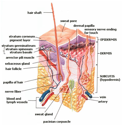
Skin layers: epidermis, dermis, and subcutis, showing a hair follicle, sweat gland & sebaceous gland.
Skin or Epithelium is one of the four basic types of animal tissue, along with connective tissue, muscle tissue and nervous tissue. Epithelial tissues line the cavities and surfaces of structures throughout the body, and also form many glands. Functions of epithelial cells include secretion, selective absorption, protection, transcellular transport and detection of sensation. In Greek "Epi" means, "on, upon," and "Theli" meaning "tissue." Epithelial layers are avascular, so they must receive nourishment via diffusion of substances from the underlying connective tissue, through the basement membrane. Epithelia can also be organized into clusters of cells that function as exocrine and endocrine glands. Exocrine and endocrine epithelial cells are highly vascular.
General structure
Cells in epithelium are very densely packed together like bricks in a wall, leaving very little intercellular space. The cells form continuous sheets which are attached to each other at many locations by tight junctions and desmosomes.[1] The epithelial tissues cover the interior and exterior part of our skin.
Basement membrane
All epithelial cells rest on a basement membrane, which acts as a scaffolding on which epithelium can grow and regenerate after injuries.[2] Epithelial tissue is innervated, but avascular. Thus epithelial tissue must be nourished by substances diffusing from the blood vessels in the underlying tissue. The basement membrane acts as a selectively permeable membrane that determines which substances will be able to enter the epithelium.[3][4]
Cell junctions
Cell junctions are especially abundant in epithelial tissues. They consist of protein complexes and provide contact between neighbouring cells, between a cell and the extracellular matrix, or they build up the paracellular barrier of epithelia and control the paracellular transport.[citation needed]
Cell junctions are the contact points between plasma membrane and tissue cells. There are mainly 5 different types of cell junctions. They are tight junctions, adherens junctions, desmosomes, hemidesmosomes, and gap junctions. Tight junctions are a pair of trans-membranar protein fused on outer plasma membrane. Adherens junctions are a plaque (protein layer on the inside plasma membrane) which attaches both protein and microfilaments. Desmosomes attach to the microfilaments of cytoskeleton made up of keratin protein. Hemidesmosomes resemble desmsomes on a section. They are made up of the integrin (a transmembraner protein) instead of cadherin. They attach the epithelial cell to the basement membrane. Gap junctions connect the cytoplasm of two cells and are made up of proteins called connexins (six of which come together to make a connexon).
Classification of epithelial tissue
Types of epithelium
Tissues are generally classified by the morphology of their cells, and the number of layers they are composed of.[1][3][5] Epithelial tissue that is only one cell thick is known as simple epithelium.[6] If it is two or more cells thick, it is known as stratified epithelium.[7] However, when taller simple epithelial cells (see columnar, below) are viewed in cross section with several nuclei appearing at different heights, they can be confused with stratified epithelia. This kind of epithelium is therefore described as "pseudostratified" epithelium.[8]
Simple epithelium
Simple epithelium is one cell thick, that is, every cell is in direct contact with the underlying basement membrane. It is generally found where absorption and filtration occur. The thinness of the epithelial barrier facilitates these processes.[1]
Simple epithelial tissues are generally classified by the shape of their cells. The four major classes of simple epithelium are: (1) simple squamous; (2) simple cuboidal; (3) simple columnar; (4) pseudostratified.[1]
(1) simple squamous; which is found lining areas where passive diffusion of gases occur. e.g walls of capillaries, linings of the pericardial, pleural,and peritoneal cavities, as well as the linings of the alveoli of the lungs.
(2) simple cuboidal: these cells may have secretory, absorptive, or excretory functions. examples include small collecting ducts of kidney,pancreas and salivary gland.
(3) simple columnar; found in areas with extremely high secretive (as in wall of the stomach), or absorptive (as in small intestine) areas. they possess cellular extensions (e.g microvilli in the small intestine, or cilia found almost exclusively in the female reproductive tract).
(4) pseudostratified epithelia; they are also called respiratory epithelium. this is due to their almost exclusive confinement to the larger respiratory airways i.e the nasal cavity, trachea, bronchi e.t.c. Template:Table of simple epithelium types
Stratified epithelium
Stratified epithelium differs from simple epithelium in that it is multilayered. It is therefore found where body linings have to withstand mechanical or chemical insult such that layers can be abraded and lost without exposing subepithelial layers. Cells flatten as the layers become more apical, though in their most basal layers the cells can be squamous, cuboidal or columnar.[citation needed]
Stratified epithelial tissue also differs from simple epithelial tissue in that stratified epithelial tissues do not contain junctional complexes, and have their cells bound together only by desmosomes.[7]
Stratified epithelia (of columnar, cuboidal or squamous type) can have the following specializations:[citation needed]
| Specialization | Description |
|---|---|
| Keratinized | In this particular case, the most apical layers (exterior) of cells are dead and lose their nucleus and cytoplasm, instead contain a tough, resistant protein called keratin. This specialization makes the epithelium waterproof, so is found in the mammalian skin. The lining of the esophagus is an example of a non-keratinized or "moist" stratified epithelium.[citation needed] |
| Transitional | Transitional epithelia are found in tissues that stretch and it can appear to be stratified cuboidal when the tissue is not stretched or stratified squamous when the organ is distended and the tissue stretches. It is sometimes called the urothelium since it is almost exclusively found in the bladder, ureters and urethra.[citation needed] |
Functions
The primary functions of epithelial tissues are: (1) to protect the tissues that lie beneath it from radiation, desiccation, toxins, and physical trauma; (2) the regulation and exchange of chemicals between the underlying tissues and a body cavity; (3) the secretion of hormones into the blood vascular system, and/or (3) the secretion of sweat, mucus, enzymes, and other products that are delived by ducts glandular epithelium.[9]
Secretory epithelia
As stated above, secretion is one major function of epithelial cells. Glands are formed from the invagination / infolding of epithelial cells and subsequent growth in the underlying connective tissue. There are two major classifications of glands: endocrine glands and exocrine glands. Endocrine glands secrete their product into the extracellular space where it is rapidly taken up by the blood vascular system. the exocrine glands secrete their products into a duct that then delivers the product to the lumen of an organ or onto the free surface of the epithelium.
Functions
Skin performs the following functions:
- Protection: an anatomical barrier between the internal and external environment in bodily defense; Langerhans cells in the skin are part of the adaptive immune system
- Sensation: contains a variety of nerve endings that react to heat and cold, touch, pressure, vibration, and tissue injury; see somatosensory system and haptics.
- Heat regulation: the skin contains a blood supply far greater than its requirements which allows precise control of energy loss by radiation, convection and conduction. Dilated blood vessels increase perfusion and heat loss while constricted vessels greatly reduce cutaneous blood flow and conserve heat. Erector pili muscles are significant in animals.
- Control of evaporation: the skin provides a relatively dry and impermeable barrier to fluid loss. Loss of this function contributes to the massive fluid loss in burns.
- Aesthetics and communication: others see our skin and can assess our mood, physical state and attractiveness.
- Storage and synthesis: acts as a storage center for lipids and water, as well as a means of synthesis of vitamin D by action of UV on certain parts of the skin.
- Excretion: sweat contains urea, however its concentration is 1/130th that of urine, hence excretion by sweating is at most a secondary function to temperature regulation.
- Absorption: Oxygen, nitrogen and carbon dioxide can diffuse into the epidermis in small amounts, some animals using their skin for their sole respiration organ. In addition, medicine can be administered through the skin, by ointments or by means of adhesive patch, such as the nicotine patch or iontophoresis. The skin is an important site of transport in many other organisms.
Hygiene
Unclean skin favors the development of pathogenic organisms – the dead cells that continually slough off of the epidermis mix with the secretions of the sweat and sebaceous glands and the dust found on the skin to form a filthy layer on its surface. If not washed away, the slurry of sweat and sebaceous secretions mixed with dirt and dead skin is decomposed by bacterial flora, producing a foul smell. Functions of the skin are disturbed when it is excessively dirty; it becomes more easily damaged, the release of antibacterial compounds decreases, and dirty skin is more prone to develop infections. Cosmetics should be used carefully because these may cause allergic reactions. Each season requires suitable clothing in order to facilitate the evaporation of the sweat. Sunlight, water and air play an important role in keeping the skin healthy.
The skin supports its own ecosystems of microorganisms, including yeasts and bacteria, which cannot be removed by any amount of cleaning. Estimates place the number of individual bacteria on the surface of one square inch (6.5 square cm) of human skin at 50 million though this figure varies greatly over the average 20 feet2 (1.9 m²) of human skin. Oily surfaces, such as the face, may contain over 500 million bacteria per square inch (6.5 cm²). Despite these vast quantities, all of the bacteria found on the skin's surface would fit into a volume the size of a pea.[10] In general, the microorganisms keep one another in check and are part of a healthy skin. When the balance is disturbed, there may be an overgrowth and infection, such as when antibiotics kill microbes, resulting in an overgrowth of yeast. The skin is continuous with the inner epithelial lining of the body at the orifices, each of which supports its own complement of microbes.
Oily skin is caused by over-active glands, that produce a substance called sebum, a naturally healthy skin lubricant.[11] When the skin produces excessive sebum, it becomes heavy and thick in texture. Oily skin is typified by shininess, blemishes and pimples.[11] The oily-skin type is not necessarily bad, since such skin is less prone to wrinkling, or other signs of aging,[11] because the oil helps to keep needed moisture locked into the epidermis (outermost layer of skin).
The negative aspect of the oily-skin type is that oily complexions are especially susceptible to clogged pores, blackheads, and buildup of dead skin cells on the surface of the skin.[11] Oily skin can be sallow and rough in texture and tends to have large, clearly visible pores everywhere, except around the eyes and neck.[11]
The goal of treating oily skin is to remove excess surface sebum without complete removal of skin lipids.[11] Severe degreasing treatment can foster an actual worsening of sebum secretion, which defeats the aim of the cleansing.[11] A method of cleansing oily skin is to wash with a solution of a mild synthetic detergent[11] (see: surfactant) containing no oils, waxes or other lipid agents that could aggravate the oily condition of the skin, sometimes combined with a toning lotion. Such a product removes the oily residue and debris from the skin surface. Some cleansing products have lower concentrations of hydroxy acids, which remove dead cells from the upper levels of the stratum corneum.[11] Those products should be used on a regular basis to work adequately.[11] A light moisturizer may be included in a product to counteract any drying effects of the cleanser.[11]
Aging
- For more details on this topic, see senescence.

A typical rash
Skin infected with Scabies
As skin ages, it becomes thinner and more easily damaged. Intensifying this effect is the decreasing ability of skin to heal itself as a person ages.
Skin ageing is caused by the fall in elasticity. Ageing skin also receives less blood flow and lower gland activity.
Disease
- For more details on this topic, see list of skin diseases.
In medicine, the branch concerned with the skin is called dermatology. The skin is subject to constant attack from without, and so can be afflicted by numerous ailments, such as these:
Tumors:
- Benign tumors of the skin such as Squamous cell papilloma
- Skin cancer
Others:
- Rashes
- Blisters
- Acne
- Keratosis pilaris
- Fungal infections such as athlete's foot and ringworm
- Microbial infections.
- Calcinosis cutis
- Sunburn
- Keloid
- Scabies
- Vitiligo
- Albinism
There are several other skin diseases as well.
Variability in skin tone
Individuals with ancestors from different parts of the world can have highly visible differences in skin pigmentation. Individuals with African ancestry (black people) tend towards darker skin, while those of Northern European descent (white people) have paler skin. Between these extremes are individuals of Asian, South-East Asian, Native American, Middle Eastern, Polynesian and Melanesian descent.
The skin of black people has more variation in color from one part of the body to another than does the skin of other racial groups, particularly the palms of the hands and soles of the feet. Part of this is the result of the variations in the thickness of the skin or different parts of the body. The thicker the skin, the more layers of cell with melanin in them, and the darker the color.[12] In addition, these parts of the body do not have melanin-producing cells.
Darker skin hinders UV A rays from penetrating. Since vitamin B folats are degraded by UV A and vitamin D is synthesised different skin tones are more likely to produce different vitamin deficiencies.
Animal skin products
The term skin refers to the covering of a small animal, such as a sheep, goat (goatskin), pig, snake (snakeskin) etc or the young of a large animal.
The term hides or rawhide refers to the covering of a large adult animal such as a cow, buffalo, horse etc.
Skins and hides from different animals are used for clothing, bags and other consumer products, usually in the form of leather, but also furs.
Skin can also be used to make products such as gelatin, glue and wool. Mucus of skin from hagfish is under research.
Skin layers
Skin is composed of three primary layers: the epidermis, which provides waterproofing and serves as a barrier to infection; the dermis, which serves as a location for the appendages of skin; and the hypodermis (subcutaneous adipose layer).
Epidermis
Epidermis, "epi" coming from the Greek meaning "over" or "upon", is the outermost layer of the skin. It forms the waterproof, protective wrap over the body's surface and is made up of stratified squamous epithelium with an underlying basal lamina.
The outermost epidermis consists of stratified squamous epithelium with an underlying connective tissue section, or dermis, and a hypodermis, or basement membrane. The epidermis contains no blood vessels, and cells in the deepest layers are nourished by diffusion from blood capillaries extending to the upper layers of the dermis. The main type of cells which make up the epidermis are keratinocytes, with melanocytes and Langerhans cells also present. The epidermis can be further subdivided into the following strata (beginning with the outermost layer): corneum, lucidum (only in palms of hands and bottoms of feet), granulosum, spinosum, basale. Cells are formed through mitosis at the basale layer. The daughter cells, (see cell division) move up the strata changing shape and composition as they die due to isolation from their blood source. The cytoplasm is released and the protein keratin is inserted. They eventually reach the corneum and slough off (desquamation). This process is called keratinization and takes place within about 30 days. This keratinized layer of skin is responsible for keeping water in the body and keeping other harmful chemicals and pathogens out, making skin a natural barrier to infection.
[also see: image rotating (1.1 mb) ]
Optical Coherence Tomography tomogram of fingertip, depicting stratum corneum (~500µm thick) with stratum disjunctum on top and stratum lucidum (connection to stratum spinosum) in the middle. At the bottom superficial parts of the dermis. Sweatducts are clearly visible.
Components
The epidermis contains no blood vessels, and is nourished by diffusion from the dermis. The main type of cells which make up the epidermis are keratinocytes, melanocytes, Langerhans cells and Merkels cells.
Layers
Epidermis is divided into several layers where cells are formed through mitosis at the innermost layers. They move up the strata changing shape and composition as they differentiate and become filled with keratin. They eventually reach the top layer called stratum corneum and become sloughed off, or desquamated. This process is called keratinization and takes place within weeks. The outermost layer of Epidermis consists of 25 to 30 layers of dead cells.
Sublayers
Epidermis is divided into the following 5 sublayers or strata:
- Stratum corneum
- Stratum lucidum
- Stratum granulosum
- Stratum spinosum
- Stratum germinativum (also called "stratum basale")
Mnemonics that are good for remembering the layers of the skin (using "stratum basale" instead of "stratum germinativum"):
- "Cher Likes Getting Skin Botoxed" (from superficial to deep)
- "Before Signing, Get Legal Counsel" (from deep to superficial)
- "Before Sex Get Latex Condoms (from deep to superficial)
Blood capillaries are found beneath the epidermis, and are linked to an arteriole and a venule. Arterial shunt vessels may bypass the network in ears, the nose and fingertips.
| Dermis | ||
|---|---|---|
| The distribution of the bloodvessels in the skin of the sole of the foot. (Corium - TA alternate term for dermis - is labeled at upper right.) | ||
| Latin | ' | |
| Gray's | subject #234 1065 | |
| System | ||
| MeSH | A17.815.180 | |
 | ||
| A diagrammatic sectional view of the skin (click on image to magnify). (Dermis labeled at center right.) | ||
Dermis
The dermis is the layer of skin beneath the epidermis that consists of connective tissue and cushions the body from stress and strain. The dermis is tightly connected to the epidermis by a basement membrane. It also harbors many nerve endings that provide the sense of touch and heat. It contains the hair follicles, sweat glands, sebaceous glands, apocrine glands, lymphatic vessels and blood vessels. The blood vessels in the dermis provide nourishment and waste removal to its own cells as well as the Stratum basale of the epidermis.
Structure
The dermis is structurally divided into two areas: a superficial area adjacent to the epidermis, called the papillary region, and a deep thicker area known as the reticular region.
Papillary region
The papillary region is composed of loose areolar connective tissue. It is named for its fingerlike projections called papillae, that extend toward the epidermis. The papillae provide the dermis with a "bumpy" surface that interdigitates with the epidermis, strengthening the connection between the two layers of skin.
In the palms, fingers, soles, and toes, the influence of the papillae projecting into the epidermis forms contours in the skin's surface. These are called friction ridges, because they help the hand or foot to grasp by increasing friction. Friction ridges occur in patterns (see: fingerprint) that are genetically and epigenetically determined and are therefore unique to the individual, making it possible to use fingerprints or footprints as a means of identification.
Reticular region
The reticular region lies deep in the papillary region and is usually much thicker. It is composed of dense irregular connective tissue, and receives its name from the dense concentration of collagenous, elastic, and reticular fibers that weave throughout it. These protein fibers give the dermis its properties of strength, extensibility, and elasticity.
Also located within the reticular region are the roots of the hair, sebaceous glands, sweat glands, receptors, nails, and blood vessels.
Tattoo ink is injected into the dermis. Stretch marks from pregnancy are also located in the dermis.
The hypodermis is not part of the skin, and lies below the dermis. Its purpose is to attach the skin to underlying bone and muscle as well as supplying it with blood vessels and nerves. It consists of loose connective tissue and elastin. The main cell types are fibroblasts, macrophages and adipocytes (the hypodermis contains 50% of body fat). Fat serves as padding and insulation for the body.
Microorganisms like Staphylococcus epidermidis colonize the skin surface. The density of skin flora depends on region of the skin. The disinfected skin surface gets recolonized from bacteria residing in the deeper areas of the hair follicle, gut and urogenital openings.
See also
- Absorption (physiological)
- Cosmetics and cosmetic surgery and cosmetic techniques
- Cutaneous structure development
- Diseases - list of skin diseases
- Dermatology - branch of medicine
- Epithelial cells
- Hair - including hair follicles in skin
- Head (anatomy)
- Skin color
- Hyperpigmentation - about excess skin color
- Meissner's corpuscle
- Nails - fingernails or toenails
- Pacinian corpuscle
- Polyphenol antioxidant
- Scalp (anatomy)
- Skin disorders
- Skin electrical properties
- Skin potential
- Skin resistance
- Skin temperature
- Sweat - description of perspiration
- Superficial fascia
References
- ↑ 1.0 1.1 1.2 1.3 Marieb, Elaine M. (1995). Human Anatomy and Physiology, 3rd, 103–104, Benjamin/Cummings.
- ↑ csxcasdfrg4y24q5qdwsedrMcConnell, Thomas H. (2006). The nature of disease: pathology for the health professions, Lippincott Williams & Wilkins.
- ↑ 3.0 3.1 (2006) Dellmann's textbook of veterinary histology, Wiley-Blackwell.
- ↑ Freshney, 2002: p. 3
- ↑ Platzer, Werner (2008). Color atlas of human anatomy: Locomotor system, Thieme.
- ↑ van Lommel, 2002: p. 94
- ↑ 7.0 7.1 van Lommel, 2002: p. 97
- ↑ (2000) Permar's oral embryology and microscopic anatomy: a textbook for students in dental hygiene, Lippincott Williams & Wilkins.
- ↑ van Lommel, 2002: p. 91
- ↑ Theodor Rosebury. Life on Man: Secker & Warburg, 1969 ISBN 0-670-42793-4
- ↑ 11.00 11.01 11.02 11.03 11.04 11.05 11.06 11.07 11.08 11.09 11.10 Cite error: Invalid
<ref>tag; no text was provided for refs namedHcare - ↑ Smith, Wilma and Burns, Catherine. (1999) "Managing the hair and skin of African American pediatric patients." Journal of Pediatric Health Care 13(2):72-8.
External links
Integumentary system | |
|---|---|
| Structures |
Skin • Sweat glands • Sebaceous glands • Hair (Hair follicle) • Nails • Scale |
| Layers |
Cutis: Epidermis (Stratum corneum, Stratum lucidum, Stratum granulosum, Stratum spinosum, Stratum germinativum/basale) • Dermis |
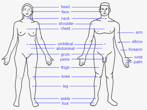 |
HEAD: Forehead – Eye – Ear – Nose – Mouth – Tongue – Teeth – Jaw – Face – Cheek – Chin TORSO: Shoulders – Spine – Chest – Breast – Ribcage – Abdomen – Belly button LIMBS: Arm – Elbow – Forearm – Wrist – Hand – Finger (Thumb - Index finger - Middle finger - Ring finger - Little finger) – Leg – Lap – Thigh – Knee – Calf – Heel – Ankle – Foot – Toe (Hallux) |
| This page uses Creative Commons Licensed content from Wikipedia (view authors). |