m (Rod cells moved to Rods (eye): Align thesaurus) |
(update) |
||
| Line 1: | Line 1: | ||
{{BioPsy}} |
{{BioPsy}} |
||
| + | {{Infobox_neuron |
||
| − | __NOTOC__ |
||
| + | |neuron_name = Rod cell |
||
| − | [[Image:Cone-response.png|right|thumb|360px|Normalised absoption spectra of human rod (R) and cone (S,M,L) cells. Note that the wavelength scale is not represented linearly.]] |
||
| + | |image_neuron = Fig_retine.png |
||
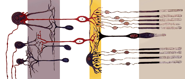
| + | |caption_neuron = Cross section of the [[retina]]. Rods are visible at far right. |
||
| + | |location = [[Retina]] |
||
| + | |function = Low light [[Photoreceptor cell|photoreceptor]] |
||
| + | |neurotranmitter = [[Glutamate]] |
||
| + | |morphology = rod shaped |
||
| + | |afferents = None |
||
| + | |efferents = [[Bipolar cell of the retina|Bipolar Cells]] and [[Horizontal cells]] |
||
| + | |NeuroLex = Rod Cell |
||
| + | |NeuroLexID = sao1458938856 |
||
| + | }} |
||
| ⚫ | '''Rod cells''', or '''rods''', are [[photoreceptor cell]]s in the [[retina]] of the [[eye]] that can function in less intense [[light]] than can the other type of photoreceptor, [[cone cell]]s. Because they are more light sensitive, rods are responsible for night vision. Named for their cylindrical shape, rods are concentrated at the outer edges of the retina and are used in [[peripheral vision]]. There are about 120 million rod cells in the human retina. |
||
| ⚫ | A rod cell is sensitive enough to respond to a single [[photon]] of light{{Fact|date=April 2009}}, and is about 100 times more sensitive to a single photon than cones. Because rods require less light to function than cones, they are therefore the primary source of visual information at night ([[scotopic vision]]). Cone cells, on the other hand, require tens to hundreds of photons to become activated. Additionally, multiple rod cells converge on a single [[interneuron]], collecting and amplifying the signals. However, this convergence comes at a cost to visual acuity (or [[image resolution]]) because the pooled information from multiple cells is less distinct than it would be if the [[visual system]] received information from each rod cell individually. The convergence of rod cells also tends to make peripheral vision very sensitive to movement, and is responsible for the phenomenon of an individual seeing something vague occur out of the corner of his or her eye. |
||
| ⚫ | '''Rod cells''', or '''rods''', are [[photoreceptor cell]]s in the [[retina]] of the [[eye]] that can function in less intense [[light]] than can the other type of photoreceptor, [[cone |
||
| + | Rods are a little narrower than cones but have the same structural basis. The pigment is on the outer side, lying on the pigment epithelium. This end contains many stacked disks, probably from the folding inward of the limiting membrane surrounding this section, allowing a higher area for visual pigment and increasing the efficiency of light absorption. Because they have only one type of light-sensitive pigment, rather than the three types that human cone cells have, rods have little, if any, role in [[color]] vision. |
||
| ⚫ | A rod cell is sensitive enough to respond to a single [[photon]] of light |
||
| − | Rod cells also respond more slowly to light than |
+ | Rod cells also respond more slowly to light than cones do, so stimuli they receive are added over about 100 milliseconds. While this makes rods more sensitive to smaller amounts of light, it also means that their ability to sense temporal changes, such as quickly changing images, is less accurate than that of cones.<ref name="Kandel"> Kandel E.R., Schwartz, J.H., Jessell, T.M. (2000). ''Principles of Neural Science'', 4th ed., pp.507-513. McGraw-Hill, New York. </ref> |
| − | Experiments by [[George Wald]] and others showed that rods are |
+ | Experiments by [[George Wald]] and others showed that rods are most sensitive to wavelengths of light around 498 nm (green-blue), and are completely insensitive to wavelengths longer than about 640 nm (red). This fact is responsible for the [[Purkinje effect]], in which blue colors appear more intense relative to reds at twilight, when rods take over as the cells responsible for vision. |
| ⚫ | Like cones, rod cells have a synaptic terminal, an inner segment, and an outer segment. The synaptic terminal forms a [[synapse]] with another neuron, for example a [[bipolar cell]]. The inner and outer segments are connected by a [[cilium]].<ref name="Kandel"> Kandel E.R., Schwartz, J.H., Jessell, T.M. (2000). ''Principles of Neural Science'', 4th ed., pp.507-513. McGraw-Hill, New York. </ref> The inner segment contains [[organelle]]s and the cell's [[nucleus (cell)|nucleus]], while the rod outer segment (abbreviated to ROS), which is pointed toward the back of the eye, contains the light-absorbing materials.<ref name="Kandel"> Kandel E.R., Schwartz, J.H., Jessell, T.M. (2000). ''Principles of Neural Science'', 4th ed., pp.507-513. McGraw-Hill, New York. </ref> |
||
| − | Because they have only one type of light sensitive pigment (rather than the three types that human cone cells have), rods have little, if any, role in [[color]] vision. |
||
| − | |||
| ⚫ | Like cones, rod cells have a synaptic terminal, an inner segment, and an outer segment. The synaptic terminal forms a [[synapse]] with another neuron, for example a [[bipolar cell]]. The inner and outer segments are connected by a [[cilium]] |
||
==Response to light== |
==Response to light== |
||
| + | [[Image:Rod&Cone.jpg|thumb|right|200px|Anatomy of a Rod Cell<ref>Human Physiology and Mechanisms of Disease by Arthur C. Guyton (1992) p.373</ref>]] |
||
| − | Activation of a photoreceptor cell is actually a [[hyperpolarization]]; when they are not being stimulated, rods and cones [[depolarization|depolarize]] and release a [[neurotransmitter]] spontaneously, and activation of photopigments by light sends a signal by preventing this. Depolarization occurs due to the fact that in the dark, cells have a relatively high concentration of [[cyclic guanosine 3'-5' monophosphate]] (cGMP), which opens [[ion channel]]s (largely [[sodium channel]]s, though [[Calcium]] can enter through these channels as well). The positive charges of the [[ions]] that enter the cell down its [[electrochemical gradient]] change the cell's [[membrane potential]], cause depolarization, and lead to the release of the neurotransmitter [[glutamate]]. Glutamate can depolarize some neurons and hyperpolarize others, allowing photoreceptors to interact in an antagonistic manner. |
||
| + | |||
| ⚫ | Activation of a single molecule of [[rhodopsin]], the photosensitive pigment in rods, can lead to a large reaction in the cell because the signal is amplified. Once activated, rhodopsin can activate hundreds of transducin molecules, each of which in turn activate a phosphodiesterase molecule, which can break down over a thousand cGMP molecules per second.<ref name="Kandel"> Kandel E.R., Schwartz, J.H., Jessell, T.M. (2000). ''Principles of Neural Science'', 4th ed., pp.507-513. McGraw-Hill, New York. </ref> Thus rods can have a large response to a small amount of light. |
||
| + | |||
| + | As the retinal component of rhodopsin is derived from vitamin A, a deficiency of vitamin A causes a deficit in the pigment needed by rod cells. Consequently, fewer rod cells are able to sufficiently respond in darker conditions, and as the cone cells are poorly adapted for sight in the dark, blindness can result. This is night-blindness. |
||
| + | |||
| + | ===Revert to the resting state=== |
||
| + | |||
| + | Rods make use of three inhibitory mechanisms (negative feedback mechanisms) to allow a rapid revert to the resting state after a flash of light. |
||
| + | |||
| + | Firstly, there exists a rhodopsin kinase (RK) which would phosphorylate the cytosolic tail of the activated rhodopsin on the multiple serines, partially inhibiting the activation of transducin. Also, an inhibitory protein - arrestin then binds to the phosphorylated rhodopsins to further inhibit the rhodopsin's activity. |
||
| + | |||
| + | While arrestin shuts off rhodopsin, an RGS protein (functioning as a GTPase-activiating proteins(GAPs)) drives the transducin (G-protein) into an "off" state by increasing the rate of hydrolysis of the bounded GTP to GDP. |
||
| + | |||
| + | Also as the cGMP sensitive channels allow not only the influx of sodium ions, but also calcium ions, with the decrease in concentration of cGMP, cGMP sensitive channels are then closed and reducing the normal influx of calcium ions. The decrease in the concentration of calcium ions stimulates the calcium ion-sensitive proteins, which would then activiate the guanylyl cyclase to replenish the cGMP, rapidly restoring its original concentration. The restoration opens the cGMP sensitive channels and causes a depolarization of the plasma membrane.<ref name="Alberts"> Bruce Alberts, Alexander Johnson, Julian Lewis, Martin Raff, Keith Roberts, Peter Walter (2008). Molecular Biology of The Cell'', 5th ed., pp.919-921. Garland Science. </ref> |
||
| + | |||
| + | ===Desensitization=== |
||
| + | When the rods are exposed to a high concentration of photons for a prolonged period, they become desensitized (adapted) to the environment. |
||
| − | When light hits photoreceptive pigments within the photoreceptor cell, the pigment changes shape. This causes it to activate a regulatory protein called [[transducin]], which leads to the activation of [[cGMP phosphodiesterase]], which breaks cGMP down into 5'-GMP. Reduction in cGMP allows the ion channels to close, preventing the influx of positive ions, hyperpolarizing the cell, and stopping the release of neurotransmitters (Kandel et al., 2000). |
||
| + | As rhodopsin is phosphorylated by rhodopsin kinase (a member of the GPCR kinases(GRKs)), it binds with high affinity to the arrestin. The bound arrestin can contribute to the desensitization process in at least two ways. First, it prevents the interaction between the G protein and the activated receptor. Second, it serves as an adaptor protein to aid the receptor to the clathrin-dependent endocytosis machinery (to induce receptor-mediated endocytosis).<ref name="Alberts"> Bruce Alberts, Alexander Johnson, Julian Lewis, Martin Raff, Keith Roberts, Peter Walter (2008). Molecular Biology of The Cell'', 5th ed., pp.919-921. Garland Science.</ref> |
||
| ⚫ | Activation of a single molecule of rhodopsin, the photosensitive pigment in rods, can lead to a large reaction in the cell because the signal is amplified. |
||
===Table=== |
===Table=== |
||
| Line 65: | Line 91: | ||
==Reference== |
==Reference== |
||
| + | <references/> |
||
* Kandel E.R., Schwartz, J.H., Jessell, T.M. (2000). ''Principles of Neural Science'', 4th ed., pp.507-513. McGraw-Hill, New York. |
* Kandel E.R., Schwartz, J.H., Jessell, T.M. (2000). ''Principles of Neural Science'', 4th ed., pp.507-513. McGraw-Hill, New York. |
||
| Line 73: | Line 100: | ||
{{Retina}} |
{{Retina}} |
||
| − | [[Category: |
+ | [[Category:Photoreceptors]] |
| − | [[Category: |
+ | [[Category:Retina]] |
| − | [[Category:Photoreceptor cells]] |
||
| + | <!-- |
||
[[da:Stav (synet)]] |
[[da:Stav (synet)]] |
||
[[de:Stäbchen (Auge)]] |
[[de:Stäbchen (Auge)]] |
||
| Line 82: | Line 109: | ||
[[nl:Staafje]] |
[[nl:Staafje]] |
||
[[pl:Pręcik (medycyna)]] |
[[pl:Pręcik (medycyna)]] |
||
| + | --> |
||
{{enWP|Rod cell}} |
{{enWP|Rod cell}} |
||
Latest revision as of 08:01, 23 May 2009
Assessment |
Biopsychology |
Comparative |
Cognitive |
Developmental |
Language |
Individual differences |
Personality |
Philosophy |
Social |
Methods |
Statistics |
Clinical |
Educational |
Industrial |
Professional items |
World psychology |
Biological: Behavioural genetics · Evolutionary psychology · Neuroanatomy · Neurochemistry · Neuroendocrinology · Neuroscience · Psychoneuroimmunology · Physiological Psychology · Psychopharmacology (Index, Outline)
| Rod cell | |
|---|---|
 | |
| Location | Retina |
| Function | Low light photoreceptor |
| Morphology | rod shaped |
| Presynaptic connections | None |
| Postsynaptic connections | Bipolar Cells and Horizontal cells |
Rod cells, or rods, are photoreceptor cells in the retina of the eye that can function in less intense light than can the other type of photoreceptor, cone cells. Because they are more light sensitive, rods are responsible for night vision. Named for their cylindrical shape, rods are concentrated at the outer edges of the retina and are used in peripheral vision. There are about 120 million rod cells in the human retina.
A rod cell is sensitive enough to respond to a single photon of light[How to reference and link to summary or text], and is about 100 times more sensitive to a single photon than cones. Because rods require less light to function than cones, they are therefore the primary source of visual information at night (scotopic vision). Cone cells, on the other hand, require tens to hundreds of photons to become activated. Additionally, multiple rod cells converge on a single interneuron, collecting and amplifying the signals. However, this convergence comes at a cost to visual acuity (or image resolution) because the pooled information from multiple cells is less distinct than it would be if the visual system received information from each rod cell individually. The convergence of rod cells also tends to make peripheral vision very sensitive to movement, and is responsible for the phenomenon of an individual seeing something vague occur out of the corner of his or her eye.
Rods are a little narrower than cones but have the same structural basis. The pigment is on the outer side, lying on the pigment epithelium. This end contains many stacked disks, probably from the folding inward of the limiting membrane surrounding this section, allowing a higher area for visual pigment and increasing the efficiency of light absorption. Because they have only one type of light-sensitive pigment, rather than the three types that human cone cells have, rods have little, if any, role in color vision.
Rod cells also respond more slowly to light than cones do, so stimuli they receive are added over about 100 milliseconds. While this makes rods more sensitive to smaller amounts of light, it also means that their ability to sense temporal changes, such as quickly changing images, is less accurate than that of cones.[1]
Experiments by George Wald and others showed that rods are most sensitive to wavelengths of light around 498 nm (green-blue), and are completely insensitive to wavelengths longer than about 640 nm (red). This fact is responsible for the Purkinje effect, in which blue colors appear more intense relative to reds at twilight, when rods take over as the cells responsible for vision.
Like cones, rod cells have a synaptic terminal, an inner segment, and an outer segment. The synaptic terminal forms a synapse with another neuron, for example a bipolar cell. The inner and outer segments are connected by a cilium.[1] The inner segment contains organelles and the cell's nucleus, while the rod outer segment (abbreviated to ROS), which is pointed toward the back of the eye, contains the light-absorbing materials.[1]
Response to light
Anatomy of a Rod Cell[2]
Activation of a single molecule of rhodopsin, the photosensitive pigment in rods, can lead to a large reaction in the cell because the signal is amplified. Once activated, rhodopsin can activate hundreds of transducin molecules, each of which in turn activate a phosphodiesterase molecule, which can break down over a thousand cGMP molecules per second.[1] Thus rods can have a large response to a small amount of light.
As the retinal component of rhodopsin is derived from vitamin A, a deficiency of vitamin A causes a deficit in the pigment needed by rod cells. Consequently, fewer rod cells are able to sufficiently respond in darker conditions, and as the cone cells are poorly adapted for sight in the dark, blindness can result. This is night-blindness.
Revert to the resting state
Rods make use of three inhibitory mechanisms (negative feedback mechanisms) to allow a rapid revert to the resting state after a flash of light.
Firstly, there exists a rhodopsin kinase (RK) which would phosphorylate the cytosolic tail of the activated rhodopsin on the multiple serines, partially inhibiting the activation of transducin. Also, an inhibitory protein - arrestin then binds to the phosphorylated rhodopsins to further inhibit the rhodopsin's activity.
While arrestin shuts off rhodopsin, an RGS protein (functioning as a GTPase-activiating proteins(GAPs)) drives the transducin (G-protein) into an "off" state by increasing the rate of hydrolysis of the bounded GTP to GDP.
Also as the cGMP sensitive channels allow not only the influx of sodium ions, but also calcium ions, with the decrease in concentration of cGMP, cGMP sensitive channels are then closed and reducing the normal influx of calcium ions. The decrease in the concentration of calcium ions stimulates the calcium ion-sensitive proteins, which would then activiate the guanylyl cyclase to replenish the cGMP, rapidly restoring its original concentration. The restoration opens the cGMP sensitive channels and causes a depolarization of the plasma membrane.[3]
Desensitization
When the rods are exposed to a high concentration of photons for a prolonged period, they become desensitized (adapted) to the environment.
As rhodopsin is phosphorylated by rhodopsin kinase (a member of the GPCR kinases(GRKs)), it binds with high affinity to the arrestin. The bound arrestin can contribute to the desensitization process in at least two ways. First, it prevents the interaction between the G protein and the activated receptor. Second, it serves as an adaptor protein to aid the receptor to the clathrin-dependent endocytosis machinery (to induce receptor-mediated endocytosis).[3]
Table
Comparison of rod and cone cells, from Kandel et al. (2000).
| Rods | Cones |
|---|---|
| used for night vision | used for day vision |
| very light sensitive; sensitive to scattered light | not very light sensitive; sensitive only to direct light |
| loss causes night blindness | loss causes legal blindness |
| low visual acuity | high visual acuity; better spacial resolution |
| not present in fovea | concentrated in fovea |
| slow response, to light, stimuli added over time | fast response to light, can perceive more rapid changes in stimuli |
| have more pigment than cones, so can detect less light | have less pigment than rods, require more light to detect images |
| stacks of membrane-enclosed disks are unattached to cell membrane | disks are attached to outer membrane |
| 20 times more rods than cones in the retina | |
| one type of photosensitive pigment | three types of photosensitive pigment in humans |
| confer achromatic vision | confer color vision |
Reference
- ↑ 1.0 1.1 1.2 1.3 Kandel E.R., Schwartz, J.H., Jessell, T.M. (2000). Principles of Neural Science, 4th ed., pp.507-513. McGraw-Hill, New York.
- ↑ Human Physiology and Mechanisms of Disease by Arthur C. Guyton (1992) p.373
- ↑ 3.0 3.1 Bruce Alberts, Alexander Johnson, Julian Lewis, Martin Raff, Keith Roberts, Peter Walter (2008). Molecular Biology of The Cell, 5th ed., pp.919-921. Garland Science. Cite error: Invalid
<ref>tag; name "Alberts" defined multiple times with different content
- Kandel E.R., Schwartz, J.H., Jessell, T.M. (2000). Principles of Neural Science, 4th ed., pp.507-513. McGraw-Hill, New York.
See also
| Sensory system - Visual system - Eye - Retina - edit |
|---|
| Photoreceptor cells (Cone cell, Rod cell) → (Horizontal cell) → Bipolar cell → (Amacrine cell) → Ganglion cell |
| This page uses Creative Commons Licensed content from Wikipedia (view authors). |