(New page: {{Biopsy}} {{Infobox Muscle | Name = Psoas major muscle | Latin = m. psoas major | GraySubject = 127 | GrayPage = 467 | Image = Anterior Hip Muscles 2....) |
|||
| Line 53: | Line 53: | ||
*[[Iliacus]] |
*[[Iliacus]] |
||
*[[Hip flexor]] |
*[[Hip flexor]] |
||
| + | *[[Locomotion]] |
||
*[[Psoas minor muscle]] |
*[[Psoas minor muscle]] |
||
| − | *[[Snapping hip syndrome| Iliopsoas tendinitis]] |
||
== References == |
== References == |
||
Latest revision as of 12:12, 2 April 2009
Assessment |
Biopsychology |
Comparative |
Cognitive |
Developmental |
Language |
Individual differences |
Personality |
Philosophy |
Social |
Methods |
Statistics |
Clinical |
Educational |
Industrial |
Professional items |
World psychology |
Biological: Behavioural genetics · Evolutionary psychology · Neuroanatomy · Neurochemistry · Neuroendocrinology · Neuroscience · Psychoneuroimmunology · Physiological Psychology · Psychopharmacology (Index, Outline)
| Psoas major muscle | ||
|---|---|---|
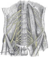
| The psoas major and nearby muscles | ||
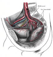
| Horizontal disposition of the peritoneum in the lower part of the abdomen. (Psoas major labeled at bottom left.) | ||
| Latin | m. psoas major | |
| Gray's | subject #127 467 | |
| Origin: | Transverse processes of T12-L5 and the lateral aspects of the discs between them | |
| Insertion: | in the lesser trochanter of the femur | |
| Blood: | lumbar branch of iliolumbar artery | |
| Nerve: | Lumbar plexus via anterior branches of L2-L4 nerves | |
| Action: | flexes and rotates laterally thigh | |
| Antagonist: | Gluteus maximus | |
| MeSH | A02.633.567.825 | |
| Dorlands/Elsevier | m_22/12550274 | |
The Psoas major is a long fusiform muscle placed on the side of the lumbar region of the vertebral column and brim of the lesser pelvis.
Location
Origin
It arises:
- (1) from the anterior surfaces of the bases and lower borders of the transverse processes of all the lumbar vertebrae
- (2) from the sides of the bodies and the corresponding intervertebral fibrocartilages of the last thoracic and all the lumbar vertebrae by five slips, each of which is attached to the adjacent upper and lower margins of two vertebrae, and to the intervertebral fibrocartilage;
- (3) from a series of tendinous arches which extend across the constricted parts of the bodies of the lumbar vertebrae between the previous slips; the lumbar arteries and veins, and filaments from the sympathetic trunk pass beneath these tendinous arches.
Insertion
The muscle proceeds downward across the brim of the lesser pelvis, and diminishing gradually in size, passes beneath the inguinal ligament and in front of the capsule of the hip-joint and ends in a tendon; the tendon receives nearly the whole of the fibers of the Iliacus and is inserted into the lesser trochanter of the femur.
A large bursa which may communicate with the cavity of the hip-joint, separates the tendon from the pubis and the capsule of the joint.
Function
It forms part of a group of muscles called the hip flexors, whose action is primarily to lift the upper leg towards the body when the body is fixed or to pull the body towards the leg when the leg is fixed.
For example, when doing a sit up that brings the torso (including the lower back) away from the ground and towards the front of the leg, the hip flexors (including the iliopsoas) will flex the spine upon the pelvis.
Indications
Crepitus, or “popping” in the lower back can indicate unequal lateral forces on the lumbar spine owing to a strength and/or laxity discrepancy between the right and left psoas major muscles.[1] Tight hip flexors (psoas, illiacus, tensor fascia latae), either unilaterally or bilaterally (one side or both sides), are a common though unsung cause of lower back pain, and can be accompanied by Iliopsoas tendinitis, or “snapping hip syndrome.” Physical therapists may recommend stretching and/or strengthening of one of both psoas muscles if lower back pain is indicated.
Stretching the psoas can be difficult because it is impossible to isolate the muscle, and thus any stretch thereof must necessarily stretch all hip flexor muscles and typically the quadriceps as well. Strengthening the psoas is also tricky, but typically involves some variation of supine leg raises, or, in a gym setting, knee raises on a levered machine. A common evaluation of psoas strength is to ask the patient to stand straight up with back against a wall, feet 2-3 inches from the wall. The patient is then instructed to raise the knee as high (close to the chest as possible) for 30 seconds. If the patient’s knee falls below horizontal (femur below 90 degrees) before 30 seconds has elapsed, weakness of the iliopsoas on the side of the knee being raised is indicated.
The Psoas is the major leg mover of gait (walking). Stretching and Chiropractic adjustments to the femur, pelvus and lumbars is often indicated in correcting the psoas imbalance and resolving low back pain.
See also
- Iliopsoas
- Iliacus
- Hip flexor
- Locomotion
- Psoas minor muscle
References
- ↑ Rolf, 1977: Rolfing, the Integration of Human Structures, pg. 118
Additional images
External links
- GPnotebook -1261436848
- LUC psmj
- SUNY Labs 40:16-0101 - "Posterior Abdominal Wall: Muscles of the Posterior Abdominal Wall"
- State University of New York (SUNY) Anatomy Image 8916
- Cross section at UV pelvis/pelvis-e12-2
This article was originally based on an entry from a public domain edition of Gray's Anatomy. As such, some of the information contained herein may be outdated. Please edit the article if this is the case, and feel free to remove this notice when it is no longer relevant.
Template:Muscles of lower limb
| This page uses Creative Commons Licensed content from Wikipedia (view authors). |


