(update wp) |
No edit summary |
||
| Line 18: | Line 18: | ||
}} |
}} |
||
| − | The '''pons''' (sometimes '''pons Varolii''' |
+ | The '''pons''' (sometimes '''pons Varolii''') is part of the [[brainstem]] that links the [[medulla oblongata]] and the [[thalamus]].<ref>O.D.E. 2005</ref> In full it is ''pons Varolii'' named using the Latin word for "bridge" and after the Italian anatomist and surgeon [[Costanzo Varolio]] (1543–75).<ref name="Gray1862">{{cite book|author=Henry Gray|title=Anatomy, descriptive and surgical|url=http://books.google.com/books?id=btSyie_UtxAC&pg=PA514|accessdate=10 November 2010|year=1862|publisher=Blanchard and Lea|pages=514–}}</ref> It is [[Anatomical terms of location|cranial]] to the medulla oblongata, [[caudal (anatomical term)|caudal]] to the [[midbrain]], and [[ventral]] to the [[cerebellum]]. In [[human]]s and other [[biped]]s, this means it is above the medulla, below the midbrain, and [[anterior]] to the cerebellum. This [[white matter]] includes tracts that conduct signals from the [[cerebrum]] down to the cerebellum and medulla, and tracts that carry the sensory signals up into the thalamus.<ref name="Saladin Kenneth S.2007">Saladin Kenneth S.(2007)</ref> |
| + | |||
| − | |||
| + | The pons in humans measures about 2.5 cm or 1 inch in length. Most of it appears as a broad anterior bulge [[Anatomical_terms_of_location#Other_directional_terms|rostral]] to the medulla. Posteriorly, it consists mainly of two pairs of thick stalks called [[Superior cerebellar peduncle|cerebellar peduncle]]s. They connect the cerebellum to the pons and midbrain.<ref name="Saladin Kenneth S.2007" /> |
||
| + | |||
==Function== |
==Function== |
||
| + | The pons contains [[Nucleus (neuroanatomy)|nuclei]] that relay signals from the forebrain to the cerebellum, along with nuclei that deal primarily with [[sleep]], [[respiration]], [[swallowing]], [[bladder control]], [[hearing]], [[equilibrium]], [[taste]], [[eye movement]], [[facial expressions]], facial sensation, and [[posture]].<ref name="Saladin Kenneth S.2007"/>. |
||
| ⚫ | |||
| + | |||
| ⚫ | |||
| + | |||
| + | Within the pons is the [[pneumotaxic center]], a nucleus that regulates the change from inspiration to expiration.<ref name="Saladin Kenneth S.2007"/> |
||
| + | |||
| + | The pons is implicated in [[sleep paralysis]], and also plays a role in generating dreams.{{Citation needed|date=March 2012}} |
||
==Anatomy of the pons== |
==Anatomy of the pons== |
||
| Line 41: | Line 49: | ||
* lower down in the pons: [[facial nerve nucleus]] (VII) |
* lower down in the pons: [[facial nerve nucleus]] (VII) |
||
* lower down in the pons: [[Vestibulocochlear nerve|vestibulocochlear]] nuclei ([[vestibular nuclei]] and [[cochlear nuclei]]) (VIII) |
* lower down in the pons: [[Vestibulocochlear nerve|vestibulocochlear]] nuclei ([[vestibular nuclei]] and [[cochlear nuclei]]) (VIII) |
||
| + | |||
| + | ==Embryonic development== |
||
| + | During embryonic development, the embryonic [[metencephalon]] develops from the [[rhombencephalon]] and gives rise to two structures: the pons and the cerebellum.<ref name="Saladin Kenneth S.2007"/> The [[alar plate]] produces sensory [[neuroblast]]s, which will give rise to the [[solitary nucleus]] and its [[special visceral afferent]] (SVA) column; the [[Cochlear nuclei|cochlear]] and [[vestibular nuclei]], which form the [[special somatic afferent]] (SSA) fibers of the [[vestibulocochlear nerve]], the spinal and principal [[trigeminal nerve nuclei]], which form the [[General somatic afferent fibers|general somatic afferent column]] (GSA) of the [[trigeminal nerve]], and the [[pontine nuclei]] which relays to the [[cerebellum]]. |
||
| + | |||
| + | [[Basal plate (neural tube)|Basal plate]] neuroblasts give rise to the [[abducens nucleus]],which forms the [[general somatic efferent fibers]] (GSE); the facial and motor trigeminal nuclei, which form the [[special visceral efferent]] (SVE) column, and the [[superior salivatory nucleus]], which forms the [[general visceral efferent fibers]] of the [[facial nerve]]. |
||
| + | |||
| + | ==Evolution== |
||
| + | |||
| + | The pons first evolved as an offshoot of the medullary [[reticular formation]].<ref name=Pritchard>[[#refPritchard|Pritchard and Alloway ''Medical Neuroscience'']]</ref> Since lampreys possess a pons, it has been argued that it must have evolved as a region distinct from the [[medulla oblongata|medulla]] by the time the first [[agnathans]] appeared, 505 million years ago.<ref name=Butler>[[#refButler|Butler and Hodos ''Comparative vertebrate neuroanatomy: evolution and adaptation'']]</ref> |
||
==Related diseases== |
==Related diseases== |
||
| Line 46: | Line 63: | ||
==See also== |
==See also== |
||
| + | *[[Parabrachial area]] |
||
*[[Raphe nuclei]] |
*[[Raphe nuclei]] |
||
| + | |||
==Additional images== |
==Additional images== |
||
| Line 81: | Line 100: | ||
{{Rhombencephalon}} |
{{Rhombencephalon}} |
||
[[Category:Brainstem]] |
[[Category:Brainstem]] |
||
| + | [[Category:pons]] |
||
[[Category:Neuroanatomy]] |
[[Category:Neuroanatomy]] |
||
Latest revision as of 12:55, 25 October 2013
Assessment |
Biopsychology |
Comparative |
Cognitive |
Developmental |
Language |
Individual differences |
Personality |
Philosophy |
Social |
Methods |
Statistics |
Clinical |
Educational |
Industrial |
Professional items |
World psychology |
Biological: Behavioural genetics · Evolutionary psychology · Neuroanatomy · Neurochemistry · Neuroendocrinology · Neuroscience · Psychoneuroimmunology · Physiological Psychology · Psychopharmacology (Index, Outline)
| Brain: Pons | ||
|---|---|---|
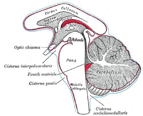 | ||
| Diagram showing the positions of the three principal subarachnoid cisternæ. (Pons visible at center.) | ||
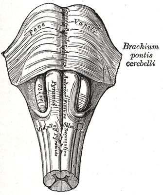 | ||
| Anteroinferior view of the medulla oblongata and pons. | ||
| Latin | {{{Latin}}} | |
| Gray's | subject #187 785 | |
| Part of | Brain stem | |
| Components | ||
| Artery | pontine arteries | |
| Vein | transverse and lateral pontine veins | |
| BrainInfo/UW | hier-538 | |
| MeSH | A08.186.211.132.810.428.600 | |
The pons (sometimes pons Varolii) is part of the brainstem that links the medulla oblongata and the thalamus.[1] In full it is pons Varolii named using the Latin word for "bridge" and after the Italian anatomist and surgeon Costanzo Varolio (1543–75).[2] It is cranial to the medulla oblongata, caudal to the midbrain, and ventral to the cerebellum. In humans and other bipeds, this means it is above the medulla, below the midbrain, and anterior to the cerebellum. This white matter includes tracts that conduct signals from the cerebrum down to the cerebellum and medulla, and tracts that carry the sensory signals up into the thalamus.[3]
The pons in humans measures about 2.5 cm or 1 inch in length. Most of it appears as a broad anterior bulge rostral to the medulla. Posteriorly, it consists mainly of two pairs of thick stalks called cerebellar peduncles. They connect the cerebellum to the pons and midbrain.[3]
Function
The pons contains nuclei that relay signals from the forebrain to the cerebellum, along with nuclei that deal primarily with sleep, respiration, swallowing, bladder control, hearing, equilibrium, taste, eye movement, facial expressions, facial sensation, and posture.[3].
In some theories, the pon has a role in dreaming.[4]
Within the pons is the pneumotaxic center, a nucleus that regulates the change from inspiration to expiration.[3]
The pons is implicated in sleep paralysis, and also plays a role in generating dreams.[citation needed]
Anatomy of the pons
The "knob-like" process (Basal pons) is 2 centimeters long and located on the anterior (front) of the brainstem. It is formed of nerves that travel from one side (left or right) to the other. Most other fibres in the brainstem travel up and down.
The posterior (back) surface of the pons forms part of the wall of the fourth ventricle of the brain.
Most blood to the pons is supplied by pontine arteries. These are small arteries that branch off the basilar artery of the Circle of Willis. Blood also comes from the anterior inferior, and superior cerebellar arteries.
There are two main domains in the pons for control of respiration:[5]
- the apneustic center - lower pons
- the pneumotaxic center - upper pons
Cranial nerve nuclei
A number of cranial nerve nuclei are present in the pons:
- mid-pons: The chief or pontine nucleus of the trigeminal nerve sensory nucleus (V)
- mid-pons: the motor nucleus for the trigeminal nerve (V)
- lower down in the pons: abducens nucleus (VI)
- lower down in the pons: facial nerve nucleus (VII)
- lower down in the pons: vestibulocochlear nuclei (vestibular nuclei and cochlear nuclei) (VIII)
Embryonic development
During embryonic development, the embryonic metencephalon develops from the rhombencephalon and gives rise to two structures: the pons and the cerebellum.[3] The alar plate produces sensory neuroblasts, which will give rise to the solitary nucleus and its special visceral afferent (SVA) column; the cochlear and vestibular nuclei, which form the special somatic afferent (SSA) fibers of the vestibulocochlear nerve, the spinal and principal trigeminal nerve nuclei, which form the general somatic afferent column (GSA) of the trigeminal nerve, and the pontine nuclei which relays to the cerebellum.
Basal plate neuroblasts give rise to the abducens nucleus,which forms the general somatic efferent fibers (GSE); the facial and motor trigeminal nuclei, which form the special visceral efferent (SVE) column, and the superior salivatory nucleus, which forms the general visceral efferent fibers of the facial nerve.
Evolution
The pons first evolved as an offshoot of the medullary reticular formation.[6] Since lampreys possess a pons, it has been argued that it must have evolved as a region distinct from the medulla by the time the first agnathans appeared, 505 million years ago.[7]
Related diseases
- Central pontine myelinosis, a demyelination disease that causes difficulty with sense of balance, walking, sense of touch, swallowing and speaking to mention just a few symptoms. In a clinical setting it is often associated with transplant. Undiagnosed it can lead to death or 'locked in' syndrome.
See also
Additional images
")
")
")
")
")
")
")
")
")
")
")
")
")
")
")
")
References
- ↑ O.D.E. 2005
- ↑ Henry Gray (1862). Anatomy, descriptive and surgical, 514–, Blanchard and Lea. URL accessed 10 November 2010.
- ↑ 3.0 3.1 3.2 3.3 3.4 Saladin Kenneth S.(2007)
- ↑ The "Science of Dreaming" in Neurontic: Psychology for the Modern Mind..
- ↑ Physiology at MCG 4/4ch6/s4ch6_10
- ↑ Pritchard and Alloway Medical Neuroscience
- ↑ Butler and Hodos Comparative vertebrate neuroanatomy: evolution and adaptation
External links
| This page uses Creative Commons Licensed content from Wikipedia (view authors). |