No edit summary |
m (Reverted edits by 75.51.177.25 (talk | block) to last version by Dr Joe Kiff) |
||
| Line 1: | Line 1: | ||
| + | {{BioPsy}} |
||
| ⚫ | |||
| + | {{Infobox Brain| |
||
| ⚫ | |||
| + | Name = {{PAGENAME}} | |
||
| + | Latin = lobus parietalis | |
||
| + | GraySubject = 189 | |
||
| + | GrayPage = 822 | |
||
| + | Map = Cerebrum map| |
||
| + | MapPos = | |
||
| + | MapCaption = Principal fissures and lobes of the [[cerebrum]] viewed laterally. (Parietal Lobe is shown in yellow) | |
||
| + | Image2 = Gray726.png | |
||
| + | Caption2 = Lateral surface of left cerebral hemisphere, viewed from the side. (Parietal Lobe is in upper right.) | |
||
| + | IsPartOf = [[Cerebrum]]| |
||
| + | Components = | |
||
| + | Artery = [[Anterior cerebral artery|Anterior cerebral]] <br/> [[Middle cerebral artery|Middle cerebral]]| |
||
| + | Vein = [[Superior sagittal sinus]]| |
||
| + | BrainInfoType = hier | |
||
| + | BrainInfoNumber = 77 | |
||
| + | MeshName = Parietal+Lobe | |
||
| + | MeshNumber = A08.186.211.730.885.213.670 | |
||
| + | }} |
||
| ⚫ | |||
| + | |||
| ⚫ | The parietal lobe integrates [[sensory]] information from different [[sensory modality|modalities]], particularly determining spatial sense and navigation. For example, it comprises [[somatosensory cortex]] and the [[dorsal stream]] of the [[visual system]]. This enables regions of the parietal cortex to map objects perceived visually into body coordinate positions. |
||
| + | |||
| + | ==Anatomy== |
||
| + | The parietal lobe is defined by four anatomical boundaries: the [[central sulcus]] separates the parietal lobe from the [[frontal lobe]]; the [[parieto-occipital sulcus]] separates the parietal and [[occipital lobe]]; the [[lateral sulcus]] (sylvian fissure) is the most lateral boundary separating it from the [[temporal lobe]]; and the [[medial longitudinal fissure]] divides the two hemispheres. |
||
| + | |||
| + | Immediately posterior to the central sulcus, and the most anterior part of the parietal lobe, is the [[postcentral gyrus]] ([[Brodmann area]] 3), the primary [[somatosensory cortex|somatosensory cortical area]]. Dividing this and the posterior parietal cortex is the [[postcentral sulcus]]. |
||
| + | |||
| + | The posterior parietal cortex can be subdivided into the [[superior parietal lobule]] (Brodmann areas [[Brodmann area 5|5]] + [[Brodmann area 7|7]]) and the [[inferior parietal lobule]] ([[Brodmann area 39|39]] + [[Brodmann area 40|40]]), separated by the [[intraparietal sulcus]] (IP). The intraparietal sulcus and adjacent [[gyrus|gyri]] are essential in guidance of limb and [[saccade|eye movement]], and based on cytoarchitectural and functional differences is further divided into medial (MIP), lateral (LIP), ventral (VIP), and anterior (AIP) areas. |
||
==Function== |
==Function== |
||
The parietal lobe plays important roles in integrating sensory information from various parts of the body, knowledge of numbers and their relations<ref>Blakemore & Frith (2005). ''The Learning Brain''. Blackwell Publishing. ISBN 1-4051-2401-6</ref>, and in the manipulation of objects. Portions of the parietal lobe are involved with visuospatial processing. Much less is known about this lobe than the other three in the cerebrum. |
The parietal lobe plays important roles in integrating sensory information from various parts of the body, knowledge of numbers and their relations<ref>Blakemore & Frith (2005). ''The Learning Brain''. Blackwell Publishing. ISBN 1-4051-2401-6</ref>, and in the manipulation of objects. Portions of the parietal lobe are involved with visuospatial processing. Much less is known about this lobe than the other three in the cerebrum. |
||
| + | |||
| + | Various studies in the 1990s found that different regions of the parietal cortex in [[Macaque]]s represent different parts of space. |
||
| + | *The lateral intraparietal (LIP) contains a 2-dimensional topographic map of retinotopically-coded space representing the saliency of spatial locations. It can be used by the oculomotor system for targeting eye movements, when appropriate. |
||
*The ventral intraparietal (VIP) area receives input from a number of senses (visual, [[somatosensory]], auditory, and [[Vestibular system|vestibular]]<ref name="avillac2005">Avillac M, Deneve S, Olivier E, Pouget A, Duhamel JR. (2005) Reference frames for representing visual and tactile locations in parietal cortex. Nat Neurosci. 8(7):941-9.</ref>). Neurons with [[tactile]] receptive fields represented space in a head-centered reference frame<ref name="avillac2005"/>. The cells with visual receptive fields also fire with head-centered reference frames<ref name="zhang2004">Zhang T, Heuer HW, Britten KH. (2004) Parietal area VIP neuronal responses to heading stimuli are encoded in head-centered coordinates. Neuron 42(6):993-1001.</ref> but possibly also with eye-centered coordinates<ref name="avillac2005"/> |
*The ventral intraparietal (VIP) area receives input from a number of senses (visual, [[somatosensory]], auditory, and [[Vestibular system|vestibular]]<ref name="avillac2005">Avillac M, Deneve S, Olivier E, Pouget A, Duhamel JR. (2005) Reference frames for representing visual and tactile locations in parietal cortex. Nat Neurosci. 8(7):941-9.</ref>). Neurons with [[tactile]] receptive fields represented space in a head-centered reference frame<ref name="avillac2005"/>. The cells with visual receptive fields also fire with head-centered reference frames<ref name="zhang2004">Zhang T, Heuer HW, Britten KH. (2004) Parietal area VIP neuronal responses to heading stimuli are encoded in head-centered coordinates. Neuron 42(6):993-1001.</ref> but possibly also with eye-centered coordinates<ref name="avillac2005"/> |
||
| + | |||
| + | *The medial intraparietal (MIP) area neurons encode the location of a reach target in eye-centered coordinates.<ref name="pesaran2006">Pesaran B, Nelson MJ, Andersen RA. (2006) Dorsal premotor neurons encode the relative position of the hand, eye, and goal during reach planning. Neuron 51(1):125-34.</ref> |
||
| + | |||
| + | *The anterior intraparietal (AIP) area contains neurons responsive to shape, size, and orientation of objects to be grasped<ref name="murata2000">Murata A, Gallese V, Luppino G, Kaseda M, Sakata H. (2000) Selectivity for the shape, size, and orientation of objects for grasping in neurons of monkey parietal area AIP. J Neurophysiol 83(5):2580. PMID 10805659</ref> as well as for manipulation of hands themselves, both to viewed<ref name="murata2000"/> and remembered stimuli. <ref name="murata1996">Murata A, Gallese V, Kaseda M, Sakata H. (1996) Parietal neurons related to memory-guided hand manipulation. J Neurophysiol 75(5):2180-6. PMID 8734616</ref> |
||
| + | |||
| + | ==Pathology== |
||
| + | |||
| + | [[Gerstmann's syndrome]] is associated with lesion to the dominant (usually left) parietal lobe. [[Balint's syndrome]] is associated with bilateral lesions. The syndrome of [[hemispatial neglect]] is usually associated with large deficits of attention of the non-dominant hemisphere. |
||
| + | |||
| + | ==Additional images== |
||
| + | <gallery> |
||
| + | Image:Illu cerebrum lobes.jpg|Lobes |
||
| + | Image:Gray1197.png|Drawing to illustrate the relations of the brain to the skull. |
||
| + | </gallery> |
||
| + | |||
== See also == |
== See also == |
||
*[[Lobes of the brain]] |
*[[Lobes of the brain]] |
||
*[[Sensory system disorders]] |
*[[Sensory system disorders]] |
||
| + | |||
| + | ==References== |
||
| + | {{reflist}} |
||
| + | |||
| + | {{Prosencephalon}} |
||
| + | |||
[[Category:Cerebrum]] |
[[Category:Cerebrum]] |
||
[[Category:Lobes of the brain]] |
[[Category:Lobes of the brain]] |
||
| + | |||
| + | <!-- |
||
| + | [[de:Parietallappen]] |
||
| + | [[el:Βρεγματικός λοβός]] |
||
| + | [[es:Lóbulo parietal]] |
||
| + | [[fa:لوب آهیانهای]] |
||
| + | [[fr:Lobe pariétal]] |
||
| + | [[nl:Pariëtale kwab]] |
||
| + | [[no:Isselapp]] |
||
| + | [[fi:Päälakilohko]] |
||
| + | [[sv:Parietallob]] |
||
| + | --> |
||
| + | |||
| + | {{enWP|Parietal lobe}} |
||
Latest revision as of 20:57, 27 May 2010
Assessment |
Biopsychology |
Comparative |
Cognitive |
Developmental |
Language |
Individual differences |
Personality |
Philosophy |
Social |
Methods |
Statistics |
Clinical |
Educational |
Industrial |
Professional items |
World psychology |
Biological: Behavioural genetics · Evolutionary psychology · Neuroanatomy · Neurochemistry · Neuroendocrinology · Neuroscience · Psychoneuroimmunology · Physiological Psychology · Psychopharmacology (Index, Outline)
| Brain: Parietal lobe | ||
|---|---|---|
| [[Image:{{{Image}}}|250px|center|]] | ||
| {{{Caption}}} | ||
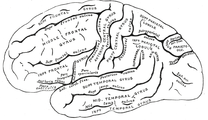 | ||
| Lateral surface of left cerebral hemisphere, viewed from the side. (Parietal Lobe is in upper right.) | ||
| Latin | lobus parietalis | |
| Gray's | subject #189 822 | |
| Part of | Cerebrum | |
| Components | ||
| Artery | Anterior cerebral Middle cerebral | |
| Vein | Superior sagittal sinus | |
| BrainInfo/UW | hier-77 | |
| MeSH | A08.186.211.730.885.213.670 | |
The parietal lobe is a lobe in the brain. It is positioned above (superior to) the occipital lobe and behind (posterior to) the frontal lobe.
The parietal lobe integrates sensory information from different modalities, particularly determining spatial sense and navigation. For example, it comprises somatosensory cortex and the dorsal stream of the visual system. This enables regions of the parietal cortex to map objects perceived visually into body coordinate positions.
Anatomy
The parietal lobe is defined by four anatomical boundaries: the central sulcus separates the parietal lobe from the frontal lobe; the parieto-occipital sulcus separates the parietal and occipital lobe; the lateral sulcus (sylvian fissure) is the most lateral boundary separating it from the temporal lobe; and the medial longitudinal fissure divides the two hemispheres.
Immediately posterior to the central sulcus, and the most anterior part of the parietal lobe, is the postcentral gyrus (Brodmann area 3), the primary somatosensory cortical area. Dividing this and the posterior parietal cortex is the postcentral sulcus.
The posterior parietal cortex can be subdivided into the superior parietal lobule (Brodmann areas 5 + 7) and the inferior parietal lobule (39 + 40), separated by the intraparietal sulcus (IP). The intraparietal sulcus and adjacent gyri are essential in guidance of limb and eye movement, and based on cytoarchitectural and functional differences is further divided into medial (MIP), lateral (LIP), ventral (VIP), and anterior (AIP) areas.
Function
The parietal lobe plays important roles in integrating sensory information from various parts of the body, knowledge of numbers and their relations[1], and in the manipulation of objects. Portions of the parietal lobe are involved with visuospatial processing. Much less is known about this lobe than the other three in the cerebrum.
Various studies in the 1990s found that different regions of the parietal cortex in Macaques represent different parts of space.
- The lateral intraparietal (LIP) contains a 2-dimensional topographic map of retinotopically-coded space representing the saliency of spatial locations. It can be used by the oculomotor system for targeting eye movements, when appropriate.
- The ventral intraparietal (VIP) area receives input from a number of senses (visual, somatosensory, auditory, and vestibular[2]). Neurons with tactile receptive fields represented space in a head-centered reference frame[2]. The cells with visual receptive fields also fire with head-centered reference frames[3] but possibly also with eye-centered coordinates[2]
- The medial intraparietal (MIP) area neurons encode the location of a reach target in eye-centered coordinates.[4]
- The anterior intraparietal (AIP) area contains neurons responsive to shape, size, and orientation of objects to be grasped[5] as well as for manipulation of hands themselves, both to viewed[5] and remembered stimuli. [6]
Pathology
Gerstmann's syndrome is associated with lesion to the dominant (usually left) parietal lobe. Balint's syndrome is associated with bilateral lesions. The syndrome of hemispatial neglect is usually associated with large deficits of attention of the non-dominant hemisphere.
Additional images
")
See also
References
- ↑ Blakemore & Frith (2005). The Learning Brain. Blackwell Publishing. ISBN 1-4051-2401-6
- ↑ 2.0 2.1 2.2 Avillac M, Deneve S, Olivier E, Pouget A, Duhamel JR. (2005) Reference frames for representing visual and tactile locations in parietal cortex. Nat Neurosci. 8(7):941-9.
- ↑ Zhang T, Heuer HW, Britten KH. (2004) Parietal area VIP neuronal responses to heading stimuli are encoded in head-centered coordinates. Neuron 42(6):993-1001.
- ↑ Pesaran B, Nelson MJ, Andersen RA. (2006) Dorsal premotor neurons encode the relative position of the hand, eye, and goal during reach planning. Neuron 51(1):125-34.
- ↑ 5.0 5.1 Murata A, Gallese V, Luppino G, Kaseda M, Sakata H. (2000) Selectivity for the shape, size, and orientation of objects for grasping in neurons of monkey parietal area AIP. J Neurophysiol 83(5):2580. PMID 10805659
- ↑ Murata A, Gallese V, Kaseda M, Sakata H. (1996) Parietal neurons related to memory-guided hand manipulation. J Neurophysiol 75(5):2180-6. PMID 8734616
| This page uses Creative Commons Licensed content from Wikipedia (view authors). |