No edit summary |
No edit summary |
||
| Line 1: | Line 1: | ||
{{BioPsy}} |
{{BioPsy}} |
||
| + | {{Infobox Anatomy | |
||
| − | The '''pancreas''' is an organ that serves two functions: |
||
| + | Name = '''Pancreas''' | |
||
| − | * [[exocrine]] - it produces pancreatic juice containing digestive [[enzyme]]s. |
||
| + | Latin = | |
||
| − | * [[endocrine system|endocrine]] - it produces several important [[hormone]]s, including [[insulin]]. |
||
| + | GraySubject = 251 | |
||
| + | GrayPage = 1199 | |
||
| + | Image = Illu pancrease.jpg | |
||
| + | Caption = | |
||
| + | Image2 = illu_pancreas_duodenum.jpg | |
||
| + | Caption2 = 1: [[Head of pancreas]]<BR>2: [[Uncinate process of pancreas]]<BR>3: [[Pancreatic notch]]<BR>4: [[Body of pancreas]]<BR>5: [[Anterior surface of pancreas]]<BR>6: [[Inferior surface of pancreas]]<BR>7: [[Superior margin of pancreas]]<BR>8: [[Anterior margin of pancreas]]<BR>9: [[Inferior margin of pancreas]]<BR>10: [[Omental tuber]]<BR>11: [[Tail of pancreas]]<BR>12: [[Duodenum]] | |
||
| + | Precursor = [[pancreatic bud]]s | |
||
| + | System = | |
||
| + | Artery = [[inferior pancreaticoduodenal artery]], [[superior pancreaticoduodenal artery]], [[splenic artery]] | |
||
| + | Vein = [[pancreaticoduodenal veins]], [[pancreatic veins]] | |
||
| + | Nerve = [[pancreatic plexus]], [[celiac ganglia]], [[vagus]]<ref>{{GeorgiaPhysiology|6/6ch2/s6ch2_30}}</ref> | |
||
| + | Lymph = | |
||
| + | MeshName = Pancreas| |
||
| + | MeshNumber = A03.734 | |
||
| + | Dorlands = six/000077678 | |
||
| + | DorlandsID = Pancreas |
||
| + | }} |
||
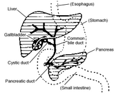
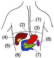
| + | The '''pancreas''' is a [[gland]] [[Organ (anatomy)|organ]] in the [[digestive system|digestive]] and [[endocrine system]] of [[vertebrate]]s. It is both an endocrine gland (producing several important [[hormone]]s, including [[insulin]], [[glucagon]], and [[somatostatin]]), as well as an [[exocrine gland]], secreting [[pancreatic juice]] containing [[Digestion|digestive]] [[enzyme]]s that pass to the [[small intestine]]. These enzymes help in the further breakdown of the [[carbohydrates]], [[protein]], and [[fat]] in the [[chyme]]. |
||
| − | ==Anatomy== |
||
| − | The pancreas is an organ located posterior to the [[stomach]] and in close association with the duodenum. |
||
| + | ==Histology== |
||
| ⚫ | |||
| + | Under a [[microscope]], stained sections of the pancreas reveal two different types of [[parenchyma]]l tissue.<ref>{{BUHistology|10404loa}}</ref> Lightly staining clusters of cells are called [[islets of Langerhans]], which produce [[hormone]]s that underlie the [[endocrine gland|endocrine]] functions of the pancreas. Darker staining cells form [[acinus|acini]] connected to [[exocrine duct|ducts]]. Acinar cells belong to the [[exocrine pancreas]] and secrete [[digestive enzyme]]s into the gut via a system of ducts. |
||
| + | {| class="wikitable" |
||
| − | In humans, the pancreas is a small elongated organ in the abdomen. It is described as having a head, body and tail. The pancreatic head abuts the second part of the [[duodenum]] while the tail extends towards the [[spleen]]. The [[pancreatic duct]] runs the length of the pancreas and empties into the second part of the duodenum at the [[ampulla of Vater]]. The [[common bile duct]] commonly joins the pancreatic duct at or near this point. |
||
| + | | '''Structure''' || '''Appearance''' || '''Function''' |
||
| + | |- |
||
| + | | [[Islets of Langerhans]] || Lightly staining, large, spherical clusters || [[Hormone]] production and secretion ([[endocrine pancreas]]) |
||
| + | |- |
||
| + | | [[Exocrine pancreas|Pancreatic acini]] || Darker staining, small, berry-like clusters || [[Digestive enzyme]] production and secretion ([[exocrine pancreas]]) |
||
| + | |} |
||
| ⚫ | |||
| − | It is supplied arterially by the [[pancreaticoduodenal arteries]], themselves branches of the [[superior mesenteric artery]]. Venous drainage is via the [[pancreaticoduodenal veins]] which end up in the [[portal vein]]. The [[splenic vein]] passes posterior to the pancreas but is said to not drain the pancreas itself. The [[portal vein]] is formed by the union of the [[superior mesenteric vein]] and [[splenic vein]] posterior to the body of the pancreas. In some people (some books say 40% of people), the [[inferior mesenteric vein]] also joins with the [[splenic vein]] behind the pancreas (in others it simply joins with the [[superior mesenteric vein]] instead). |
||
| + | The pancreas is a dual-function gland, having features of both [[endocrine gland|endocrine]] and [[exocrine gland]]s. |
||
| ⚫ | |||
| − | <div> |
||
| + | {{main|Endocrine pancreas}} |
||
| − | [[Image:Pancreas.jpg|thumb|250px|The duodenum and pancreas (stomach removed).]]</div> |
||
| + | |||
| + | The part of the pancreas with endocrine function is made up of a million<ref>{{cite journal |author=Hellman B, Gylfe E, Grapengiesser E, Dansk H, Salehi A |title=[Insulin oscillations--clinically important rhythm. Antidiabetics should increase the pulsative component of the insulin release] |language=Swedish |journal=Lakartidningen |volume=104 |issue=32-33 |pages=2236–9 |year=2007 |pmid=17822201 |doi=}}</ref> cell clusters called [[islets of Langerhans]]. There are four main cell types in the islets. They are relatively difficult to distinguish using standard staining techniques, but they can be classified by their secretion: α cells secrete [[glucagon]], β cells secrete [[insulin]], δ cells secrete [[somatostatin]], and PP cells secrete [[pancreatic polypeptide]].<ref>BRS physiology 4th edition ,page 255-256, Linda S. Constanzo, Lippincott publishing</ref> |
||
| + | |||
| + | The islets are a compact collection of endocrine cells arranged in clusters and cords and are crisscrossed by a dense network of capillaries. The capillaries of the islets are lined by layers of [[endocrine]] cells in direct contact with vessels, and most endocrine cells are in direct contact with blood vessels, by either [[cytoplasm]]ic processes or by direct apposition. According to the volume ''The Body,'' by [[Alan E. Nourse]],<ref>''The Body,'' by [[Alan E. Nourse]], in the Time-Life Science Library Series (op. cit., p. 171.) </ref> the islets are "busily manufacturing their hormone and generally disregarding the pancreatic cells all around them, as though they were located in some completely different part of the body." |
||
| ⚫ | |||
| − | The pancreas produces enzymes that break down all categories of digestible foods (exocrine pancreas) and secretes hormones that affect carbohydrate metabolism (endocrine pancreas). |
||
===Exocrine=== |
===Exocrine=== |
||
| + | {{main|Exocrine pancreas}} |
||
| − | The pancreas is covered in a tissue capsule that partitions the gland into lobules. The bulk of the pancreas is composed of pancreatic exocrine cells, whose ducts are arranged in clusters called ''acini'' (singular ''acinus''). The cells are filled with secretory granules containing the pre-cursor digestive enzymes (mainly [[trypsinogen]], [[chymotrypsinogen]], [[pancreatic lipase]], and [[amylase]]) that are secreted into the [[lumen]] of the acinus. These granules are termed zymogen granules (zymogen referring to the inactive precursor enzymes). |
||
| + | In contrast to the endocrine pancreas, which secretes hormones into the blood, the exocrine pancreas produces [[digestive enzyme]]s and an alkaline fluid, and secretes them into the [[small intestine]] through a system of [[exocrine duct]]s in response to the small intestine hormones [[secretin]] and [[cholecystokinin]]. Digestive enzymes include [[trypsin]], [[chymotrypsin]], [[pancreatic lipase]], and [[pancreatic amylase]], and are produced and secreted by [[acinar cell]]s of the exocrine pancreas. Specific cells that line the pancreatic ducts, called [[centroacinar cell]]s, secrete a [[bicarbonate]]- and [[salt]]-rich solution into the small intestine.<ref>{{cite book |
||
| − | Zymogen granules are localized to the subapical area of pancreatic acinar cells. After fusion with the apical membrane, they are flushed into the duodenum, where enterokinases (bound to enterocytes but facing the lumen of the duodenum) catalyze the activation of trypsinogen into trypsin. Trypsin, an endopeptidase, cleaves amino acids from chymotrypsinogen to produce an active endopeptidase, chymotrypsin. These in turn can 'chop up' polypeptides, released from stomach, into absorbable units. They also activate the other enzymes released. It is important to synthesize inactive enzymes in the pancreas to avoid autodegradation, which can lead to pancreatitis. |
||
| + | | last = Maton |
||
| + | | first = Anthea |
||
| + | | authorlink = |
||
| + | | coauthors = Jean Hopkins, Charles William McLaughlin, Susan Johnson, Maryanna Quon Warner, David LaHart, Jill D. Wright |
||
| + | | title = Human Biology and Health |
||
| + | | publisher = Prentice Hall |
||
| + | | date = 1993 |
||
| + | | location = Englewood Cliffs, New Jersey, USA |
||
| + | | pages = |
||
| + | | url = |
||
| + | | doi = |
||
| + | | id = |
||
| + | | isbn = 0-13-981176-1 |
||
| + | | oclc = 32308337}}</ref> |
||
| + | === Regulation === |
||
| − | The pancreas is the main source of enzymes for digesting fats (lipids) and proteins - the intestinal walls have enzymes that will digest polysaccharides. Pancreatic secretions from ductal cells contain [[bicarbonate]] ions and are [[alkaline]] in order to neutralize the acidic [[chyme]] that the stomach churns out. |
||
| + | The pancreas receives regulatory innervation via [[hormone]]s in the blood and through the [[autonomic nervous system]]. These two inputs regulate the secretory activity of the pancreas. |
||
| − | Control of the exocrine function of the pancreas are via the hormones [[gastrin]], [[cholecystokinin]] and [[secretin]], which are [[hormone]]s secreted by cells in the [[stomach]] and [[duodenum]], in response to distension and/or food and which cause secretion of pancreatic juices. |
||
| + | {| class="wikitable" |
||
| − | The two major proteases the pancreas secretes are [[trypsinogen]] and [[chymotrypsinogen]]. These [[zymogen|zymogens]] are inactivated forms of [[trypsin]] and [[chymotrypsin]]. Once released in the intestine, the enzyme [[enterokinase]] present in the intestinal [[mucosa]] activates trypsinogen by cleaving it to form trypsin. The free trypsin then cleaves the rest of the trypsinogen and chymotrypsinogen to their active forms. |
||
| + | | '''[[Sympathetic nervous system|Sympathetic]]''' ([[adrenergic]]) || '''[[Parasympathetic]]''' ([[muscarinic]]) |
||
| + | |- |
||
| + | | α2: decreases secretion from [[beta cell]]s, increases secretion from [[alpha cell]]s || M3<ref>{{cite journal |author=Verspohl EJ, Tacke R, Mutschler E, Lambrecht G |title=Muscarinic receptor subtypes in rat pancreatic islets: binding and functional studies |journal=Eur. J. Pharmacol. |volume=178 |issue=3 |pages=303–11 |year=1990 |pmid=2187704| doi = 10.1016/0014-2999(90)90109-J <!--Retrieved from CrossRef by DOI bot-->}}</ref> increases stimulation from [[alpha cell]]s and [[beta cell]] |
||
| + | |} |
||
| + | ==Diseases of the pancreas==<!-- This section is linked from [[Pancreatic dysfunction]]. See [[WP:MOS#Section management]] --> |
||
| − | Pancreatic secretions accumulate in intralobular ducts that drain to the main pancreatic duct, which drains directly into the duodenum. |
||
| + | {{main|Pancreatic disease}} |
||
| + | Because the pancreas is a storage depot for digestive enzymes, injury to the pancreas is potentially very dangerous. A puncture of the pancreas generally requires prompt and experienced medical intervention. |
||
| ⚫ | |||
| − | Due to the potency of its enzyme contents, it is a very dangerous organ to injure and a puncture of the pancreas tends to require careful medical intervention. |
||
| + | The pancreas was first identified by [[Herophilus]] (335-280 BC), a [[Greeks|Greek]] [[anatomist]] and [[surgery|surgeon]]. Only a few hundred years later, [[Ruphos]], another Greek anatomist, gave the pancreas its name. The term "pancreas" is derived from the [[Greek language|Greek]] ''pan'', "all", and ''kreas'', "flesh", probably referring to the organ's homogeneous appearance.<ref>{{cite web |last=Harper |first=Douglas |title=Pancreas |work=Online Etymology Dictionary |url=http://www.etymonline.com/index.php?term=pancreas |accessdate=2007-04-04}}</ref> |
||
| + | ==Embryological development== |
||
| ⚫ | |||
| − | Embedded throughout the exocrine tissue are small clusters of cells called the [[Islets of Langerhans]], which are the [[endocrine system|endocrine]] cells of the pancreas and secrete [[insulin]], [[glucagon]], and several other hormones. The islets contain three major types of cells — [[alpha cell]]s, [[beta cell]]s, and [[delta cell]]s. The largest number of cells are, by far, the [[beta cell]]s which produce [[insulin]]. The [[alpha cell]]s produce [[glucagon]] and the [[delta cell]]s produce [[somatostatin]], which lead to both decreased glucagon and insulin levels. There are also the [[PP cell]]s and the D1 cells, about which little is known. |
||
| + | [[Image:Suckale08FBS fig1 pancreas development.jpeg|thumbnail|300px|Schematic illustrating the development of the [[pancreas]] from a [[dorsal]] and a [[ventral]] bud. During maturation the ventral bud flips to the other side of the gut tube (arrow) where it typically fuses with the dorsal lobe. An additional ventral lobe which usually regress during development is omitted.]] |
||
| − | ==Edibility== |
||
| − | Pancreas comes from the Greek ''pankreas'' (a combination of ''pan'' and ''kreas'') which means 'all meat'. ''Kreas'' in Homeric literature meant edible animal flesh. An example of one such food that can be made from the pancreas of a calf, lamb or pig is [[sweetbread]]. |
||
| + | The pancreas forms from the embryonic [[foregut]] and is therefore of [[endoderm]]al origin. Pancreatic development begins the formation of a ventral and dorsal anlage (or buds). Each structure communicates with the foregut through a duct. The ventral pancreatic bud becomes the head and unciate process, and comes from the hepatic diverticulum. |
||
| − | == Diseases of the pancreas == |
||
| + | |||
| − | *[[Benign tumours]] |
||
| + | Differential rotation and fusion of the ventral and dorsal pancreatic buds results in the formation of the definitive pancreas.<ref name="carlson">{{cite book |author=Carlson, Bruce M. |title=Human embryology and developmental biology |publisher=Mosby |location=St. Louis |year=2004 |pages=372–4 |isbn=0-323-01487-9 |oclc= |doi=}}</ref> As the duodenum rotates to the right, it carries with it the ventral pancreatic bud and common bile duct. Upon reaching its final destination, the ventral pancreatic bud fuses with the much larger dorsal pancreatic bud. At this point of fusion, the main ducts of the ventral and dorsal pancreatic buds fuse, forming the [[duct of Wirsung]], the main pancreatic duct. |
||
| − | *[[Carcinoma of pancreas]] |
||
| + | |||
| − | *[[Cystic fibrosis]] |
||
| + | Differentiation of cells of the pancreas proceeds through two different pathways, corresponding to the dual endocrine and exocrine functions of the pancreas. In progenitor cells of the exocrine pancreas, important molecules that induce differentiation include [[follistatin]], [[fibroblast growth factor]]s, and activation of the [[Notch receptor]] system.<ref name="carlson"/> Development of the exocrine acini progresses through three successive stages. These include the predifferentiated, protodifferentiated, and differentiated stages, which correspond to undetectable, low, and high levels of digestive enzyme activity, respectively. |
||
| − | *[[Diabetes]] |
||
| + | |||
| − | *[[Exocrine pancreatic insufficiency]] |
||
| + | Progenitor cells of the endocrine pancreas arise from cells of the protodifferentiated stage of the exocrine pancreas.<ref name="carlson"/> Under the influence of [[neurogenin-3]] and [[Isl-1]], but in the absence of Notch receptor signaling, these cells differentiate to form two lines of committed endocrine precursor cells. The first line, under the direction of [[Pax-6]], forms α- and γ- cells, which produce the peptides [[glucagon]] and [[pancreatic polypeptide]], respectively. The second line, influenced by [[Pax-4]], produces β- and δ-cells, which secrete [[insulin]] and [[somatostatin]], respectively. |
||
| − | *[[Pancreatitis]] |
||
| + | |||
| − | **[[Acute pancreatitis]] |
||
| + | Insulin and glucagon can be detected in the fetal circulation by the fourth or fifth month of fetal development.<ref name="carlson"/> |
||
| − | **[[Chronic pancreatitis]] |
||
| + | |||
| − | *[[Pancreatic pseudocyst]] |
||
| + | ==Additional images== |
||
| + | <gallery> |
||
| ⚫ | |||
| + | Image:BauchOrgane wn.png|Digestive organs. |
||
| + | Image:Gray533.png|The celiac artery and its branches; the stomach has been raised and the peritoneum removed. |
||
| + | Image:Gray614.png|Lymphatics of stomach, etc. The stomach has been turned upward. |
||
| + | Image:Gray1097.png|Transverse section through the middle of the first lumbar vertebra, showing the relations of the pancreas. |
||
| + | Image:Gray1098.png|The duodenum and pancreas. |
||
| + | Image:Gray1100.png|The pancreatic duct. |
||
| + | Image:Gray1101.png|Pancreas of a human embryo of five weeks. |
||
| + | Image:Gray1102.png|Pancreas of a human embryo at end of sixth week. |
||
| + | Image:Gray1225.png|Front of abdomen, showing surface markings for duodenum, pancreas, and kidneys. |
||
| + | </gallery> |
||
| ⚫ | |||
| − | The pancreas was discovered by [[Herophilus]], a [[Greeks|Greek]] [[anatomist]] and [[surgery|surgeon]]. Only a few hundred years later, [[Ruphos]], another Greek anatomist, gave the pancreas its name. |
||
==See also== |
==See also== |
||
| + | * [[Endocrine glands]] |
||
| − | *[[Pancreas transplantation]] |
||
| + | * [[Gastrointestinal system]] |
||
| + | |||
| ⚫ | |||
| + | {{reflist}} |
||
| + | {{Digestive glands}} |
||
| ⚫ | |||
| + | {{endocrine pancreas}} |
||
| − | http://www.randomhouse.com/wotd/index.pperl?date=20010222 Review 2005-03-10 |
||
| + | * ''[http://www.britannica.com/eb/article-9058232/pancreas Pancreas entry in Britannica]'' |
||
| − | {{digestive_system}} |
||
| + | [[category:Gastrointestinal system]] |
||
| − | [[Category:Abdomen]] |
||
| − | [[Category: |
+ | [[Category:Glands]] |
| − | [[Category: |
+ | [[Category:Pancreas| ]] |
| − | [[Category:Diabetes]] |
||
| + | <!-- |
||
| + | [[af:Pankreas]] |
||
[[ar:بنكرياس]] |
[[ar:بنكرياس]] |
||
| + | [[bn:অগ্ন্যাশয়]] |
||
| ⚫ | |||
| + | [[bs:Gušterača]] |
||
| + | [[bg:Панкреас]] |
||
| + | [[ca:Pàncrees]] |
||
| ⚫ | |||
[[da:Bugspytkirtlen]] |
[[da:Bugspytkirtlen]] |
||
| − | [[de: |
+ | [[de:Bauchspeicheldrüse]] |
| + | [[et:Kõhunääre]] |
||
| + | [[el:Πάγκρεας]] |
||
[[es:Páncreas]] |
[[es:Páncreas]] |
||
[[eo:Pankreato]] |
[[eo:Pankreato]] |
||
| + | [[eu:Pankrea]] |
||
| + | [[fa:لوزالمعده]] |
||
[[fr:Pancréas]] |
[[fr:Pancréas]] |
||
| + | [[gl:Páncreas]] |
||
| + | [[ko:이자 (기관)]] |
||
| + | [[hr:Gušterača]] |
||
| + | [[id:Pankreas]] |
||
| + | [[is:Bris]] |
||
[[it:Pancreas]] |
[[it:Pancreas]] |
||
[[he:לבלב]] |
[[he:לבלב]] |
||
| + | [[jv:Pankreas]] |
||
| + | [[ku:Pankreas]] |
||
| + | [[la:Pancreas]] |
||
| + | [[lv:Aizkuņģa dziedzeris]] |
||
[[lt:Kasa]] |
[[lt:Kasa]] |
||
| + | [[hu:Hasnyálmirigy]] |
||
| + | [[mk:Панкреас]] |
||
[[nl:Alvleesklier]] |
[[nl:Alvleesklier]] |
||
[[ja:膵臓]] |
[[ja:膵臓]] |
||
| − | [[no: |
+ | [[no:Bukspyttkjertelen]] |
[[nn:Bukspyttkjertelen]] |
[[nn:Bukspyttkjertelen]] |
||
[[pl:Trzustka]] |
[[pl:Trzustka]] |
||
[[pt:Pâncreas]] |
[[pt:Pâncreas]] |
||
| + | [[ro:Pancreas]] |
||
| + | [[qu:Suyk'upin]] |
||
| + | [[ru:Поджелудочная железа]] |
||
| + | [[sq:Pankreasi]] |
||
| + | [[scn:Pancreas]] |
||
| + | [[simple:Pancreas]] |
||
[[sk:Podžalúdková žľaza]] |
[[sk:Podžalúdková žľaza]] |
||
| + | [[sl:Trebušna slinavka]] |
||
| + | [[so:Ganac]] |
||
[[sr:Гуштерача]] |
[[sr:Гуштерача]] |
||
[[fi:Haima]] |
[[fi:Haima]] |
||
| + | [[sv:Bukspottkörtel]] |
||
| + | [[ta:கணையம்]] |
||
| + | [[te:క్లోమము]] |
||
[[vi:Tụy]] |
[[vi:Tụy]] |
||
| + | [[tr:Pankreas]] |
||
| + | [[uk:Підшлункова залоза]] |
||
| + | [[yi:פאנקרעאס]] |
||
[[zh:胰脏]] |
[[zh:胰脏]] |
||
| + | --> |
||
{{enWP|Pancreas}} |
{{enWP|Pancreas}} |
||
Latest revision as of 22:41, 16 December 2008
Assessment |
Biopsychology |
Comparative |
Cognitive |
Developmental |
Language |
Individual differences |
Personality |
Philosophy |
Social |
Methods |
Statistics |
Clinical |
Educational |
Industrial |
Professional items |
World psychology |
Biological: Behavioural genetics · Evolutionary psychology · Neuroanatomy · Neurochemistry · Neuroendocrinology · Neuroscience · Psychoneuroimmunology · Physiological Psychology · Psychopharmacology (Index, Outline)
| '''Pancreas''' | ||
|---|---|---|
| Latin | ' | |
| Gray's | subject #251 1199 | |
| System | ||
| MeSH | A03.734 | |
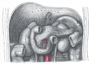
| 1: Head of pancreas 2: Uncinate process of pancreas 3: Pancreatic notch 4: Body of pancreas 5: Anterior surface of pancreas 6: Inferior surface of pancreas 7: Superior margin of pancreas 8: Anterior margin of pancreas 9: Inferior margin of pancreas 10: Omental tuber 11: Tail of pancreas 12: Duodenum | ||
The pancreas is a gland organ in the digestive and endocrine system of vertebrates. It is both an endocrine gland (producing several important hormones, including insulin, glucagon, and somatostatin), as well as an exocrine gland, secreting pancreatic juice containing digestive enzymes that pass to the small intestine. These enzymes help in the further breakdown of the carbohydrates, protein, and fat in the chyme.
Histology
Under a microscope, stained sections of the pancreas reveal two different types of parenchymal tissue.[1] Lightly staining clusters of cells are called islets of Langerhans, which produce hormones that underlie the endocrine functions of the pancreas. Darker staining cells form acini connected to ducts. Acinar cells belong to the exocrine pancreas and secrete digestive enzymes into the gut via a system of ducts.
| Structure | Appearance | Function |
| Islets of Langerhans | Lightly staining, large, spherical clusters | Hormone production and secretion (endocrine pancreas) |
| Pancreatic acini | Darker staining, small, berry-like clusters | Digestive enzyme production and secretion (exocrine pancreas) |
Function
The pancreas is a dual-function gland, having features of both endocrine and exocrine glands.
Endocrine
- Main article: Endocrine pancreas
The part of the pancreas with endocrine function is made up of a million[2] cell clusters called islets of Langerhans. There are four main cell types in the islets. They are relatively difficult to distinguish using standard staining techniques, but they can be classified by their secretion: α cells secrete glucagon, β cells secrete insulin, δ cells secrete somatostatin, and PP cells secrete pancreatic polypeptide.[3]
The islets are a compact collection of endocrine cells arranged in clusters and cords and are crisscrossed by a dense network of capillaries. The capillaries of the islets are lined by layers of endocrine cells in direct contact with vessels, and most endocrine cells are in direct contact with blood vessels, by either cytoplasmic processes or by direct apposition. According to the volume The Body, by Alan E. Nourse,[4] the islets are "busily manufacturing their hormone and generally disregarding the pancreatic cells all around them, as though they were located in some completely different part of the body."
Exocrine
- Main article: Exocrine pancreas
In contrast to the endocrine pancreas, which secretes hormones into the blood, the exocrine pancreas produces digestive enzymes and an alkaline fluid, and secretes them into the small intestine through a system of exocrine ducts in response to the small intestine hormones secretin and cholecystokinin. Digestive enzymes include trypsin, chymotrypsin, pancreatic lipase, and pancreatic amylase, and are produced and secreted by acinar cells of the exocrine pancreas. Specific cells that line the pancreatic ducts, called centroacinar cells, secrete a bicarbonate- and salt-rich solution into the small intestine.[5]
Regulation
The pancreas receives regulatory innervation via hormones in the blood and through the autonomic nervous system. These two inputs regulate the secretory activity of the pancreas.
| Sympathetic (adrenergic) | Parasympathetic (muscarinic) |
| α2: decreases secretion from beta cells, increases secretion from alpha cells | M3[6] increases stimulation from alpha cells and beta cell |
Diseases of the pancreas
- Main article: Pancreatic disease
Because the pancreas is a storage depot for digestive enzymes, injury to the pancreas is potentially very dangerous. A puncture of the pancreas generally requires prompt and experienced medical intervention.
History
The pancreas was first identified by Herophilus (335-280 BC), a Greek anatomist and surgeon. Only a few hundred years later, Ruphos, another Greek anatomist, gave the pancreas its name. The term "pancreas" is derived from the Greek pan, "all", and kreas, "flesh", probably referring to the organ's homogeneous appearance.[7]
Embryological development
Schematic illustrating the development of the pancreas from a dorsal and a ventral bud. During maturation the ventral bud flips to the other side of the gut tube (arrow) where it typically fuses with the dorsal lobe. An additional ventral lobe which usually regress during development is omitted.
The pancreas forms from the embryonic foregut and is therefore of endodermal origin. Pancreatic development begins the formation of a ventral and dorsal anlage (or buds). Each structure communicates with the foregut through a duct. The ventral pancreatic bud becomes the head and unciate process, and comes from the hepatic diverticulum.
Differential rotation and fusion of the ventral and dorsal pancreatic buds results in the formation of the definitive pancreas.[8] As the duodenum rotates to the right, it carries with it the ventral pancreatic bud and common bile duct. Upon reaching its final destination, the ventral pancreatic bud fuses with the much larger dorsal pancreatic bud. At this point of fusion, the main ducts of the ventral and dorsal pancreatic buds fuse, forming the duct of Wirsung, the main pancreatic duct.
Differentiation of cells of the pancreas proceeds through two different pathways, corresponding to the dual endocrine and exocrine functions of the pancreas. In progenitor cells of the exocrine pancreas, important molecules that induce differentiation include follistatin, fibroblast growth factors, and activation of the Notch receptor system.[8] Development of the exocrine acini progresses through three successive stages. These include the predifferentiated, protodifferentiated, and differentiated stages, which correspond to undetectable, low, and high levels of digestive enzyme activity, respectively.
Progenitor cells of the endocrine pancreas arise from cells of the protodifferentiated stage of the exocrine pancreas.[8] Under the influence of neurogenin-3 and Isl-1, but in the absence of Notch receptor signaling, these cells differentiate to form two lines of committed endocrine precursor cells. The first line, under the direction of Pax-6, forms α- and γ- cells, which produce the peptides glucagon and pancreatic polypeptide, respectively. The second line, influenced by Pax-4, produces β- and δ-cells, which secrete insulin and somatostatin, respectively.
Insulin and glucagon can be detected in the fetal circulation by the fourth or fifth month of fetal development.[8]
Additional images
See also
References
- ↑ Histology at Boston University 10404loa
- ↑ Hellman B, Gylfe E, Grapengiesser E, Dansk H, Salehi A (2007). [Insulin oscillations--clinically important rhythm. Antidiabetics should increase the pulsative component of the insulin release]. Lakartidningen 104 (32-33): 2236–9.
- ↑ BRS physiology 4th edition ,page 255-256, Linda S. Constanzo, Lippincott publishing
- ↑ The Body, by Alan E. Nourse, in the Time-Life Science Library Series (op. cit., p. 171.)
- ↑ Maton, Anthea; Jean Hopkins, Charles William McLaughlin, Susan Johnson, Maryanna Quon Warner, David LaHart, Jill D. Wright (1993). Human Biology and Health, Englewood Cliffs, New Jersey, USA: Prentice Hall.
- ↑ Verspohl EJ, Tacke R, Mutschler E, Lambrecht G (1990). Muscarinic receptor subtypes in rat pancreatic islets: binding and functional studies. Eur. J. Pharmacol. 178 (3): 303–11.
- ↑ Harper, Douglas Pancreas. Online Etymology Dictionary. URL accessed on 2007-04-04.
- ↑ 8.0 8.1 8.2 8.3 Carlson, Bruce M. (2004). Human embryology and developmental biology, 372–4, St. Louis: Mosby.
Anatomy of torso, digestive system: Digestive glands | |
|---|---|
| Organs |
Pancreas: by region (Tail, Body, Head, Uncinate process) - by function (Islets of Langerhans, Exocrine pancreas) Liver: by region (Caudate lobe, Quadrate lobe, Right lobe, Left lobe) - by function (Hepatocyte, Space of Disse, Kupffer cell, Liver sinusoid, Ito cell, Hepatic lobule) |
| Ducts |
Bile ducts: (Bile canaliculus, Common hepatic duct, Cystic duct, Common bile duct) |
Islets of Langerhans: alpha cell (glucagon) • beta cell (insulin) • delta cell (somatostatin) • PP cell (pancreatic polypeptide) • epsilon cell (ghrelin)
| This page uses Creative Commons Licensed content from Wikipedia (view authors). |


