Assessment |
Biopsychology |
Comparative |
Cognitive |
Developmental |
Language |
Individual differences |
Personality |
Philosophy |
Social |
Methods |
Statistics |
Clinical |
Educational |
Industrial |
Professional items |
World psychology |
Biological: Behavioural genetics · Evolutionary psychology · Neuroanatomy · Neurochemistry · Neuroendocrinology · Neuroscience · Psychoneuroimmunology · Physiological Psychology · Psychopharmacology (Index, Outline)
Opthalmology
The optic cup is the white, cup-like area in the center of the optic disc. The ratio of the size of the optic cup to the optic disc (or cup-to-disc ratio) is measured to diagnose glaucoma.
Embryology
| Embryology: Optic cup | ||
|---|---|---|
| Transverse section of head of chick embryo of forty-eight hours’ incubation. (Margin of optic cup labeled at upper right.) | ||
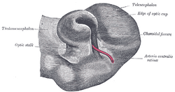
| Optic cup and choroidal fissure seen from below, from a human embryo of about four weeks. (Edge of optic cup labeled at upper right.) | ||
| Latin | c. ophthalmicus | |
| Gray's | subject #224 1001 | |
| System | ||
| Carnegie stage | 13 | |
| Days | {{{Days}}} | |
| Precursor | ||
| Gives rise to | ||
| MeSH | [1] | |
| Dorlands/Elsevier | c_03/12205940 | |
During embryonic development of the eye, the outer wall of the bulb of the optic vesicles becomes thickened and invaginated, and the bulb is thus converted into a cup, the optic cup (or ophthalmic cup), consisting of two strata of cells). These two strata are continuous with each other at the cup margin, which ultimately overlaps the front of the lens and reaches as far forward as the future aperture of the pupil.
External links
This article was originally based on an entry from a public domain edition of Gray's Anatomy. As such, some of the information contained herein may be outdated. Please edit the article if this is the case, and feel free to remove this notice when it is no longer relevant.
Neural development/Neurulation - Neurula - Neural folds - Neural groove - Neural tube - Neural crest - Neuromere (Rhombomere) - Notochord - Neural plate
Eye development - Optic vesicles - Optic stalk - Optic cup - Auditory vesicle - Auditory pit
| This page uses Creative Commons Licensed content from Wikipedia (view authors). |

