No edit summary |
|||
| Line 1: | Line 1: | ||
| − | |||
| − | |||
[[Image:PurkinjeCell.jpg|thumb|250px| Drawing by [[Santiago Ramón y Cajal]] of cells in the pigeon cerebellum. (A) Denotes [[Purkinje cell]]s, an example of a bipolar neuron. (B) Denotes [[granule cells]] which are multipolar.]] |
[[Image:PurkinjeCell.jpg|thumb|250px| Drawing by [[Santiago Ramón y Cajal]] of cells in the pigeon cerebellum. (A) Denotes [[Purkinje cell]]s, an example of a bipolar neuron. (B) Denotes [[granule cells]] which are multipolar.]] |
||
'''Neurons''' (also spelled '''neurones''' or called '''nerve cells''') are a major class of [[cell (biology)|cells]] in the [[nervous system]]. |
'''Neurons''' (also spelled '''neurones''' or called '''nerve cells''') are a major class of [[cell (biology)|cells]] in the [[nervous system]]. |
||
| Line 32: | Line 30: | ||
''Afferent'' and ''efferent'' can also refer to neurons which convey information from one region of the brain to another. |
''Afferent'' and ''efferent'' can also refer to neurons which convey information from one region of the brain to another. |
||
| + | |||
| − | {{BioPsy}} |
||
'''Structural classification''' |
'''Structural classification''' |
||
Most neurons can be anatomically characterized into one of three categories: |
Most neurons can be anatomically characterized into one of three categories: |
||
Revision as of 09:03, 10 February 2007
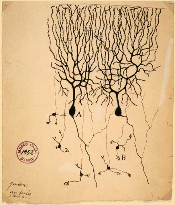
Drawing by Santiago Ramón y Cajal of cells in the pigeon cerebellum. (A) Denotes Purkinje cells, an example of a bipolar neuron. (B) Denotes granule cells which are multipolar.
Neurons (also spelled neurones or called nerve cells) are a major class of cells in the nervous system. In vertebrates, they are found in the brain, the spinal cord and in the nerves and ganglia of the peripheral nervous system, and their primary role is to process and transmit neural information. One important characteristic of neurons is that they have excitable membranes which allow them to generate and propagate electrical signals.
The concept of a neuron as the primary computational unit of the nervous system was devised by Spanish anatomist Santiago Ramón y Cajal. Cajal proposed that neurons were discrete cells which communicated with each other via specialized junctions. This became known as the Neuron Doctrine, one of the central tenets of modern neuroscience.
Anatomy and histology
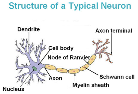
Many highly-specialized types of neurons exist, and these differ widely in appearance. Neurons have cellular extensions known as processes which they use to send and receive information. Neurons are highly asymmetric in shape, and consist of:
- The soma, or cell-body, is the central part of the cell between the dendrites and the axon. It is where the nucleus is located and is where most protein synthesis occurs.
- The dendrite, a branching arbor of cellular extensions. Most neurons have multiple dendrites with profuse dendritic branches. The overall shape and structure of a neuron's dendrites is called its dendritic tree. The dendritic tree form has traditionally been thought to be the main information receiving network for the neuron. However information outflow (i.e. from dendrites to other neurons) can also occur.
- The axon, a much finer, cable-like projection which may extend tens, hundreds, or even tens of thousands of times the diameter of the soma in length. This is the structure that carries nerve signals away from the neuron. Neurons have only one axon, but this axon may - and usually will - undergo extensive branching, enabling communication with many target cells. The part of the axon where it emerges from the soma is called the 'axon hillock'. Besides being an anatomical structure, the axon hillock is also the part of the neuron that has the greatest density of voltage-dependent sodium channels. Thus it has the most hyperpolarized action potential threshold of any part of the neuron. In other words, it is the most easily-excitable part of the neuron, and thus serves as the spike initiation zone for the axon. While the axon and axon hillock are generally considered places of information outflow, this region can receive input from other neurons as well.
- The axon terminal, a specialized structure at the end of the axon that is used to release neurotransmitter and communicate with target neurons.
Although the canonical view of the neuron is to assign strictly defined and dedicated functions to its various anatomical components, the fact that dendrites and axons very often act contrary to their so-called main function is but one small glimpse into the complex integrative capacity of every nerve cell. Nervous systems bear little resemblance to simple feed-forward Input/Output circuits, and this understanding begins by appreciating the global signaling capacity of individual neurons.
Axons and dendrites in the central nervous system are typically only about a micrometer thick, while some of those in the peripheral nervous system are much thicker. The soma is usually about 10–25 micrometers in diameter and not much larger than the cell nucleus it contains. The longest axon of a human motoneuron can be over a meter long, reaching from the base of the spine to the toes, while giraffes have single axons running along the whole length of their necks, several meters in length. Much of what we currently know about axonal function comes from studying the squid giant axon, an ideal experimental preparation for research due to its relatively immense size (0.5–1 millimeters thick, several centimeters long).
Classes
Functional classification There are three functional classes of neurons: afferent neurons, efferent neurons, and interneurons.
- Afferent neurons convey information from tissues and organs into the central nervous system.
- Efferent neurons transmit signals from the central nervous system to the effector cells.
- Interneurons connect neurons within specific regions of the central nervous system.
Afferent and efferent can also refer to neurons which convey information from one region of the brain to another.
Structural classification Most neurons can be anatomically characterized into one of three categories:
- Unipolar or Pseudounipolar- dendrite and axon emerging from same process.
- Bipolar - single axon and single dendrite on oposite ends of the soma.
- Multipolar - more than two dendrites
Connectivity
Neurons communicate with one another and to other cells through synapses, where the axon terminal of one cell impinges upon a dendrite or soma of another, or less commonly to an axon. Neurons such as the Purkinje cells in the cerebellum, can have over 1000 dendrites each, enabling connections with tens of thousands of other cells. Synapses can either be excitatory or inhibitory and will either respectively increase or decrease activity in the target neuron. Neurons can also communicate via electrical synapses, which are direct, electrically-conductive junctions between cells.
The human brain has a gigantic number of synapses. Each of 100 billion neurons has on average 7,000 synaptic connections to other neurons. Most authorities estimate total number of synapses at 1,000 trillion for a three-year-old child. This number declines and with age, stabilizing by adulthood. Estimates vary for an adult from 100 to 500 trillion synapses. [[1]]
Adaptations to carrying action potentials
The cell membrane in the axon and soma contain voltage-gated ion channels which allow the neuron to generate and propagate an electrical impulse known as an action potential.
Substantial early knowledge of neuron electrical activity came from experiments with squid giant axons. In 1937, John Zachary Young suggested that the giant squid axon might be used to better understand nerve cells [2]. Since they are much larger than human neurons, but similar in nature, it was easier to study them with less advanced technology at that time. By inserting electrodes into the giant squid axons, accurate measurements could be made of the membrane potential.
Electrical activity can be produced in neurons by a number of stimuli. Pressure, stretch, chemical transmitters, and electrical current passing across the nerve membrane as a result of a potential difference in voltage all can initiate nerve activity [3].
The narrow cross-section of axons lessens the metabolic expense of carrying action potentials, however thicker axons convey the impulses more rapidly. In order to minimize metabolic expense yet maintain a rapid conduction velocity, many neurons have insulating sheaths of myelin around their axons. The sheaths are formed by glial cells: oligodendrocytes in the central nervous system and Schwann cells in the peripheral nervous system. The sheath enables the action potentials to travel faster than in unmyelinated axons of the same diameter whilst simultaneously spending less energy to "recharge" the action potential after. The myelin sheath in peripheral nerves normally runs along the axon in sections about 1 mm long, punctuated by unsheathed nodes of Ranvier which contain a high density of voltage-gated ion channels. Multiple sclerosis is a neurological disorder which results from abnormal demyelination of peripheral nerves. Neurons with demyelinated axons do not conduct electrical signals properly.
Histology and internal structure
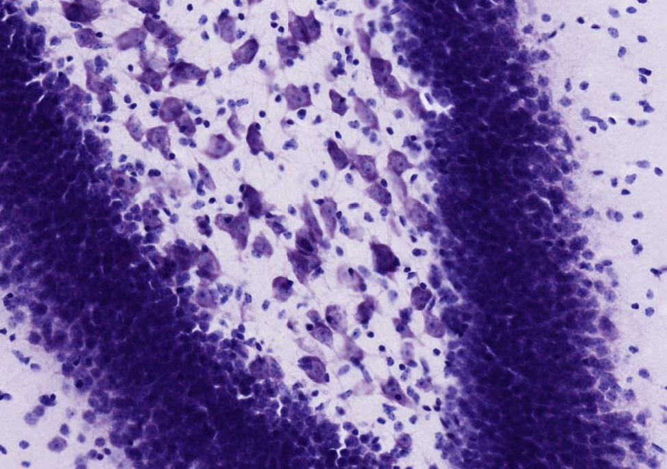
Image of a Nissl-stained histological section through the rodent hippocampus showing various classes of cells.
Nerve cell bodies stained with basophilic dyes will show numerous microscopic clumps of Nissl substance (named after German psychiatrist and neuropathologist Franz Nissl, 1860–1919), which consists of rough endoplasmic reticulum and associated ribosomes. The prominence of the Nissl substance can be explained by the fact that nerve cells are metabolically very active, and hence are involved in large amounts of protein synthesis.
The cell body of a neuron is supported by a complex meshwork of structural proteins called neurofilaments, which are assembled into larger neurofibrils. Some neurons also contain pigment granules, such as neuromelanin (a brownish-black pigment, byproduct of synthesis of catecholamines) and lipofuscin (yellowish-brown pigment that accumulates with age).
Challenges to the neuron doctrine
While the neuron doctrine has remained a central tenet of modern neuroscience, recent studies challenging this view have suggested that the narrow confines of this doctrine need to be expanded. Among the most serious challenges to the neuron doctrine is the fact that electrical synapses are more common in the central nervous system than previously thought. This means that rather than functioning as individual units, in some parts of the brain large ensembles of neurons may be active together in order to process neural information. A second challenge comes from the fact that dendrites, like axons, also have voltage-gated ion channels and can generate electrical potentials which convey information to and from the soma. This challenges the view that dendrites are simply passive recipients of information and axons the sole transmitters. It also suggests that the neuron is not simply active as a single element, but that complex computations can occur within a single neuron. Finally, the role of glia in processing neural information has begun to be appreciated. Neurons and glia make up the two chief cell types of the central nervous system. There are far more glial cells than neurons: It has been estimated that glial cells outnumber neurons by as many as 50:1. Recent experimental results have suggested that glial cells play a vital role in information processing among neurons, indicating that neurons may not be the sole information processing cells in the nervous system.
Neurons in the brain
The number of neurons contained within the brain varies dramatically across species. For example the human brain has about 100 billion () neurons and 100 trillion () connections (synapses) between them. In contrast, the nematode worm (Caenorhabditis elegans) has 302 neurons. Scientists have mapped all of the nematode's neurons. As a result, such worms are ideal candidates for neurobiological experiments and tests. Many properties of neurons, ranging from the type of neurotransmitter used to ion channel composition are maintained across species, allowing scientists to study processes occurring in more complex organisms in much simpler experimental systems.


See also
Sources
- Kandel E.R., Schwartz, J.H., Jessell, T.M. 2000. Principles of Neural Science, 4th ed., McGraw-Hill, New York.
- Bullock, T.H., Bennett, M.V.L., Johnston, D., Josephson, R., Marder, E., Fields R.D. 2005. The Neuron Doctrine, Redux, Science, V.310, p. 791-793.
- Ramón y Cajal, S. 1933 Histology, 10th ed., Wood, Baltimore.
External links
- Cell Centered Database UC San Diego images of neurons.
bg:Неврон ca:Neurona cs:Neuron da:Neuron de:Nervenzelle eo:Neŭrono es:Neurona fi:Neuroni fr:Neurone he:נוירון io:Neurono is:Taugafruma lt:Neuronas mk:Неврон nl:Zenuwcel no:Nevron pt:Neurónio ru:Нейроны simple:Neuron sv:Neuron uk:Нейрон zh:神经元
| This page uses Creative Commons Licensed content from Wikipedia (view authors). |