(new article) |
No edit summary |
||
| (4 intermediate revisions by 2 users not shown) | |||
| Line 18: | Line 18: | ||
MeshNumber = A08.186.211.730.885.213.863 | |
MeshNumber = A08.186.211.730.885.213.863 | |
||
}} |
}} |
||
| − | The '''temporal |
+ | The '''medial temporal lobe''' is the inner surface of the [[temporal lobe]]. It includes the [[hippocampal formation]], the [[parahippocampal gyrus]] and the [[entorhinal cortex]] |
| − | |||
| − | The temporal lobe is involved in [[auditory]] processing and is home to the [[primary auditory cortex]]. It is also heavily involved in [[semantics]] both in [[Speech communication|speech]] and [[Visual perception|vision]]. The temporal lobe contains the [[hippocampus]] and is therefore involved in [[memory]] formation as well. |
||
==Function== |
==Function== |
||
| + | The medial temporal lobes (near the Sagittal plane that divides left and right [[cerebral hemisphere]]s) consists of structures that are vital for declarative or long-term memory. [[Declarative_memory|Declarative]] (denotative) or [[Explicit_memory|explicit]] memory is conscious memory divided into [[semantic memory]] (facts) and [[episodic memory]] (events).<ref name=smith07 />{{rp|194|date=December 2012}} Medial temporal lobe structures that are critical for long-term memory include the [[amygdala]], [[brainstem]], and hippocampus, along with the surrounding [[Hippocampal_formation|hippocampal region]] consisting of the [[Perirhinal_cortex|perirhinal]], [[Parahippocampal_gyrus|parahippocampal]], and [[Entorhinal_cortex|entorhinal]] neocortical regions.<ref name=smith07 />{{rp|196|date=December 2012}} The hippocampus is critical for memory formation, and the surrounding medial temporal cortex is currently theorized to be critical for memory storage.<ref name=smith07 />{{rp|21|date=December 2012}} The [[prefrontal cortex|prefrontal]] and visual cortices are also involved in explicit memory.<ref name=smith07 />{{rp|21|date=December 2012}} |
||
| + | Research has shown that lesions in the hippocampus of monkeys results in limited impairment of function, whereas extensive lesions that include the hippocampus and the medial temporal cortex result in severe impairment.<ref>{{cite journal|last=Squire|first=LR|last2=Stark|first2=CE|last3=Clark|first3= RE|title=The medial temporal lobe|journal=Annual Review of Neuroscience|date=2004|volume=27|pages=279–306|pmid=15217334|url=http://whoville.ucsd.edu/PDFs/383_Squire_etal_%20AnnRevNeurosci2004.pdf|doi=10.1146/annurev.neuro.27.070203.144130}}</ref> |
||
| − | The [[superior temporal gyrus]] includes an area (within the Sylvian fissure) where auditory signals from the [[cochlea]] (relayed via several subcortical nuclei) first reach the [[cerebral cortex]]. This part of the cortex ([[primary auditory cortex]]) is involved in hearing. Adjacent areas in the superior, posterior and lateral parts of the temporal lobes are involved in high-level auditory processing. In humans this includes speech, for which the left temporal lobe in particular seems to be specialized. [[Wernicke's area]], which spans the region between temporal and parietal lobes, plays a key role (in tandem with [[Broca's area]], which is in the frontal lobe). The functions of the left temporal lobe are not limited to low-level perception but extend to comprehension, naming, [[verbal memory]] and other language functions. Sound processing is controlled by the temporal lobes- in the Broca’s area and Wernicke’s area. |
||
| + | ==Additional images== |
||
| − | The underside (ventral) part of the temporal cortices appear to be involved in high-level visual processing of complex stimuli such as [[face perception|faces]] ([[fusiform gyrus]]) and scenes ([[parahippocampal gyrus]]). Anterior parts of this [[ventral stream]] for [[visual processing]] are involved in object perception and recognition. |
||
| − | The medial temporal lobes (near the Sagittal plane that divides left and right [[cerebral hemisphere]]s) are thought to be involved in [[episodic memory|episodic]]/[[declarative memory]]. Deep inside the medial temporal lobes, the [[hippocampus|hippocampi]] seem to be particularly important for memory function - particularly transference from short to long term memory and control of spatial memory and behavior. |
||
| − | == |
+ | ==See also== |
| − | <gallery> |
||
| − | Image:Illu cerebrum lobes.jpg|Lobes |
||
| − | Image:Gray724.png|Base of brain. |
||
| − | Image:Gray743.png|Coronal section through anterior cornua of lateral ventricles. |
||
| − | Image:Gray1197.png|Drawing to illustrate the relations of the brain to the skull. |
||
| − | Image:Ventral-dorsal streams.svg|The [[dorsal stream]] (green) and [[ventral stream]] (purple) in the temporal lobe are shown. |
||
| − | </gallery> |
||
| − | |||
| − | ==See Also== |
||
*[[Lobes of the brain]] |
*[[Lobes of the brain]] |
||
| − | *[[Area TG of Bonin-1947]] |
||
| − | *[[Brain]] |
||
| − | *[[lobe (anatomy)]] |
||
*[[Hippocampus]] |
*[[Hippocampus]] |
||
| − | *[[ |
+ | *[[Medial temporal lobe epilepsy]] |
| + | |||
| + | ==Further reading== |
||
| + | * Kensinger, E. A., Ullman, M. T., & Corkin, S. (2001). Bilateral medial temporal lobe damage does not affect lexical or grammatical processing: Evidence from amnesic patient H.M. ''Hippocampus, 11,'' 337-346. [http://web.mit.edu/bnl/pdf/Kensinger%20et%20al.,%202001.pdf Full text] |
||
==External links== |
==External links== |
||
| Line 52: | Line 41: | ||
{{Prosencephalon}} |
{{Prosencephalon}} |
||
| − | |||
[[Category:Cerebrum]] |
[[Category:Cerebrum]] |
||
| + | [[Category:Temporal lobe]] |
||
Latest revision as of 22:35, 2 July 2013
| Brain: Medial temporal lobe | ||
|---|---|---|
| [[Image:{{{Image}}}|250px|center|]] | ||
| {{{Caption}}} | ||
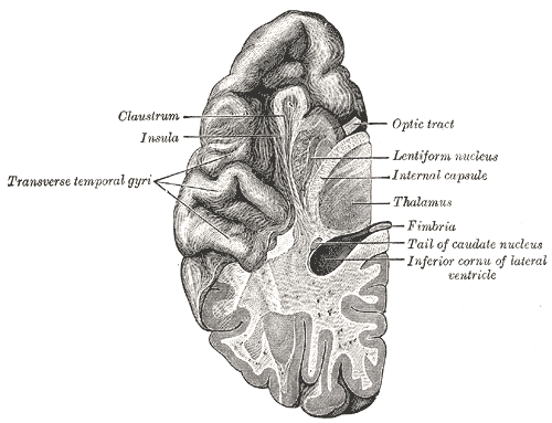 | ||
| Section of brain showing upper surface of temporal lobe. | ||
| Latin | lobus temporalis | |
| Gray's | subject #189 823 | |
| Part of | Brain | |
| Components | ||
| Artery | Middle cerebral and Posterior cerebral | |
| Vein | ||
| BrainInfo/UW | hier-107 | |
| MeSH | A08.186.211.730.885.213.863 | |
The medial temporal lobe is the inner surface of the temporal lobe. It includes the hippocampal formation, the parahippocampal gyrus and the entorhinal cortex
Function
The medial temporal lobes (near the Sagittal plane that divides left and right cerebral hemispheres) consists of structures that are vital for declarative or long-term memory. Declarative (denotative) or explicit memory is conscious memory divided into semantic memory (facts) and episodic memory (events).[1]:194 Medial temporal lobe structures that are critical for long-term memory include the amygdala, brainstem, and hippocampus, along with the surrounding hippocampal region consisting of the perirhinal, parahippocampal, and entorhinal neocortical regions.[1]:196 The hippocampus is critical for memory formation, and the surrounding medial temporal cortex is currently theorized to be critical for memory storage.[1]:21 The prefrontal and visual cortices are also involved in explicit memory.[1]:21
Research has shown that lesions in the hippocampus of monkeys results in limited impairment of function, whereas extensive lesions that include the hippocampus and the medial temporal cortex result in severe impairment.[2]
Additional images
See also
Further reading
- Kensinger, E. A., Ullman, M. T., & Corkin, S. (2001). Bilateral medial temporal lobe damage does not affect lexical or grammatical processing: Evidence from amnesic patient H.M. Hippocampus, 11, 337-346. Full text
External links