No edit summary |
|||
| (One intermediate revision by the same user not shown) | |||
| Line 21: | Line 21: | ||
}} |
}} |
||
The '''medial lemniscus''', also known as '''Reil's band''' or '''Reil's ribbon''', is a pathway in the [[brainstem]] that carries sensory information from the [[gracile nucleus|gracile]] and [[cuneate nucleus|cuneate nuclei]] to the [[thalamus]]. It forms part og the [[lemniscal system]] |
The '''medial lemniscus''', also known as '''Reil's band''' or '''Reil's ribbon''', is a pathway in the [[brainstem]] that carries sensory information from the [[gracile nucleus|gracile]] and [[cuneate nucleus|cuneate nuclei]] to the [[thalamus]]. It forms part og the [[lemniscal system]] |
||
| + | |||
| + | The term applies to one of two bundles of nerve fibers of the [[lemniscus]] which ascend through the [[medulla oblongata]] and [[pons]] and go on up to the [[somatosensory cortex]] in each hemisphere<ref>Coleman,A F (2006). Oxford Dictionary of Psychology, 2nd ed. Oxford:OUP. </ref>. |
||
==Path== |
==Path== |
||
| − | After neurons carrying proprioceptive or touch information [[synapse]] at the gracile and cuneate nuclei, axons from secondary neurons decussate at the level of the medulla and travel up the brainstem as the medial lemniscus on the contralateral (opposite) side. It is part of the [[posterior column-medial lemniscus system]], which transmits touch, vibration sense, as well as the [[pathway for proprioception]]. |
+ | After neurons carrying [[proprioceptive]] or [[touch]] information [[synapse]] at the gracile and cuneate nuclei, axons from secondary neurons decussate at the level of the medulla and travel up the brainstem as the medial lemniscus on the contralateral (opposite) side. It is part of the [[posterior column-medial lemniscus system]], which transmits touch, vibration sense, as well as the [[pathway for proprioception]]. |
The medial lemniscus axons from most of the body synapse at the [[ventral posterolateral nucleus]] of the [[thalamus]]. The axons transmitting information from the [[trigeminal nerve]] synapse at the [[ventral posteromedial nucleus]] of the [[thalamus]]. |
The medial lemniscus axons from most of the body synapse at the [[ventral posterolateral nucleus]] of the [[thalamus]]. The axons transmitting information from the [[trigeminal nerve]] synapse at the [[ventral posteromedial nucleus]] of the [[thalamus]]. |
||
| − | == |
+ | ==Location of the medial lemniscus through the brainstem== |
* The cuneate and gracile nuclei reside at the ''closed (lower) [[medulla oblongata|medulla]]'', so the lemniscus isn't formed at this level. Fibres from these nuclei will pass to the contralateral side of the brainstem, as the [[internal arcuate fibres]]. |
* The cuneate and gracile nuclei reside at the ''closed (lower) [[medulla oblongata|medulla]]'', so the lemniscus isn't formed at this level. Fibres from these nuclei will pass to the contralateral side of the brainstem, as the [[internal arcuate fibres]]. |
||
| Line 38: | Line 40: | ||
==See also== |
==See also== |
||
| − | * [[Posterior column-medial lemniscus pathway]] |
+ | * [[Posterior column-medial lemniscus pathway]] (aka Medial lemniscus tracts) |
==Additional images== |
==Additional images== |
||
| Line 48: | Line 50: | ||
Image:Gray713.png|Scheme showing the course of the fibers of the lemniscus; medial lemniscus in blue, lateral in red. |
Image:Gray713.png|Scheme showing the course of the fibers of the lemniscus; medial lemniscus in blue, lateral in red. |
||
</gallery> |
</gallery> |
||
| + | |||
| + | ==References== |
||
| + | <references/> |
||
==External links== |
==External links== |
||
Latest revision as of 22:48, 2 July 2013
Assessment |
Biopsychology |
Comparative |
Cognitive |
Developmental |
Language |
Individual differences |
Personality |
Philosophy |
Social |
Methods |
Statistics |
Clinical |
Educational |
Industrial |
Professional items |
World psychology |
Biological: Behavioural genetics · Evolutionary psychology · Neuroanatomy · Neurochemistry · Neuroendocrinology · Neuroscience · Psychoneuroimmunology · Physiological Psychology · Psychopharmacology (Index, Outline)
| Brain: Medial lemniscus | ||
|---|---|---|
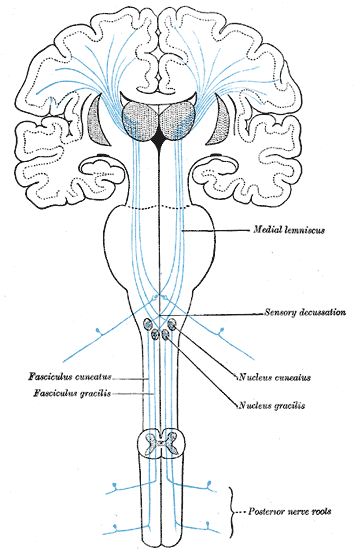 | ||
| The sensory tract. (Medial lemniscus labeled at center right.) | ||
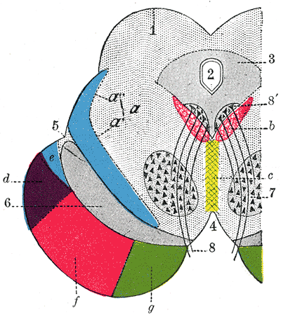 | ||
| Coronal section through mid-brain. ("e" is Portion of medial lemniscus, which runs to the lentiform nucleus and insula.) | ||
| Latin | lemniscus medialis | |
| Gray's | subject #188 803 | |
| Part of | ||
| Components | ||
| Artery | ||
| Vein | ||
| BrainInfo/UW | ancil-736 | |
| MeSH | [1] | |
The medial lemniscus, also known as Reil's band or Reil's ribbon, is a pathway in the brainstem that carries sensory information from the gracile and cuneate nuclei to the thalamus. It forms part og the lemniscal system
The term applies to one of two bundles of nerve fibers of the lemniscus which ascend through the medulla oblongata and pons and go on up to the somatosensory cortex in each hemisphere[1].
Path
After neurons carrying proprioceptive or touch information synapse at the gracile and cuneate nuclei, axons from secondary neurons decussate at the level of the medulla and travel up the brainstem as the medial lemniscus on the contralateral (opposite) side. It is part of the posterior column-medial lemniscus system, which transmits touch, vibration sense, as well as the pathway for proprioception.
The medial lemniscus axons from most of the body synapse at the ventral posterolateral nucleus of the thalamus. The axons transmitting information from the trigeminal nerve synapse at the ventral posteromedial nucleus of the thalamus.
Location of the medial lemniscus through the brainstem
- The cuneate and gracile nuclei reside at the closed (lower) medulla, so the lemniscus isn't formed at this level. Fibres from these nuclei will pass to the contralateral side of the brainstem, as the internal arcuate fibres.
- At the open medulla (further up the brainstem), the medial lemniscus contains axons from the trigeminal nerve (which supplies the head region), as well as the arms and legs. It sits very close to the midline, at the same orientation of the midline, with head fibres more dorsal (closer to the back), towards the fourth ventricle.
- By mid-pons, the medial lemniscus has rotated. Fibres from the head are medial, fibres from the leg are lateral.
- The orientation in the midbrain is similar to that in the pons.
See also
- Posterior column-medial lemniscus pathway (aka Medial lemniscus tracts)
Additional images
")
")
")
")
")
References
- ↑ Coleman,A F (2006). Oxford Dictionary of Psychology, 2nd ed. Oxford:OUP.
External links
| Mesencephalon (midbrain) |
|
cerebral peduncle: midbrain tegmentum (periaqueductal gray, ventral tegmentum, nucleus raphe dorsalis), pretectum, substantia nigra, red nucleus, pedunculopontine nucleus, medial longitudinal fasciculus, medial lemniscus, rubrospinal tract, lateral lemniscus tectum: corpora quadrigemina, inferior colliculi, superior colliculi cerebral aqueduct: oculomotor nucleus, trochlear nucleus, Edinger-Westphal nucleus |
| Nervous system, receptors: somatosensory system |
|---|
| Medial lemniscus: Touch/mechanoreceptors: Pacinian corpuscles - Meissner's corpuscles - Merkel's discs - Ruffini endings - Free nerve endings - Hair cells - Baroreceptor Proprioception: Golgi organ - Muscle spindle (Intrafusal muscle fiber) Spinothalamic tract: Pain: Nociceptors Temperature: Thermoreceptors |
| This page uses Creative Commons Licensed content from Wikipedia (view authors). |