No edit summary |
No edit summary |
||
| Line 4: | Line 4: | ||
The '''macula''' or '''macula lutea''' (from [[Latin]] ''macula'', "spot" + ''lutea'', "yellow") is an oval [[yellow]] spot near the center of the [[retina]] of the human [[eye]]. It has a diameter of about 1.5 mm and is often histologically defined as having two or more layers of [[ganglion cell]]s. Near its center is the [[fovea]], a small pit that contains the largest concentration of [[cone cell]]s in the eye and is responsible for central vision. |
The '''macula''' or '''macula lutea''' (from [[Latin]] ''macula'', "spot" + ''lutea'', "yellow") is an oval [[yellow]] spot near the center of the [[retina]] of the human [[eye]]. It has a diameter of about 1.5 mm and is often histologically defined as having two or more layers of [[ganglion cell]]s. Near its center is the [[fovea]], a small pit that contains the largest concentration of [[cone cell]]s in the eye and is responsible for central vision. |
||
| − | Whereas loss of [[peripheral vision]] may go unnoticed for some time, damage to the macula will result in loss of central vision, which is usually immediately obvious. |
+ | It is specialized for high [[Visual acuity|acuity]] vision. Within the macula are the fovea and foveola which contain a high density of [[cones]] (photoreceptors with high acuity). Whereas loss of [[peripheral vision]] may go unnoticed for some time, damage to the macula will result in loss of central vision, which is usually immediately obvious. |
| − | The progressive destruction of the macula is a |
+ | The progressive destruction of the macula is a [[disease]] known as [[macular degeneration]]. |
| + | |||
| + | Visual input to the macula occupies a substantial portion of the brain's visual capacity. As a result, some forms of [[Visual field#Visual field loss|visual field loss]] can occur without involving the macula; this is termed '''macular sparing'''. (For example, [[visual field testing]] might demonstrate ''[[homonymous hemianopsia]] with macular sparing''.) This finding can be very informative for the [[ophthalmologist]]. |
||
==See also== |
==See also== |
||
| − | *[[Visual acuity]] |
||
*[[Cystoid macular edema]] |
*[[Cystoid macular edema]] |
||
| + | *[[Intermediate Uveitis]] |
||
| + | *[[Macula]] |
||
| + | |||
| + | ==Additional images== |
||
| + | <gallery> |
||
| + | Image:Gray1206.png|The interior of the posterior half of the left eyeball. |
||
| + | </gallery> |
||
{{Eye}} |
{{Eye}} |
||
| Line 16: | Line 24: | ||
[[category:eye]] |
[[category:eye]] |
||
| + | <!-- |
||
[[de:Gelber Fleck (Auge)]] |
[[de:Gelber Fleck (Auge)]] |
||
| + | [[es:Mácula]] |
||
| ⚫ | |||
| + | [[fr:Macula]] |
||
| ⚫ | |||
| + | [[nl:Gele vlek]] |
||
| + | [[no:Macula lutea]] |
||
| + | [[pt:Mácula]] |
||
| + | [[ru:Жёлтое пятно]] |
||
[[nl:Gele vlek]] |
[[nl:Gele vlek]] |
||
[[ru:Жёлтое пятно]] |
[[ru:Жёлтое пятно]] |
||
| + | ---> |
||
{{enWP|Macula}} |
{{enWP|Macula}} |
||
Latest revision as of 23:57, 30 March 2007
Assessment |
Biopsychology |
Comparative |
Cognitive |
Developmental |
Language |
Individual differences |
Personality |
Philosophy |
Social |
Methods |
Statistics |
Clinical |
Educational |
Industrial |
Professional items |
World psychology |
Biological: Behavioural genetics · Evolutionary psychology · Neuroanatomy · Neurochemistry · Neuroendocrinology · Neuroscience · Psychoneuroimmunology · Physiological Psychology · Psychopharmacology (Index, Outline)
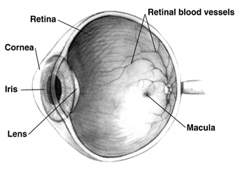
Human eye cross-sectional view.
The macula or macula lutea (from Latin macula, "spot" + lutea, "yellow") is an oval yellow spot near the center of the retina of the human eye. It has a diameter of about 1.5 mm and is often histologically defined as having two or more layers of ganglion cells. Near its center is the fovea, a small pit that contains the largest concentration of cone cells in the eye and is responsible for central vision.
It is specialized for high acuity vision. Within the macula are the fovea and foveola which contain a high density of cones (photoreceptors with high acuity). Whereas loss of peripheral vision may go unnoticed for some time, damage to the macula will result in loss of central vision, which is usually immediately obvious. The progressive destruction of the macula is a disease known as macular degeneration.
Visual input to the macula occupies a substantial portion of the brain's visual capacity. As a result, some forms of visual field loss can occur without involving the macula; this is termed macular sparing. (For example, visual field testing might demonstrate homonymous hemianopsia with macular sparing.) This finding can be very informative for the ophthalmologist.
See also
- Cystoid macular edema
- Intermediate Uveitis
- Macula
Additional images
")
| Sensory system - Visual system - Eye - edit |
|---|
| Anterior chamber | Aqueous humour | Blind spot | Choroid | Ciliary body | Conjunctiva | Cornea | Iris | Lens | Macula | Optic disc | Optic fovea | Posterior chamber | Pupil | Retina | Schlemm's canal | Sclera | Tapetum lucidum | Trabecular meshwork | Vitreous humour |
| Sensory system - Visual system - edit |
|---|
| Eye | Optic nerve | Optic chiasm | Optic tract | Lateral geniculate nucleus | Optic radiation | Visual cortex |
| This page uses Creative Commons Licensed content from Wikipedia (view authors). |