Education
 

Internal arcuate fibers

From Psychology Wiki

(Redirected from Internal arcuate fibres)

Community portal · Tasks to do · News · Help

Clinical · Educational · Ind&Org · Other fields · Professional · Transpersonal · World

Assessment | Biopsychology | Comparative | Cognitive | Developmental | Language
Personality | Philosophy | Research Methods | Social | Statistics

Biological: Behavioural genetics · Evolutionary psychology · Neuroanatomy · Neurochemistry · Neuroendocrinology · Psychoneuroimmunology · Physiological Psychology · Psychopharmacology


Internal arcuate fibers
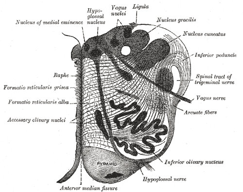
Diagram showing the course of the arcuate fibers. (Testut.) 1. Medulla oblongata anterior surface. 2. Anterior median fissure. 3. Fourth ventricle. 4. Inferior olivary nucleus, with the accessory olivary nuclei. 5. Gracile nucleus. 6. Cuneate nucleus. 7. Trigeminal. 8. Inferior peduncles, seen from in front. 9. Posterior external arcuate fibers. 10. Anterior external arcuate fibers. 11. Internal arcuate fibers. 12. Peduncle of inferior olivary nucleus. 13. Nucleus arcuatus. 14. Vagus. 15. Hypoglossal.
Latin fibrae arcuatae internae
Gray's subject #187 782
System
MeSH [1]
Section of the medulla oblongata at about the middle of the olive. (Arcuate fibers labeled at center right.)

Internal arcuate fibers are the axons of second-order neurons contained within the gracile and cuneate nuclei of the medulla oblongata.

These fibers cross (decussate) from one side of the medulla to the other to form the medial lemniscus.

Part of the dorsal column-medial lemniscus system (second neuron), the internal arcuate fibers are important for relaying the sensation of fine touch and proprioception to the thalamus and ultimately to the cerebral cortex.

[edit] External links


|- style="white-space:nowrap;background:#ddddff;text-align:right;background-color: LightYellow" ! Myelencephalon/
medulla | colspan="1" style="text-align:left;width:100%;font-size:95%;background:#f7f7f7;" | {| class="navbox collapsible nowraplinks" style="margin:auto; background:white;background:white;" |- style="text-align:center;width:100%;"

!colspan="2" |
[[[:Template:Fullurl:Template:]] v]·[[[:Template:Fullurl:Template talk:]] d]·[[[:Template:Fullurl:Template:]] e]

|- |- style="white-space:nowrap;background:#ddddff;text-align:right;padding-left:0em;padding-right:0em;background-color: LightYellow"

!
Dorsal

| colspan="1" style="text-align:left;width:100%;font-size:95%;" | surface: Posterior median sulcus - Postero-lateral sulcus - Area postrema

cranial nuclei: GVA: VII,IX,X: Solitary/tract • SVA: Gustatory nucleus • GSE: XII  • GVE: IX,X,XI: Ambiguus • SVE: X: Dorsal • IX: Inferior salivatory nucleus - MLF, III, IV and VI

sensory/ascending: Gracile nucleus • Cuneate nucleus (Accessory cuneate nucleus) • Sensory decussation  • Medial lemniscus

motor/descending: Dorsal respiratory group
|- style="white-space:nowrap;background:#ddddff;text-align:right;padding-left:0em;padding-right:0em;background-color: LightYellow"

!
Ventral

| colspan="1" style="text-align:left;width:100%;font-size:95%;background:#f7f7f7;" | motor/descending: Ventral respiratory group - Pyramid (Motor decussation) - Inferior olivary nucleus (Olivocerebellar tract, Rubro-olivary tract)
surface: Anterior median fissure - Antero-lateral sulcus - Arcuate nucleus of medulla - Olivary body
|- style="white-space:nowrap;background:#ddddff;text-align:right;padding-left:0em;padding-right:0em;background-color: LightYellow"

!
Raphe/reticular

| colspan="1" style="text-align:left;width:100%;font-size:95%;" | Reticular formation (Gigantocellular, Parvocellular, Ventral, Lateral, Paramedian) • Raphe nuclei (Obscurus, Magnus, Pallidus) |} |}

Smallwikipedialogo.png This page uses content from the English-language version of Wikipedia. The original article was at Internal arcuate fibers. The list of authors can be seen in the page history. As with Psychology Wiki, the text of Wikipedia is available under the GNU Free Documentation License.