Assessment |
Biopsychology |
Comparative |
Cognitive |
Developmental |
Language |
Individual differences |
Personality |
Philosophy |
Social |
Methods |
Statistics |
Clinical |
Educational |
Industrial |
Professional items |
World psychology |
Biological: Behavioural genetics · Evolutionary psychology · Neuroanatomy · Neurochemistry · Neuroendocrinology · Neuroscience · Psychoneuroimmunology · Physiological Psychology · Psychopharmacology (Index, Outline)
| Brain: Inferior cerebellar peduncles | ||
|---|---|---|
| Scheme showing the connections of the several parts of the brain. (Inferior peduncle labeled at bottom right.) | ||
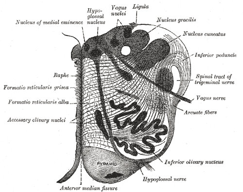 | ||
| Section of the medulla oblongata at about the middle of the olive. (Inferior peduncle labeled at upper right. | ||
| Latin | pedunculus cerebellaris inferior | |
| Gray's | subject #187 775 | |
| Part of | ||
| Components | ||
| Artery | ||
| Vein | ||
| BrainInfo/UW | hier-778 | |
| MeSH | [1] | |
The upper part of the posterior district of the medulla oblongata is occupied by the inferior peduncle, a thick rope-like strand situated between the lower part of the fourth ventricle and the roots of the glossopharyngeal and vagus nerves.
The inferior peduncles connect the medulla spinalis and medulla oblongata with the cerebellum, and are sometimes named the restiform bodies.
Function
The inferior cerebellar peduncle carries many types of input and output fibers that are mainly concerned with integrating proprioceptive sensory input with motor vestibular functions such as balance and posture maintenance.
Proprioceptive information from the body is carried to the cerebellum via the posterior spinocerebellar tract.
This tract passes through the inferior cerebellar peduncle and synapses within the paleocerebellum.
Vestibular information projects onto the archicerebellum.
This peduncle also carries information directly from the Purkinje cells to the vestibular nuclei in the dorsal brainstem located at the junction between the pons and medulla.
See also
Additional images
")
")
")
")
")
")
")
External links
This article was originally based on an entry from a public domain edition of Gray's Anatomy. As such, some of the information contained herein may be outdated. Please edit the article if this is the case, and feel free to remove this notice when it is no longer relevant.
| This page uses Creative Commons Licensed content from Wikipedia (view authors). |