(update wp) |
mNo edit summary |
||
| Line 19: | Line 19: | ||
MeshNumber = A08.186.211.730.385.357 | |
MeshNumber = A08.186.211.730.385.357 | |
||
}} |
}} |
||
| − | The '''hypothalamus''' (from Greek ὑποθαλαμος = under the thalamus) is a region of the |
+ | The '''hypothalamus''' (from Greek ὑποθαλαμος = under the thalamus) is a region of the mammalian [[brain]] located below the [[thalamus]], forming the major portion of the ventral region of the [[diencephalon]] and functioning to regulate certain [[metabolic]] processes and other [[autonomic]] activities. The hypothalamus links the [[nervous system]] to the [[endocrine system]] via the [[pituitary gland]], also known as the "master gland," by synthesizing and secreting [[hormone|neurohormones]], often called ''releasing hormones,'' as needed that control the secretion of [[hormones]] from the [[anterior pituitary gland]] — among them, [[gonadotropin-releasing hormone]] (GnRH). The [[neurons]] that secrete GnRH are linked to the [[limbic system]], which is primarily involved in the control of [[emotion]]s and [[sex]]ual activity. The hypothalamus also controls [[body temperature]], [[hunger]], [[thirst]], and [[Circadian rhythm|circadian cycles]]. |
==Hormones of the hypothalamus== |
==Hormones of the hypothalamus== |
||
Revision as of 21:41, 25 October 2006
Assessment |
Biopsychology |
Comparative |
Cognitive |
Developmental |
Language |
Individual differences |
Personality |
Philosophy |
Social |
Methods |
Statistics |
Clinical |
Educational |
Industrial |
Professional items |
World psychology |
Biological: Behavioural genetics · Evolutionary psychology · Neuroanatomy · Neurochemistry · Neuroendocrinology · Neuroscience · Psychoneuroimmunology · Physiological Psychology · Psychopharmacology (Index, Outline)
| Brain: Hypothalamus | ||
|---|---|---|
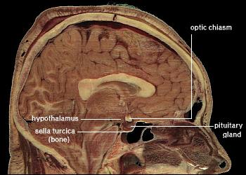 | ||
| Location of the human hypothalamus | ||
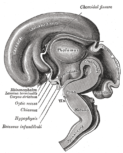 | ||
| Median sagittal section of brain of human embryo of three months. (Hypothalamus visible at center.) | ||
| Latin | hypothalamus | |
| Gray's | subject #189 812 | |
| Part of | ||
| Components | ||
| Artery | ||
| Vein | ||
| BrainInfo/UW | hier-358 | |
| MeSH | A08.186.211.730.385.357 | |
The hypothalamus (from Greek ὑποθαλαμος = under the thalamus) is a region of the mammalian brain located below the thalamus, forming the major portion of the ventral region of the diencephalon and functioning to regulate certain metabolic processes and other autonomic activities. The hypothalamus links the nervous system to the endocrine system via the pituitary gland, also known as the "master gland," by synthesizing and secreting neurohormones, often called releasing hormones, as needed that control the secretion of hormones from the anterior pituitary gland — among them, gonadotropin-releasing hormone (GnRH). The neurons that secrete GnRH are linked to the limbic system, which is primarily involved in the control of emotions and sexual activity. The hypothalamus also controls body temperature, hunger, thirst, and circadian cycles.
Hormones of the hypothalamus
- Corticotropin-releasing hormone (CRH)
- Dopamine
- Gonadotropin-releasing hormone (GnRH)
- Growth hormone releasing hormone (GHRH)
- Somatostatin
- Thyrotropin-releasing hormone (TRH)
- Hypocretin
Boundaries
The anatomical boundaries of the hypothalamus are:
- rostral, the lamina terminalis.
- caudal, the posterior margin of the mamillary bodies.
- dorsal, the hypothalamic sulcus.
- medial, the third ventricle.
- lateral, the subthalamus and internal capsule.
- ventral, the optic chiasm, tuber cinereum, mammillary bodies, and posterior pituitary.
Hypothalamic nuclei

Hypothalamic nuclei
| Region | Medial Area | Lateral Area |
| Anterior |
Medial preoptic nucleus |
Lateral preoptic nucleus |
| Tuberal |
Lateral nucleus | |
| Posterior |
Mammillary nuclei (part of mammillary bodies) |
Lateral nucleus |
- See also: ventrolateral preoptic nucleus
Inputs to the hypothalamus


Dienchephalon
The hypothalamus is a very complex region, and even small nuclei within the hypothalamus are involved in many different functions. The paraventricular nucleus for instance contains oxytocin and vasopressin neurons which project to the posterior pituitary, but also contains neurons that regulate ACTH and TSH secretion (which project to the anterior pituitary), gastric reflexes, maternal behavior, blood pressure, feeding, immune responses, and temperature.
The hypothalamus co-ordinates many seasonal and circadian rhythms, complex patterns of neuroendocrine outputs, complex homeostatic mechanisms, and many important stereotyped behaviours. The hypothalamus must therefore respond to many different signals, some of which are generated externally and some internally. The hypothalamus is thus richly connected with many parts of the CNS, including the brainstem reticular formation and autonomic zones, the limbic forebrain (particularly the amygdala, septum, diagonal band of Broca, and the olfactory bulbs, and the cerebral cortex).
The hypothalamus is responsive to:
- Light: daylength and photoperiod for generating circadian and seasonal rhythms
- Olfactory stimuli, including pheromones
- Steroids, including gonadal steroids and corticosteroids
- Neurally transmitted information arising in particular from the heart, the stomach, and the reproductive tract
- Autonomic inputs
- Blood-borne stimuli, including leptin, ghrelin, angiotensin, insulin, pituitary hormones, cytokines, plasma concentrations of glucose and osmolarity etc
- Invading microorganisms by increasing body temperature, resetting the bodys thermostat upward.
Olfactory stimuli
Olfactory stimuli are important for reproduction and neuroendocrine function in many species. For instance, if a pregnant mouse is exposed to the urine of a 'strange' male during a critical period after coitus then the pregnancy fails (the Bruce effect). Thus during coitus, a female mouse forms a precise 'olfactory memory' of her partner which persists for several days. Pheromonal cues aid synchronisation of oestrus in many species; in women, synchronised menstruation may also arise from pheromonal cues.
Blood-borne stimuli
Peptide hormones have important influences upon the hypothalamus, and to do so they must evade the blood-brain barrier. The hypothalamus is bounded in part by specialized brain regions that lack an effective blood-brain barrier; the capillary endothelium at these sites is fenestrated to allow free passage of even large proteins and other molecules. Some of these sites are the sites of neurosecretion - the neurohypophysis and the median eminence. However others are sites at which the brain samples the composition of the blood. Two of these sites, the subfornical organ and the OVLT (organum vasculosum of the lamina terminalis) are so-called circumventricular organs, where neurons are in intimate contact with both blood and CSF. These structures are densely vascularized, and contain osmoreceptive and sodium-receptive neurons which control drinking, vasopressin release, sodium excretion, and sodium appetite. They also contain neurons with receptors for angiotensin, atrial natriuretic factor, endothelin and relaxin, each of which is important in the regulation of fluid and electrolyte balance. Neurons in the OVLT and SFO project to the supraoptic nucleus and paraventricular nucleus, and also to preoptic hypothalamic areas. The circumventricular organs may also be the site of action of interleukins to elicit both fever and ACTH secretion, via effects on paraventricular neurons.
It is not clear how all peptides that influence hypothalamic activity gain the necessary access. In the case of prolactin and leptin, there is evidence of active uptake at the choroid plexus from blood into CSF. Some pituitary hormones have a negative feedback influence upon hypothalamic secretion; for example, growth hormone feeds back on the hypothalamus, but how it enters the brain is not clear. There is also evidence for central actions of prolactin and TSH.
Steroids
The hypothalamus contains neurons that are sensitive to gonadal steroids and glucocorticoids – (the steroid hormones of the adrenal gland, released in response to ACTH). It also contains specialised glucose-sensitive neurons (in the arcuate nucleus and ventromedial hypothalamus), which are important for appetite. The preoptic area contains thermosensitive neurons; these are important for TRH secretion.
Neural inputs
The hypothalamus receives many inputs from the brainstem; notably from the nucleus of the solitary tract, the locus coeruleus, and the ventrolateral medulla. Oxytocin secretion in response to suckling or vagino-cervical stimulation is mediated by some of these pathways; vasopressin secretion in response to cardiovascular stimuli arising from chemoreceptors in the carotid sinus and aortic arch, and from low-pressure atrial volume receptors, is mediated by others. In the rat, stimulation of the vagina also causes prolactin secretion, and this results in pseudo-pregnancy following an infertile mating. In the rabbit, coitus elicits reflex ovulation. In the sheep, cervical stimulation in the presence of high levels of estrogen can induce maternal behaviour in a virgin ewe. These effects are all mediated by the hypothalamus, and the information is carried mainly by spinal pathways that relay in the brainstem. Stimulation of the nipples stimulates release of oxytocin and prolactin and suppresses the release of LH and FSH. Cardiovascular stimuli are carried by the vagus nerve, but the vagus also conveys a variety of visceral information, including for instance signals arising from gastric distension to suppress feeding. Again this information reaches the hypothalamus via relays in the brainstem.
Projections
Most fiber systems of the hypothalamus run in two ways (bidirectional). Projections to areas caudal to the hypothalamus go through the medial forebrain bundle, the mammillotegmental tract and the dorsal longitudinal fasciculus. Projections to areas rostral to the hypothalamus are carried by the mammillothalamic tract, the fornix and stria terminalis.
Sexual dimorphism
The hypothalamus is sexually dimorphic, i.e. there are clear differences in both structure and function between males and females. Some differences are apparent even in gross neuroanatomy: most notable is the sexually dimorphic nucleus within the preoptic area, which is present only in males. However most of the differences are subtle changes in the connectivity and chemical sensitivity of particular sets of neurons. The importance of these changes can be recognised by functional differences between males and females. For instance, the pattern of secretion of growth hormone is sexually dimorphic, and this is one reason why in many species, adult males are much larger than females. Other striking functional dimorphisms are in the behavioral responses to ovarian steroids of the adult. Males and females respond differently to ovarian steroids, partly because the expression of estrogen-sensitive neurons in the hypothalamus is sexually dimorphic, i.e. estrogen receptors are expressed in different sets of neurons.
Estrogen and progesterone act by influencing gene expression in particular neurons. To influence gene expression, estrogen binds to an intracellular receptor, and this complex is translocated to the cell nucleus where it interacts with regions of the DNA known as estrogen regulatory elements (EREs). Increased protein synthesis may follow as soon as 30 min later. Thus, for estrogen to influence the expression of a particular gene in a particular cell, the following must occur:
- the cell must be exposed to estrogen
- the cell must express estrogen receptors
- the gene must be one that is regulated by an ERE.
Male and female brains differ in the distribution of estrogen receptors, and this difference is an irreversible consequence of neonatal steroid exposure. Estrogen receptors (and progesterone receptors) are found mainly in neurons in the anterior and mediobasal hypothalamus, notably:
- the preoptic area (where LHRH neurons are located)
- the periventricular nucleus (where somatostatin neurons are located)
- the ventromedial hypothalamus (which is important for sexual behavior).
In neonatal life, gonadal steroids influence the development of the neuroendocrine hypothalamus. For instance, they determine the ability of females to exhibit a normal reproductive cycle, and of males and females to display appropriate reproductive behaviors in adult life. Thus, if a female rat is injected once with testosterone in the first few days of postnatal life (during the "critical period" of sex-steroid influence), the hypothalamus is irreversibly masculinized; the adult rat will be incapable of generating an LH surge in response to estrogen (a characteristic of females), but will be capable of exhibiting male sexual behaviors (mounting a sexually-receptive female). By contrast, a male rat castrated just after birth will be feminized, and the adult will show female sexual behavior in response to estrogen (sexual receptivity, lordosis}.
In primates, the developmental influence of androgens is less clear, and the consequences are less complete. 'Tomboyism' in girls might reflect the effects of androgens on the fetal brain, but the sex of rearing during the first 2-3 years is believed by many to be the most important determinant of gender identity.
The paradox is that the masculinizing effects of testosterone are mediated by estrogen. Within the brain, testosterone is aromatized to (estradiol), which is the principal active hormone for developmental influences. The human testis secretes high levels of testosterone from about week 8 of fetal life until 5-6 months after birth (a similar perinatal surge in testosterone is observed in many species), a process that appears to underlie the male phenotype. Estrogen from the maternal circulation is relatively ineffective, partly because of the high circulating levels of steroid-binding proteins in pregnancy.
Sex steroids are not the only important influences upon hypothalamic development; stress in early life determines the capacity of the adult hypothalamus to respond to an acute stressor. Unlike gonadal steroid receptors, glucocorticoid receptors are very widespread throughout the brain; in the paraventricular nucleus, they mediate negative feedback control of CRF synthesis and secretion, but elsewhere their role is not well understood.
See also
External links
- Endocrine system and hypothalamus
- High-Resolution Cytoarchitectural Primate Brain Atlases
- The Hypothalamus and Pituitary at endotexts.org
- Diagram of Nuclei (psycheducation.org)
- Diagram of Nuclei (universe-review.ca)
- Diagram of Nuclei (utdallas.edu)
Human anatomy, endocrine system: endocrine glands | |||||||||||||||||||||||||
|---|---|---|---|---|---|---|---|---|---|---|---|---|---|---|---|---|---|---|---|---|---|---|---|---|---|
| Hypothalamic/ pituitary axes |
| ||||||||||||||||||||||||
| Pineal gland |
Pinealocyte · Corpora arenacea | ||||||||||||||||||||||||
| Islets of pancreas |
Alpha cell · Beta cell · Delta cell · PP cell · Epsilon cell | ||||||||||||||||||||||||
| Human brain: Limbic system | |
| Amygdala - Cingulate gyrus - Fornicate gyrus - Hippocampus - Hypothalamus - Mammillary body - Nucleus accumbens - Orbitofrontal cortex - Parahippocampal gyrus |
ca:Hipotàlem cs:Hypothalamus da:Hypothalamus de:Hypothalamus es:Hipotálamo fr:Hypothalamus he:היפותלמוס nl:Hypothalamus no:Hypothalamus pt:Hipotálamo ru:Гипоталамус sk:Podlôžko
| This page uses Creative Commons Licensed content from Wikipedia (view authors). |