Assessment |
Biopsychology |
Comparative |
Cognitive |
Developmental |
Language |
Individual differences |
Personality |
Philosophy |
Social |
Methods |
Statistics |
Clinical |
Educational |
Industrial |
Professional items |
World psychology |
Biological: Behavioural genetics · Evolutionary psychology · Neuroanatomy · Neurochemistry · Neuroendocrinology · Neuroscience · Psychoneuroimmunology · Physiological Psychology · Psychopharmacology (Index, Outline)
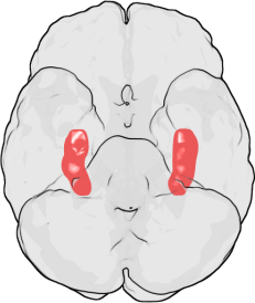
The location of the hippocampus in the human brain.
The hippocampus is a part of the brain located inside the temporal lobe (humans have two hippocampi, one in each side of the brain). It forms a part of the limbic system and plays a part in memory and navigation. The name derives from its curved shape, which supposedly resembles that of a seahorse (Greek: hippocampus).
In Alzheimer's disease, the hippocampus becomes one of the first regions of the brain to suffer damage; memory problems and disorientation appear amongst the first symptoms. Damage to the hippocampus can also result from oxygen starvation (anoxia) and encephalitis.
In the anatomy of animals, the hippocampus is among the phylogenetically oldest parts of the brain. The hippocampal emergence from the archipallium is most pronounced in primates and Cetacean sea mammals. Nonetheless, in primates, the hippocampus occupies less of the telencephalon in proportion to cerebral cortex among the youngest species, especially humans. The significant development of hippocampal volume in primates correlates more with overall increase of brain mass than with neocortical development.
Anatomy
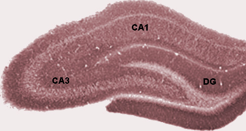
Diagram of hippocampal regions. DG: Dentate gyrus.
Although there is a lack of consensus relating to terms describing the hippocampus and the adjacent cortex, the term hippocampal formation generally applies to the dentate gyrus, fields CA1-CA3 (or CA4, frequently called the hilus and considered part of the dentate gyrus), and the subiculum (see also Cornu ammonis). The CA1 and CA3 fields make up the hippocampus proper.
Information flow through the hippocampus proceeds from dentate gyrus to CA3 to CA1 to the subiculum, with additional input information at each stage and outputs at each of the two final stages. CA2 represents only a very small portion of the hippocampus and its presence is often ignored in accounts of hippocampal function, though it is notable that this small region seems unusually resistant to conditions that usually cause large amounts of cellular damage, such as epilepsy.
The perforant path, which brings information primarily from entorhinal cortex (but also perirhinal cortex, among others), is generally considered the main source of input to the hippocampus. Layer II of entorhinal cortex (EC) brings input to the dentate gyrus and field CA3, while EC layer III brings input to field CA1 and the subiculum. The main output pathways of the hippocampus are the perforant path, the cingulum bundle, and the fimbria/fornix, which all arise from field CA1 and the subiculum.
Perforant path input from EC layer II enters the dentate gyrus and is relayed to region CA3 (and to mossy cells, located in the hilus of the dentate gyrus, which then send information to distant portions of the dentate gyrus where the cycle is repeated). Region CA3 combines this input with signals from EC layer II and sends extensive connections within the region and also sends connections to region CA1 through a set of fibers called the Schaffer collaterals. Region CA1 receives input from region CA3 as well as EC layer III and then projects to the subiculum as well as sending information along the aforementioned output paths of the hippocampus. The subiculum is the final stage in the pathway, combining information from the CA1 projection and EC layer III to also send information along the output pathways of the hippocampus.
It is widely accepted that each of these regions has a unique functional role in the information processing of the hippocampus, but to date the specific contribution of each region is poorly understood.

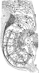
Golgi-stained neurons in the rodent hippocampus.
Role in memory
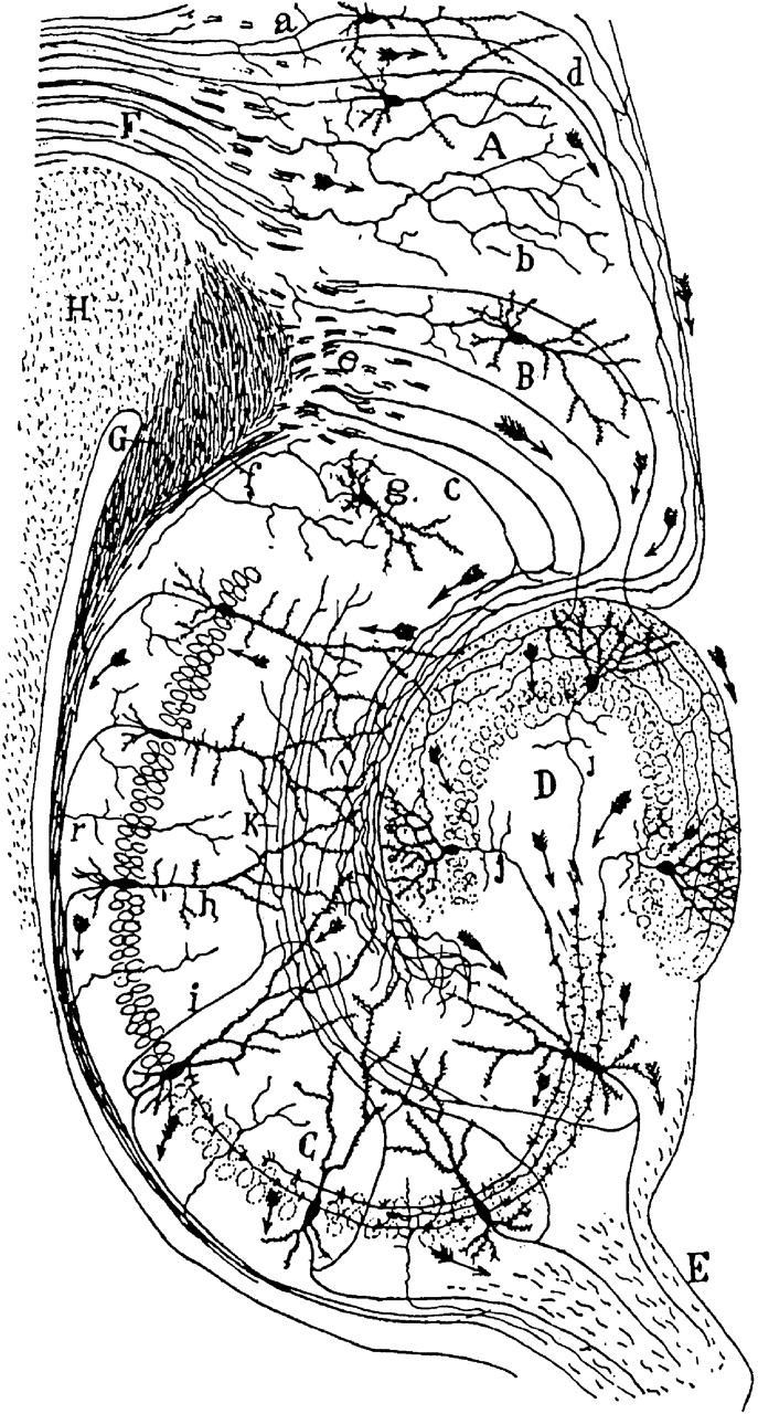
Drawing of the neural circuitry of the rodent hippocampus. S. Ramón y Cajal, 1911.
Psychologists and neuroscientists dispute the precise role of the hippocampus, but generally agree that it has an essential role in the formation of new memories about personally experienced events (episodic or autobiographical memory). Some researchers prefer to consider the hippocampus as part of a larger medial temporal lobe memory system responsible for general declarative memory (memories which can be explicitly verbalized—these would include, for example, memory for facts in addition to episodic memory).
- Main article: Role of the hippocampus in memory
Some evidence implicates the hippocampus in storing and processing spatial information. Studies in rats have shown that neurons in the hippocampus have spatial firing fields. These cells are called place cells. Some cells fire when the animal finds itself in a particular location, regardless of direction of travel, while most are at least partially sensitive to head direction and direction of travel.
- Main article: Role of the hippocampus in spatial memory and navigation
Hippocampus and depression
- Hippocampal volume loss,perhaps reflecting the effects of excessive glucocorticoids on
neurogenesis has been identified in groups of depressed people in comparison with controls.
- Main article: Depression and the hippocampus
History
The anatomist Giulio Cesare Aranzi (circa 1564) first used the term hippocampus to describe the cerebral organ because of its visual resemblance to a seahorse. This organ was initially connected with the sense of smell, rather than with its known function in memory acquisition. The Russian Vladimir Bekhterev noted the role of the hippocampus in memory around 1900, based on observations of a patient with profound memory disturbances. However for many years, the conventional view of the hippocampus was that, like the rest of the limbic system, it was responsible for emotion.
The importance of the hippocampus in memory was brought to the attention of researchers by patient HM. HM suffered from a number of anterograde and temporally graded retrograde memory impairments following the bilateral removal of various medial-temporal lobe structures (including bilateral ablation of his hippocampi) to relieve frequent epileptic seizures. Of particular importance is that HM was still able to learn procedural tasks (which are associated with the Striatum) and had an above average IQ. HM demonstrated a striking single-dissociation between intelligence and declarative memory. The relative size of the hippocampal formation in relation with the total volume of the brain is often conserved in most of the mammalian species. Nevertheless, it has been found that these areas are relatively
See also
References & Bibliography
Key texts
Books
O'Keefe, J. and Nadel, L. (1978) The Hippocampus as a Cognitive Map, Oxford: Clarendon Press.
Papers
- Amaral DG and Cowan WM. 1980. Subcortical afferents to the hippocampal formation in the monkey. Journal of Comparative Neurology. Feb 15; 189(4):573-91.
- Duvernoy, H. (2005) The Human Hippocampus, 3rd ed. Berlin: Springer-Verlag.
Additional material
Books
Papers
External links
References
External links
- http://www.psycheducation.org/emotion/hippocampus.htm Great brain tours!!
- BrainMaps at UCDavis hippocampus
- Artificial Hippocampus
- BrainInfo at the University of Washington hier-164
| Human brain: Limbic system | |
| Amygdala - Cingulate gyrus - Fornicate gyrus - Hippocampus - Hypothalamus - Mammillary body - Nucleus accumbens - Orbitofrontal cortex - Parahippocampal gyrus |
| This page uses Creative Commons Licensed content from Wikipedia (view authors). |