mNo edit summary |
|||
| (11 intermediate revisions by 3 users not shown) | |||
| Line 2: | Line 2: | ||
[[Image:hippocampus.png|thumb|128px|The location of the hippocampus in the human brain.]] |
[[Image:hippocampus.png|thumb|128px|The location of the hippocampus in the human brain.]] |
||
| + | :''For the journal of the same name, see [[Hippocampus (journal)]]'' |
||
| − | The '''hippocampus''' is a part of the [[brain]] located inside the [[temporal lobe]] (humans have two hippocampi, one in each side of the brain). It forms a part of the [[limbic system]] and plays a part in [[memory]] and [[navigation]]. The name derives from its curved shape, which supposedly resembles that of a seahorse ([[Greek language|Greek]]: ''hippocampus''). |
||
| + | The '''hippocampus''' is a part of the [[forebrain]], located in the [[medial temporal lobe]]. It belongs to the [[limbic system]] and plays major roles in [[long term memory]] and spatial [[navigation]]. Humans and other mammals have two hippocampi, one in each side of the brain. In rodents, where it has been studied most extensively, the hippocampus is shaped something like a banana. In humans, it has a curved and convoluted shape that reminded early anatomists of a seahorse. The name, in fact, derives from the Greek word for [[seahorse (fish)|seahorse]] ([[Greek language|Greek]]: ''hippos'' = ''horse'', ''campos'' = ''sea''). |
||
| − | In [[Alzheimer's disease]], the hippocampus becomes one of the first regions of the brain to suffer damage; memory problems and disorientation appear amongst the first symptoms. Damage to the hippocampus can also result from oxygen starvation ([[anoxia]]) and [[encephalitis]]. |
||
| + | In [[Alzheimer's disease]], the hippocampus is one of the first regions of the brain to suffer damage; memory problems and disorientation appear among the first symptoms. Damage to the hippocampus can also result from oxygen starvation ([[anoxia]]), [[encephalitis]] or mesial temporal lobe epilepsy. People with extensive hippocampal damage may experience [[amnesia]], that is, inability to form or retain new memories. |
||
| − | In the [[anatomy]] of [[animal]]s, the hippocampus is among the [[phylogeny|phylogenetically]] oldest parts of the brain. The hippocampal emergence from the [[archipallium]] is most pronounced in [[primate]]s and [[Cetacea|Cetacean]] sea mammals. Nonetheless, in [[primate]]s, the hippocampus occupies less of the [[telencephalon]] in proportion to [[cerebral cortex]] among the [[phylogeny|youngest species]], especially [[human]]s. The significant development of hippocampal volume in primates correlates more with overall increase of brain mass than with [[neocortex|neocortical]] development. |
||
| + | == Functions of the hippocampus == |
||
| − | == Anatomy == |
||
| − | [[Image:HippocampalRegions.jpg|thumb|250px|left|Diagram of hippocampal regions. DG: [[Dentate gyrus]].]] |
||
| − | Although there is a lack of consensus relating to terms describing the hippocampus and the adjacent [[cerebral cortex|cortex]], the term hippocampal formation generally applies to the [[dentate gyrus]], fields CA1-CA3 (or CA4, frequently called the [[hilus]] and considered part of the dentate gyrus), and the [[subiculum]] (see also ''[[Cornu ammonis]]''). The CA1 and CA3 fields make up the hippocampus proper. |
||
| + | Perhaps the earliest idea was that the hippocampus is involved in olfaction: this seems to have been suggested mainly by its location in the brain, next to the olfactory cortex. There continues to be some interest in hippocampal olfactory responses, but almost nobody now believes that the primary function of the hippocampus is olfactory. |
||
| − | Information flow through the hippocampus proceeds from dentate gyrus to CA3 to CA1 to the subiculum, with additional input information at each stage and outputs at each of the two final stages. CA2 represents only a very small portion of the hippocampus and its presence is often ignored in accounts of hippocampal function, though it is notable that this small region seems unusually resistant to conditions that usually cause large amounts of [[cell (biology) | cellular]] damage, such as [[epilepsy]]. |
||
| + | Over the years, three main ideas of hippocampal function have dominated the literature: inhibition, memory, and space. The behavioral inhibition theory (caricatured by O'Keefe and Nadel as "step on the brakes!") was very popular up to the 1960s. It derived much of its force from two observations: first, animals with hippocampal damage tend to be hyperactive; second, animals with hippocampal damage often have difficulty learning to inhibit responses that they have previously been taught. Jeffrey Gray developed this line of thought into a full-fledged theory of the role of the hippocampus in anxiety<ref>[[#refGray2000|Gray and McNaughton, 2000]]</ref>. The inhibition theory is not, however, very popular at present. |
||
| − | The [[perforant path]], which brings information primarily from [[entorhinal cortex]] (but also [[perirhinal cortex]], among others), is generally considered the main source of input to the hippocampus. Layer II of entorhinal cortex (EC) brings input to the dentate gyrus and field CA3, while EC layer III brings input to field CA1 and the subiculum. The main output pathways of the hippocampus are the perforant path, the [[cingulum]] bundle, and the fimbria/[[fornix]], which all arise from field CA1 and the subiculum. |
||
| + | The second important line of thought relates the hippocampus to memory. Although it had precursors, this idea derived its main force from a very well-known report by Scoville and Milner<ref name="Scoville">[[#refScoville1957|Scoville and Milner, 1957]]</ref> of the results of surgical destruction of the hippocampus (in an attempt to relieve epileptic seizures), in a patient known as H.M. The unexpected outcome was severe amnesia: H.M. was unable to consciously remember events that occurred after his surgery or for several years before it. This case occasioned such enormous interest that H.M. is now said to be the most intensively studied medical case in history. In the ensuing years, other patients with similar levels of hippocampal damage and amnesia (caused by accident or disease) have been studied as well, and literally thousands of experiments have studied the physiology of neural plasticity in the hippocampus. There is now almost universal agreement that the hippocampus plays some sort of important role in memory; however, the precise nature of this role remains widely debated<ref name="Squire">[[#refSquire1992|Squire, 1992]]</ref><ref name="Eichenbaum">[[#refEichenbaum1993|Eichenbaum and Cohen, 1993]]</ref>.[[Image:Gyrus Dentatus 40x.jpg|thumb|250px|Golgi-stained neurons in the rodent [[hippocampus]].]] |
||
| − | Perforant path input from EC layer II enters the dentate gyrus and is relayed to region CA3 (and to mossy cells, located in the hilus of the dentate gyrus, which then send information to distant portions of the dentate gyrus where the cycle is repeated). Region CA3 combines this input with signals from EC layer II and sends extensive connections within the region and also sends connections to region CA1 through a set of fibers called the [[Schaffer collateral]]s. Region CA1 receives input from region CA3 as well as EC layer III and then projects to the subiculum as well as sending information along the aforementioned output paths of the hippocampus. The subiculum is the final stage in the pathway, combining information from the CA1 projection and EC layer III to also send information along the output pathways of the hippocampus. |
||
| − | |||
| − | It is widely accepted that each of these regions has a unique functional role in the information processing of the hippocampus, but to date the specific contribution of each region is poorly understood. |
||
== Role in memory == |
== Role in memory == |
||
| − | |||
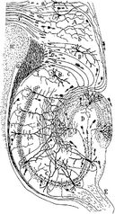
[[Image:CajalHippocampus.jpeg|thumb|128px|Drawing of the neural circuitry of the rodent hippocampus. [[S. Ramón y Cajal]], 1911.]] |
[[Image:CajalHippocampus.jpeg|thumb|128px|Drawing of the neural circuitry of the rodent hippocampus. [[S. Ramón y Cajal]], 1911.]] |
||
| Line 27: | Line 22: | ||
{{Main|Role of the hippocampus in memory}} |
{{Main|Role of the hippocampus in memory}} |
||
| + | |||
| + | == Role in spatial memory and navigation == |
||
| + | |||
| + | Some evidence implicates the hippocampus in storing and processing spatial information. Studies in rats have shown that [[neurons]] in the hippocampus have spatial firing fields. These cells are called ''[[place cell]]s''. Some cells fire when the animal finds itself in a particular location, regardless of direction of travel, while most are at least partially sensitive to head direction and direction of travel. |
||
| + | |||
| + | {{Main|Role of the hippocampus in spatial memory and navigation}} |
||
==Hippocampus and depression== |
==Hippocampus and depression== |
||
| Line 32: | Line 33: | ||
[[neurogenesis]] has been identified in groups of depressed people in comparison with controls. |
[[neurogenesis]] has been identified in groups of depressed people in comparison with controls. |
||
| − | {{Main |
+ | {{Main|Depression and the hippocampus}} |
== History == |
== History == |
||
| − | The anatomist Giulio Cesare Aranzi (circa 1564) first used the term |
+ | The anatomist [[Giulio Cesare Aranzi]] (circa 1564) first used the term '''hippocampus''' to describe the cerebral organ because of its visual resemblance to a seahorse. This organ was initially connected with the sense of smell, rather than with its known function in memory acquisition. The Russian [[Vladimir Bekhterev]] noted the role of the hippocampus in memory around 1900, based on observations of a patient with profound memory disturbances. However, for many years, the conventional view of the hippocampus was that, like the rest of the [[limbic system]], it was responsible for emotion. |
| − | The Russian [[Vladimir Bekhterev]] noted the role of the hippocampus in memory around 1900, based on observations of a patient with profound memory disturbances. However for many years, the conventional view of the hippocampus was that, like the rest of the [[limbic system]], it was responsible for [[emotion]]. |
||
| − | The importance of the hippocampus in memory was brought to the attention of researchers by patient [[HM (patient)|HM]]. HM suffered from a number of anterograde and temporally |
+ | The importance of the hippocampus in memory was brought to the attention of researchers by patient [[HM (patient)|HM]]. HM suffered from a number of anterograde and temporally-graded retrograde memory impairments (such impairments are the subject of the movie ''[[Memento (film)|Memento]]'') following the bilateral removal of various medial-temporal lobe structures (including bilateral ablation of his hippocampi) to relieve frequent [[epileptic seizure]]s. Of particular importance is that HM was still able to learn procedural tasks (which are associated with the [[Striatum|striatum]]) and had an above-average IQ. HM demonstrated a striking single-dissociation between intelligence and declarative memory. The relative size of the hippocampal formation in relation with the total volume of the brain is often conserved in most of the mammalian species. Nevertheless, it has been found that these areas are relatively [[Wiktionary:Hypotrophy|hypotrophic]] in [[cetaceans]]. |
| + | ==Anatomy== |
||
| + | {{main|Hippocampus anatomy}} |
||
| + | [[Image:Gray739-emphasizing-hippocampus.png|thumb|300px|right|Human hippocampus.]] |
||
| − | ==See also== |
||
| + | [[Image:Brainmaps-macaque-hippocampus.jpg|thumb|300px|right|Nissl-stained coronal section of the brain of a macaque monkey, showing hippocampus (circled). Source: brainmaps.org]] |
||
| + | |||
| + | Anatomically, the hippocampus is an elaboration of the edge of the |
||
| + | cortex. It can be distinguished as a zone where the cortex narrows into a single layer of very |
||
| + | densely packed neurons, which curls into a tight S shape. The structures that line the edge of the cortex make up the so-called [[Limbic_system|limbic system]] (Latin ''limbus'' = |
||
| + | ''border''): these include the hippocampus, cingulate cortex, olfactory |
||
| + | cortex, and amygdala. [[Paul_D._MacLean|Paul MacLean]] once suggested, as |
||
| + | part of his [[Triune_brain|triune brain]] theory, that the limbic structures comprise |
||
| + | the neural basis of emotion. Most neuroscientists no longer believe |
||
| + | that the concept of a unified "limbic system" is valid, though. |
||
| + | |||
| + | The hippocampus, as a whole, |
||
| + | ends up looks something like a curved tube, which has been |
||
| + | analogized variously to a seahorse, or a ram's horn (''Cornu Ammonis''), or a banana. |
||
| + | This general layout holds across the full range of mammalian species, |
||
| + | from hedgehog to human, although the details vary. In the rat, the |
||
| + | two hippocampi look astonishingly like a pair of bananas, joined at |
||
| + | the stem. In human or monkey brains, the portion of the hippocampus |
||
| + | down at the bottom, near the base of the temporal lobe, is much |
||
| + | broader than the part at the top. One of the consequences of this complex |
||
| + | geometry is that cross-sections through the hippocampus can show a |
||
| + | bewildering variety of shapes, depending on the angle and location of |
||
| + | the cut. |
||
| + | |||
| + | The strongest connections of the hippocampus are with the entorhinal cortex (EC), which lies next to it in the temporal lobe. The superficial layers of the EC provide the most numerous inputs to the hippocampus, and the deep layers of the EC receive the most numerous outputs. The EC, in turn, is strongly, and reciprocally, connected with many other parts of the cortex. The hippocampus also receives a very important projection from the medial septal area. Destruction of the septal area abolishes the hippocampal theta rhythm, and severely impairs certain types of memory. (So-called "date rape" drugs are thought to exert their amnestic effects at least partly by antagonizing the cholinergic projection from the medial septum to the hippocampus.) |
||
| + | |||
| + | == Physiology == |
||
| + | |||
| + | [[Image:Rat-hippocampal-activity-modes.png|thumb|350px|right|Examples of rat hippocampal EEG and CA1 neural activity in the theta (awake/behaving) and LIA (slow-wave sleep) modes. Each plot show 20 seconds of data, with a hippocampal EEG trace at the top, spike rasters from 40 simultaneously recorded CA1 pyramidal cells in the middle (each raster line represents a different cell), and a plot of running speed at the bottom. The top plot represents a time period during which the rat was actively searching for scattered food pellets. For the bottom plot, the rat was asleep.]] |
||
| + | |||
| + | The hippocampus shows two major "modes" of activity, each associated |
||
| + | with a distinct pattern of EEG waves and neural population activity. |
||
| + | These modes are named after the EEG patterns associated with them: |
||
| + | ''theta'' and ''large irregular activity'' (LIA). Here are some of |
||
| + | their main characteristics in the rat, the animal that has been most |
||
| + | extensively studied:<ref>[[#refBuzsaki2006|Buzsaki, 2006]]</ref> |
||
| + | |||
| + | The '''theta''' mode appears during states of active, alert behavior |
||
| + | (especially locomotion), and also during REM (dreaming) sleep. In the |
||
| + | theta mode, the EEG is dominated by large regular waves with a |
||
| + | frequency range of 6-9 Hz, and the main groups of hippocampal neurons |
||
| + | (pyramidal cells and granule cells) show sparse population activity, |
||
| + | which means that in any short time interval, the great majority of |
||
| + | cells are silent, while the small remaining fraction fire at |
||
| + | relatively high rates, up to 50 spikes in one second for the most |
||
| + | active of them. An active cell typically stays active for from |
||
| + | half a second to a few seconds. As the rat behaves, the active cells |
||
| + | fall silent and new cells become active, but the overall percentage of |
||
| + | active cells remains more or less constant. In many situations, cell |
||
| + | activity is determined largely by the spatial location of the animal, |
||
| + | but other behavioral variables also clearly influence it. |
||
| + | |||
| + | The '''LIA''' mode appears during slow-wave (non-dreaming) sleep, and also |
||
| + | during states of waking immobility, such as resting or eating. In the |
||
| + | LIA mode, the EEG is dominated by sharp waves, which are |
||
| + | randomly-timed large deflections of the EEG signal lasting for 200-300 |
||
| + | msec. These sharp waves also determine the population neural activity |
||
| + | patterns. Between them, pyramidal cells and granule cells are very |
||
| + | quiet (but not silent). During a sharp wave, as many as 5-10% of the |
||
| + | population may emit action potentials during a period of 50 msec; many |
||
| + | of these cells emit not one but a burst of spikes. |
||
| + | |||
| + | These two hippocapampal activity modes can be seen in primates as well |
||
| + | as rats, with the important exception that it has been difficult to |
||
| + | see robust theta rhythmicity in the primate hippocampus. There are, |
||
| + | however, qualitatively similar sharp waves, and similar |
||
| + | state-dependent changes in neural population |
||
| + | activity.<ref>[[#refSkaggs2007|Skaggs et al., 2007]]</ref>. |
||
| + | |||
| + | === The theta rhythm === |
||
| + | |||
| + | Because of its densely packed neural layers, the hippocampus generates |
||
| + | some of the largest EEG signals of any brain structure. In some |
||
| + | situations the EEG is dominated by regular waves, often continuing for |
||
| + | many seconds. This EEG pattern is known as the theta rhythm. It was |
||
| + | one of the earliest EEG phenomena to be discovered: the first |
||
| + | description came from Jung and Kornmuller, in 1938. It was not until |
||
| + | 1954, however, with the publication by Green and Arduini of a long and |
||
| + | thorough study of theta rhythm in rabbits, cats, and monkeys, that |
||
| + | interest really took off.<ref>[[#refGreen1954|Green and Arduini, 1954]]</ref> |
||
| + | Perhaps largely because they related the |
||
| + | theta rhythm to arousal, which was the hot topic of the day, their |
||
| + | paper provoked a flood of followup studies, resulting in the |
||
| + | publication of literally hundreds of studies of the physiology and |
||
| + | pharmacology of theta during the 1950s and 1960s. In spite of this |
||
| + | rather daunting body of work, many questions remained unanswered, |
||
| + | especially the question of function. Even at present this most |
||
| + | critical of questions has not yet been convincingly answered. |
||
| + | |||
| + | Theta rhythmicity is very obvious in rabbits and rodents, and also |
||
| + | clearly present in cats and dogs. Whether theta can be seen in |
||
| + | primates is a vexing question. Green and Arduini reported only very |
||
| + | short bursts of rather irregular rhythmicity in monkeys, and most |
||
| + | later studies have seen little more. However, variations in |
||
| + | methodology have made it difficult to draw strong conclusions.<ref>[[#refCantero2003|Cantero et al., 2003]]</ref> |
||
| + | |||
| + | In rats (the animals that have been by far the most extensively |
||
| + | studied), theta is seen mainly in two conditions: first, when an |
||
| + | animal is walking or in some other way actively interacting with its |
||
| + | surroundings; second, during REM sleep.<ref>[[#refVanderwolf1969|Vanderwolf, 1969]]</ref> |
||
| + | The frequency increases as a |
||
| + | function of running speed, starting at about 6.5 Hz on the low end, |
||
| + | and increasing to about 9 Hz on the high end, although higher |
||
| + | frequencies are sometimes seen for dramatic movements such as jumps |
||
| + | across wide gaps. In other, larger, species of animals, theta |
||
| + | frequencies are generally a bit lower. The behavioral dependency also |
||
| + | seems to vary by species: in cats and rabbits, theta is often |
||
| + | observed during states of motionless alertness. This has been |
||
| + | reported for rats as well, but only when they are severely frightened.<ref>[[#refSainsbury1987|Sainsbury et al., 1987]]</ref> |
||
| + | |||
| + | Theta is not just confined to the hippocampus. In rats, it can be |
||
| + | observed in many parts of the brain, including nearly all that |
||
| + | interact strongly with the hippocampus. The pacemaker for the rhythm |
||
| + | is thought to lie within the medial septal area: this area projects |
||
| + | to all of the regions that show theta rhythmicity, and destruction of |
||
| + | it eliminates theta throughout the brain. (There may be one |
||
| + | exception, a small area in the hypothalamus called the supramamillary |
||
| + | nucleus, which seems to be capable of sustaining theta independently |
||
| + | of the septum in some situations.<ref>[[#refKirk1991|Kirk and McNaughton, 1991]]</ref>) |
||
| + | |||
| + | The function of theta, presuming it has one, has not yet been |
||
| + | convincingly explained, although numerous theories have been proposed.<ref>[[#refBuzsaki2006|Buzsáki, 2006]]</ref> |
||
| + | The most popular trend has been to relate it to learning and memory. |
||
| + | It is well established that lesions of the medial septum---the central |
||
| + | node of the theta system---cause severe disruptions of memory. |
||
| + | However, the medium septum is more than just the controller of theta, |
||
| + | it is also the main source of cholinergic projections to the |
||
| + | hippocampus. It has not been established that septal lesions exert |
||
| + | their effects specifically by eliminating theta. |
||
| + | |||
| + | === Sharp waves === |
||
| + | |||
| + | During sleep, or during waking states when an animal is resting or |
||
| + | otherwise not engaged with its surroundings, the hippocampal EEG shows |
||
| + | a pattern of irregular slow waves, somewhat larger in amplitude than |
||
| + | theta waves. This pattern is occasionally interrupted by large surges |
||
| + | called '''sharp waves'''. These events are associated with bursts of |
||
| + | spike activity, lasting 50-100 msec, in pyramidal cells of CA3 and |
||
| + | CA1. They are also associated with short-lasting high-frequency EEG |
||
| + | oscillations called "ripples". Ripples, with frequencies in the range |
||
| + | 150-200 Hz in rats, can usually be detected only by electrodes located |
||
| + | either inside, or very close to, the CA1 cell body layer. In |
||
| + | contrast, electrodes located anywhere inside the hippocampus, or even |
||
| + | in neighboring brain structures, will often pick up sharp waves as |
||
| + | large slow EEG deflections, lasting 200-400 msec. |
||
| + | |||
| + | In rats, sharp waves are most robust during sleep, when they occur at |
||
| + | an average rate around 1 per second, but in a very irregular temporal |
||
| + | pattern. Sharp waves also occur during inactive waking states, but |
||
| + | they are less frequent then and usually smaller. Sharp waves have |
||
| + | also been observed in the human temporal lobe and monkey hippocampus. |
||
| + | In monkeys, sharp waves are quite robust, but do not occur nearly as |
||
| + | frequently as in rats. |
||
| + | |||
| + | One of the most interesting aspects of sharp waves is that they appear |
||
| + | to be associated with memory. Wilson and McNaughton 1994, and |
||
| + | numerous later studies, reported that when hippocampal place cells |
||
| + | have overlapping spatial firing fields (and therefore often fire in |
||
| + | near-simultaneity), they tend to show correlated activity during sleep |
||
| + | following the behavioral session. This enhancement of correlation, |
||
| + | commonly known as '''reactivation''', has been found to be confined |
||
| + | mainly to sharp waves. It has been proposed that sharp waves are, in |
||
| + | fact, reactivations of neural activity patterns that were memorized |
||
| + | during behavior, driven by strengthening of synaptic connections |
||
| + | within the hippocampus. This idea forms a key component of the |
||
| + | "two-stage memory" theory, advocated by Buzsaki and others, which |
||
| + | proposes that memories are stored within the hippocampus during |
||
| + | behavior, and then later transferred to the neocortex during sleep: |
||
| + | sharp waves are suggested to drive Hebbian synaptic changes in the |
||
| + | neocortical targets of hippocampal output pathways. |
||
| + | |||
| + | ==Evolution== |
||
| + | |||
| + | The hippocampus has a generally similar appearance across the range of |
||
| + | mammal species, from basal ones such as the hedgehog to the most |
||
| + | "advanced" ones such as humans<ref>[[#refWest1990|West, 1990]]</ref>. |
||
| + | The hippocampal-size-to-body-size ratio broadly increases, being about |
||
| + | twice as large for primates as for the hedgehog. It does not, |
||
| + | however, increase at anywhere close to the rate of the neocortex-to-body-size ratio. |
||
| + | Thus, the hippocampus takes up a much larger volume of the cortical |
||
| + | mantle in rodents than in primates. |
||
| + | |||
| + | There is also a general relationship between the size of the |
||
| + | hippocampus and spatial memory: when comparisons are made between |
||
| + | similar species, ones that have a greater capacity for spatial memory |
||
| + | tend to have larger hippocampal volumes.<ref name="Jacobs2003">[[#refJacobs2003|Jacobs, |
||
| + | 2003]]</ref>. This relationship also extends to sex differences: in |
||
| + | species where males and females show strong differences in spatial |
||
| + | memory ability, they also tend to show corresponding differences in |
||
| + | hippocampal volume<ref>[[#refJacobs1990|Jacobs et al., 1990]]</ref> |
||
| + | |||
| + | Non-mammalian species do not have a brain structure that looks like |
||
| + | the mammalian hippocampus, but they have one that is considered |
||
| + | [[Homology_(biology)|homologous]] to it. The hippocampus, as pointed out above, |
||
| + | is essentially the medial edge of the cortex. Only mammals have a |
||
| + | fully developed cortex, but the structure it evolved from, called the |
||
| + | [[pallium]], is present in all vertebrates, even the most primitive |
||
| + | ones such as the lamprey or hagfish<ref>[[#refAboitiz2003|Aboitiz et |
||
| + | al., 2003]]</ref>. The pallium is usually divided into three |
||
| + | zones: medial, lateral, and dorsal. The medial pallium forms the |
||
| + | precursor of the hippocampus. It does not resemble the hippocampus |
||
| + | visually, because the layers are not warped into an S shape or |
||
| + | enwrapped by the dentate gyrus, but the homology in indicated by |
||
| + | strong chemical and functional affinities. There is now evidence that |
||
| + | these hippocampal-like stuctures are involved in spatial cognition in |
||
| + | birds, reptiles, and fish.<ref>[[#refRodriguez2002|Rodríguez et al., 2002]]</ref> |
||
| + | |||
| + | In birds, the correspondence is sufficiently well established that |
||
| + | most anatomists refer to the medial pallial zone as the "avian |
||
| + | hippocampus".<ref>[[#refColombo2000|Colombo and Broadbent, |
||
| + | 2000]]</ref> Numerous species of birds have strong spatial skills, |
||
| + | particularly those that cache food. There is evidence that |
||
| + | food-caching birds have a larger hippocampus than other types of |
||
| + | birds, and that damage to the hippocampus causes impairments in |
||
| + | spatial memory.<ref>[[#refShettleworth2003|Shettleworth, |
||
| + | 2003]]</ref>. |
||
| + | |||
| + | The story for fish is more complex. In [[teleost]] fish (which make |
||
| + | up the great majority of existing species), the forebrain is weirdly |
||
| + | distorted in comparison to other types of vertebrates. Most |
||
| + | neuroanatomists believe that the teleost forebrain is essentially |
||
| + | everted, like a sock turned inside-out, so that structures that lie in |
||
| + | the interior, next to the ventricles, for most vertebrates, are found |
||
| + | on the outside in teleost fish, and vice |
||
| + | versa.<ref>[[#refNieuwenhuys1982|Nieuwenhuys, 1982]]</ref> One of the |
||
| + | consequences of this is that the medial pallium ("hippocampal" zone) |
||
| + | of a typical vertebrate is thought to correspond to the lateral |
||
| + | pallium of a typical fish. Several types of fish (particularly |
||
| + | goldfish) have been show experimentally to have strong spatial memory |
||
| + | abilities, even forming "cognitive maps" of the areas they |
||
| + | inhabit.<ref name="Jacobs2003" /> There is evidence |
||
| + | that damage to the lateral pallium impairs spatial |
||
| + | memory.<ref>[[#refPortavella2002|Portavella et al., 2002]]</ref> |
||
| + | <ref>[[#refVargas2006|Vargas et al., 2006]]</ref> |
||
| + | (Long-distance navigation, such as homing by salmon, seems to rely on |
||
| + | different mechanisms, however.) |
||
| + | |||
| + | Thus, the role of the hippocampal region in navigation appears to begin |
||
| + | far back in vertebrate evolution, predating splits that occurred |
||
| + | hundreds of millions of years ago.<ref>[[#refBroglio2005|Broglio et |
||
| + | al., 2005]]</ref> It is not yet known whether the medial pallium plays |
||
| + | a similar role in even more primitive vertebrates, such as sharks and |
||
| + | rays, or even lampreys and hagfish. Some types of insects, and |
||
| + | molluscs such as the octopus, also have strong spatial learning and |
||
| + | navigation abilities, but these appear to work differently from the |
||
| + | mammalian spatial system, so there is as yet no good reason to think |
||
| + | that they have a common evolutionary origin; nor is there sufficient |
||
| + | similarity in brain structure to enable anything resembling a |
||
| + | "hippocampus" to be identified in these species. |
||
| + | |||
| + | |||
| + | |||
| + | |||
| + | |||
| + | ==See also== |
||
| + | *[[Amygdalohippocampectomy]] |
||
| + | *[[Median forebrain bundle]] |
||
| + | *[[Septal nuclei]] |
||
==References & Bibliography== |
==References & Bibliography== |
||
| + | {{Reflist|2}} |
||
| + | |||
| + | |||
==Key texts== |
==Key texts== |
||
===Books=== |
===Books=== |
||
| + | *<cite id=Aboitiz2003>{{cite journal |
||
| − | O'Keefe, J. and Nadel, L. (1978) The Hippocampus as a Cognitive Map, Oxford: Clarendon Press. |
||
| + | | last =Aboitiz |
||
| + | | first =F |
||
| + | | coauthors = Morales D, Montiel J |
||
| + | | title =The evolutionary origin of the mammalian isocortex: Towards an integrated developmental and functional approach |
||
| + | |journal =Behav. Brain Sciences |
||
| + | | volume = 26 |
||
| + | | pages = 535-552 |
||
| + | | date =2003 |
||
| + | | pmid=15179935 |
||
| + | }}</cite> |
||
| + | |||
| + | *<cite id=Amaral2006>{{cite book |
||
| + | | last=Amaral |
||
| + | | first=D |
||
| + | | coauthors=Lavenex P |
||
| + | | editor=Andersen P, Morris R, Amaral D, Bliss T, O'Keefe J |
||
| + | | title=The Hippocampus Book |
||
| + | | year=2006 |
||
| + | | chapter=Ch 3. Hippocampal Neuroanatomy |
||
| + | | publisher=Oxford University Press |
||
| + | | isbn=9780195100273}}</cite> |
||
| + | |||
| + | *<cite id=Broglio2005>{{cite journal |
||
| + | | last =Broglio |
||
| + | | first =C |
||
| + | | coauthors = Gómez A, Durán E, Ocaña FM, Jiménez-Moya F, Rodríguez F, Salas C |
||
| + | | title =Hallmarks of a common forebrain vertebrate plan: Specialized pallial areas for spatial, temporal and emotional memory in actinopterygian fish |
||
| + | |journal =Brain Res. Bull. |
||
| + | | volume = 57 |
||
| + | | pages = 397-399 |
||
| + | | date =2002 |
||
| + | | pmid =16144602 |
||
| + | }}</cite> |
||
| + | |||
| + | *<cite id=Buzsaki2002>{{cite journal |
||
| + | | last =Buzsáki |
||
| + | | first =G |
||
| + | | title =Theta oscillations in the hippocampus |
||
| + | | journal =Neuron |
||
| + | | volume = 33 |
||
| + | | pages = 325-340 |
||
| + | | date =2002 |
||
| + | | pmid =11832222 |
||
| + | | url =http://osiris.rutgers.edu/BuzsakiHP/Publications/PDFs/BuzsakiTheta.pdf |
||
| + | }}</cite> |
||
| + | |||
| + | *<cite id=Buzsaki2006>{{cite journal |
||
| + | | last =Buzsáki |
||
| + | | first =G |
||
| + | | title =Rhythms of the Brain |
||
| + | | publisher =Oxford University Press |
||
| + | | date =2006 |
||
| + | | isbn =0195301064 |
||
| + | | url =http://books.google.com/books?id=yVz4d4d9ZzsC |
||
| + | }}</cite> |
||
| + | |||
| + | *<cite id=Cantero2003>{{cite journal |
||
| + | | last =Cantero |
||
| + | | first =JL |
||
| + | | coauthors =Atienza M, Stickgold R, Kahana MJ, Madsen JR, Kocsis B |
||
| + | | title =Sleep-dependent theta oscillations in the human hippocampus and neocortex |
||
| + | | journal =J. Neurosci. |
||
| + | | volume = 23 |
||
| + | | pages = 10897-10903 |
||
| + | | date =2003 |
||
| + | | pmid =14645485 |
||
| + | | url =http://www.jneurosci.org/cgi/content/full/23/34/10897 |
||
| + | }}</cite> |
||
| + | |||
| + | *<cite id=Colombo2000>{{cite journal |
||
| + | | last =Colombo |
||
| + | | first =M |
||
| + | | coauthors = Broadbent N |
||
| + | | title =Is the avian hippocampus a functional homologue of the mammalian hippocampus? |
||
| + | |journal =Neurosci. Biobehav. Rev. |
||
| + | | volume = 24 |
||
| + | | pages = 465-484 |
||
| + | | date =2000 |
||
| + | | pmid=10817844}}</cite> |
||
| + | |||
| + | *<cite id=Eichenbaum1993>{{cite book |
||
| + | | last =Eichnbaum |
||
| + | | first =H |
||
| + | | coauthors =Cohen NJ |
||
| + | | title = Memory, Amnesia, and the Hippocampal System |
||
| + | | date =1993 | publisher =MIT Press |
||
| + | }}</cite> |
||
| + | |||
| + | *<cite id=Ekstrom2003>{{cite journal |
||
| + | | last =Ekstrom |
||
| + | | first =AD |
||
| + | | coauthors =Kahana MJ, Caplan JB, Fields TA, Isham EA, Newman EL, Fried I |
||
| + | | title =Cellular networks underlying human spatial navigation |
||
| + | | journal =Nature |
||
| + | | volume =425 |
||
| + | | pages =184-188 |
||
| + | | date =2003 |
||
| + | | url =http://memory.psych.upenn.edu/publications/files/EkstEtal03.pdf |
||
| + | | pmid =12968182 |
||
| + | }} </cite> |
||
| + | |||
| + | *<cite id=Gray2000>{{cite book |
||
| + | | last =Gray |
||
| + | | first =JA |
||
| + | | coauthors =McNaughton N |
||
| + | | title = The Neuropsychology of Anxiety: An Enquiry into the Functions of the Septo-Hippocampal System |
||
| + | | date =2000 |
||
| + | | publisher =Oxford University Press |
||
| + | }}</cite> |
||
| + | |||
| + | *<cite id=Green1954>{{cite journal |
||
| + | | last =Green |
||
| + | | first =JD |
||
| + | | coauthors = Arduini AA |
||
| + | | title =Hippocampal electrical activity in arousal |
||
| + | | journal =J. Neurophysiol. |
||
| + | | volume = 17 |
||
| + | | pages = 533-557 |
||
| + | | date =1954 |
||
| + | | pmid =13212425 |
||
| + | | url =http://jn.physiology.org/cgi/content/citation/17/6/533 |
||
| + | }}</cite> |
||
| + | |||
| + | *<cite id=Jacobs1990>{{cite journal |
||
| + | | last =Jacobs |
||
| + | | first =LF |
||
| + | | coauthors = Gaulin SJ, Sherry DF, Hoffman GE |
||
| + | | title =Evolution of spatial cognition: sex-specific patterns of spatial behavior predict hippocampal size |
||
| + | |journal =PNAS |
||
| + | | volume = 87 |
||
| + | | pages = 6349-6352 |
||
| + | | date =1990 |
||
| + | | url=http://www.pnas.org/cgi/reprint/87/16/6349 |
||
| + | | pmid=2201026}}</cite> |
||
| + | |||
| + | *<cite id=Jacobs2003>{{cite journal |
||
| + | | last =Jacobs |
||
| + | | first =LF |
||
| + | | title =The Evolution of the Cognitive Map |
||
| + | |journal =Brain Behav. Evol. |
||
| + | | volume = 62 |
||
| + | | pages = 128-139 |
||
| + | | date =2003 |
||
| + | | doi=10.1159/000072443 |
||
| + | | pmid=12937351}}</cite> |
||
| + | |||
| + | *<cite id=Kirk1991>{{cite journal |
||
| + | | last =Kirk |
||
| + | | first =IJ |
||
| + | | coauthors =McNaughton N |
||
| + | | title =Supramammillary cell firing and hippocampal rhythmical slow activity |
||
| + | | journal =Neuroreport |
||
| + | | volume = 11 |
||
| + | | pages = 723-725 |
||
| + | | date =1991 |
||
| + | | pmid =1810464 |
||
| + | }}</cite> |
||
| + | |||
| + | *<cite id=MaguireFrith2000>{{cite journal |
||
| + | | last =Maguire |
||
| + | | first =EA | authorlink = |
||
| + | | coauthors =Gadian DG, Johnsrude IS, Good CD, Ashburner J, Frackowiak RS, Frith CD |
||
| + | | title = Navigation-related structural change in the hippocampi of taxi drivers |
||
| + | | journal =PNAS |
||
| + | | volume = 97 |
||
| + | | pages =4398-4403 |
||
| + | | date =2000 |
||
| + | | url =http://www.pnas.org/cgi/content/full/97/8/4398 |
||
| + | | doi =10.1073/pnas.070039597 |
||
| + | | pmid =10716738 |
||
| + | }}</cite> |
||
| + | |||
| + | *<cite id=McNaughton2006>{{cite journal |
||
| + | | last =McNaughton |
||
| + | | first =BL |
||
| + | | coauthors =Battaglia FP, Jensen O, Moser EI, Moser MB |
||
| + | | title =Path integration and the neural basis of the 'cognitive map' |
||
| + | | journal =Nat. Rev. Neurosci. |
||
| + | | volume = 7 |
||
| + | | pages = 663-678 |
||
| + | | date =2006 |
||
| + | | url =http://www.nature.com/nrn/journal/v7/n8/abs/nrn1932.html |
||
| + | | pmid =16858394 |
||
| + | }}</cite> |
||
| + | |||
| + | *<cite id=Moser2008>{{cite journal |
||
| + | | last =Moser |
||
| + | | first =EI |
||
| + | | coauthors = Kropf E, Moser M-B |
||
| + | | title =Place Cells, Grid Cells, and the Brain's Spatial Representation System |
||
| + | | journal =Ann. Rev. Neurosci. |
||
| + | | volume = 31 |
||
| + | | date =2008 |
||
| + | | doi =10.1146/annurev.neuro.31.061307.090723 |
||
| + | }}</cite> |
||
| + | |||
| + | *<cite id=Nieuwenhuys1982>{{cite journal |
||
| + | | last =Nieuwenhuys |
||
| + | | first =R |
||
| + | | title =An Overview of the Organization of the Brain of Actinopterygian Fishes |
||
| + | |journal =Am. Zool. |
||
| + | | volume = 22 |
||
| + | | pages = 287-310 |
||
| + | | date =1982 |
||
| + | | doi =10.1093/icb/22.2.287 |
||
| + | }}</cite> |
||
| + | |||
| + | *<cite id=OKane2004>{{cite journal |
||
| + | | last =O'Kane |
||
| + | | first =G |
||
| + | | coauthors = Kensinger EA, Corkin S |
||
| + | | title =Evidence for semantic learning in profound amnesia: An investigation with patient H.M. |
||
| + | | journal =Hippocampus |
||
| + | | volume = 14 |
||
| + | | pages = 417-425 |
||
| + | | date =2004 |
||
| + | | url =http://www3.interscience.wiley.com/cgi-bin/abstract/108562086/ABSTRACT |
||
| + | | pmid =15224979 |
||
| + | }}</cite> |
||
| + | |||
| + | *<cite id=OKeefe1978>{{cite book |
||
| + | | last =O'Keefe |
||
| + | | first =J |
||
| + | | coauthors =Nadel L |
||
| + | | title = The Hippocampus as a Cognitive Map |
||
| + | | date =1978 | publisher =Oxford University Press |
||
| + | | url = http://www.cognitivemap.net/HCMpdf/HCMChapters.html |
||
| + | }}</cite> |
||
| + | |||
| + | *<cite id=Portavella2002>{{cite journal |
||
| + | | last =Portavella |
||
| + | | first =M |
||
| + | | coauthors = Vargas JP, Torres B, Salas C |
||
| + | | title =The effects of telencephalic pallial lesions on spatial, temporal, and emotional learning in goldfish |
||
| + | |journal =Brain Res. Bull. |
||
| + | | volume = 57 |
||
| + | | pages = 397-399 |
||
| + | | date =2002 |
||
| + | | pmid =11922997 |
||
| + | }}</cite> |
||
| + | |||
| + | *<cite id=Rodriguez2002>{{cite journal |
||
| + | | last =Rodríguez |
||
| + | | first =F |
||
| + | | coauthors = Lópeza JC, Vargasa JP, Broglioa C, Gómeza Y, Salas C |
||
| + | | title =Spatial memory and hippocampal pallium through vertebrate evolution: insights from reptiles and teleost fish |
||
| + | |journal =Brain Res. Bull. |
||
| + | | volume = 57 |
||
| + | | pages = 499-503 |
||
| + | | date =2002 |
||
| + | | pmid=11923018}}</cite> |
||
| + | |||
| + | *<cite id=Sainsbury1987>{{cite journal |
||
| + | | last =Sainsbury |
||
| + | | first =RS |
||
| + | | coauthors =Heynen A, Montoya CP |
||
| + | | title =Behavioral correlates of hippocampal type 2 theta in the rat |
||
| + | | journal =Physiol. Behav. |
||
| + | | volume = 39 |
||
| + | | pages = 513-519 |
||
| + | | date =1987 |
||
| + | | pmid =3575499 |
||
| + | }}</cite> |
||
| + | |||
| + | *<cite id=Scoville1957>{{cite journal |
||
| + | | last =Scoville |
||
| + | | first =WB |
||
| + | | authorlink = |
||
| + | | coauthors =Milner B |
||
| + | | title = Loss of Recent Memory After Bilateral Hippocampal Lesions |
||
| + | | journal =J. Neurol. Neurosurg. Psych. |
||
| + | | volume = 20 |
||
| + | | pages =11-21 |
||
| + | | date =1957 |
||
| + | | url = http://neuro.psychiatryonline.org/cgi/content/full/12/1/103 |
||
| + | }}</cite> |
||
| + | |||
| + | *<cite id=Shettleworth2003>{{cite journal |
||
| + | | last =Shettleworth |
||
| + | | first =SJ |
||
| + | | title =Memory and Hippocampal Specialization in Food-Storing Birds: Challenges for Research on Comparative Cognition |
||
| + | |journal =Brain Behav. Evol. |
||
| + | | volume = 62 |
||
| + | | pages = 108-116 |
||
| + | | date =2003 |
||
| + | | pmid=12937349}}</cite> |
||
| + | |||
| + | *<cite id=Skaggs1996>{{cite journal |
||
| + | | last =Skaggs |
||
| + | | first =WE |
||
| + | | coauthors = McNaughton BL, Wilson MA, Barnes CA |
||
| + | | title =Theta phase precession in hippocampal neuronal populations and the compression of temporal sequences |
||
| + | | journal =Hippocampus |
||
| + | | volume = 6 |
||
| + | | pages = 149-176 |
||
| + | | date =1996 |
||
| + | | url =http://www3.interscience.wiley.com/cgi-bin/abstract/72392/ABSTRACT |
||
| + | | pmid=8797016}}</cite> |
||
| + | |||
| + | *<cite id=Squire1992>{{cite journal |
||
| + | | last =Squire |
||
| + | | first =LR |
||
| + | | title = Memory and the hippocampus: a synthesis from findings with rats, monkeys, and humans |
||
| + | | journal =Psych. Rev. |
||
| + | | volume = 99 |
||
| + | | pages =195-231 |
||
| + | | date =1992 |
||
| + | }}</cite> |
||
| + | |||
| + | *<cite id=Squire2002>{{cite book |
||
| + | | last =Squire |
||
| + | | first =LR |
||
| + | | coauthors =Schacter DL |
||
| + | | title = The Neuropsychology of Memory |
||
| + | | date =2002 |
||
| + | | publisher =Guilford Press |
||
| + | }}</cite> |
||
| + | |||
| + | *<cite id=Vanderwolf1969>{{cite journal |
||
| + | | last =Vanderwolf |
||
| + | | first =CH |
||
| + | | title =Hippocampal electrical activity and voluntary movement in the rat |
||
| + | | journal =EEG & Clin. Neurophysiol. |
||
| + | | volume = 26 |
||
| + | | pages = 407-418 |
||
| + | | date =1969 |
||
| + | | pmid =4183562 |
||
| + | | url =http://psycnet.apa.org/?fa=main.doiLanding&uid=1970-20296-001 |
||
| + | }}</cite> |
||
| + | |||
| + | *<cite id=Vargas2006>{{cite journal |
||
| + | | last =Vargas |
||
| + | | first =JP |
||
| + | | coauthors = Bingman VP, Portavella M, López JC |
||
| + | | title =Telencephalon and geometric space in goldfish |
||
| + | | journal =Eur. J. Neurosci. |
||
| + | | volume = 24 |
||
| + | | pages = 2870-2878 |
||
| + | | date =2006 |
||
| + | | pmid =17156211 |
||
| + | }}</cite> |
||
| + | |||
| + | *<cite id=West1990>{{cite journal |
||
| + | | last =West |
||
| + | | first =MJ |
||
| + | | title =Stereological studies of the hippocampus: a comparison of the hippocampal subdivisions of diverse species including hedgehogs, laboratory rodents, wild mice and men. |
||
| + | |journal =Prog. Brain Res. |
||
| + | | volume = 83 |
||
| + | | pages = 13-36 |
||
| + | | date =1990 |
||
| + | | pmid =2203095}}</cite> |
||
| + | |||
| + | |||
| + | |||
| + | |||
| + | |||
===Papers=== |
===Papers=== |
||
*Amaral DG and Cowan WM. 1980. Subcortical afferents to the hippocampal formation in the monkey. ''Journal of Comparative Neurology''. Feb 15; 189(4):573-91. |
*Amaral DG and Cowan WM. 1980. Subcortical afferents to the hippocampal formation in the monkey. ''Journal of Comparative Neurology''. Feb 15; 189(4):573-91. |
||
| − | |||
*Duvernoy, H. (2005) ''The Human Hippocampus'', 3rd ed. Berlin: Springer-Verlag. |
*Duvernoy, H. (2005) ''The Human Hippocampus'', 3rd ed. Berlin: Springer-Verlag. |
||
| Line 60: | Line 678: | ||
===Papers=== |
===Papers=== |
||
*[http://scholar.google.com/scholar?sourceid=mozclient&num=50&scoring=d&ie=utf-8&oe=utf-8&q=Hippocampus Google Scholar] |
*[http://scholar.google.com/scholar?sourceid=mozclient&num=50&scoring=d&ie=utf-8&oe=utf-8&q=Hippocampus Google Scholar] |
||
| − | |||
| − | ==External links== |
||
| − | |||
| − | |||
| − | ==References== |
||
| − | |||
==External links== |
==External links== |
||
| Line 72: | Line 684: | ||
* [http://www.newscientist.com/news/news.jsp?id=ns99993488 Artificial Hippocampus] |
* [http://www.newscientist.com/news/news.jsp?id=ns99993488 Artificial Hippocampus] |
||
* {{BrainInfo|hier|164}} |
* {{BrainInfo|hier|164}} |
||
| + | * [http://www.cognitivemap.net/HCMpdf/HCMChapters.html John O'Keefe & Lynn Nadel (1978) The Hippocampus as a Cognitive Map , Oxford University Press. Full Text] |
||
| + | |||
| + | ==Additional images== |
||
| + | <gallery> |
||
| + | Image:Gray682.png|Superficial dissection of brain-stem. Lateral view. |
||
| + | Image:Gray717.png|Coronal section of brain immediately in front of pons. |
||
| + | Image:Gray739.png|Posterior and inferior cornua of left lateral ventricle exposed from the side. |
||
| + | Image:Gray740.png|Inferior and posterior cornua, viewed from above. |
||
| + | Image:Gray748.png|The fornix and corpus callosum from below. |
||
| + | Image:Human brain frontal (coronal) section description 2.JPG|Human brain frontal (coronal) section |
||
| + | Image:Human brain right dissected lateral view description.JPG|Human brain right dissected lateral view |
||
| + | Image:Hippocampus (brain).jpg||Diagram of the humanhippocampus |
||
| + | Image:Dopamineseratonin.gif|Dopamine and serotonin pathways |
||
| + | Image:HippocampalRegions.jpg|Hippocampal areas in a Nissl-stained coronal section of the rat brain. DG: [[Dentate gyrus]].]] |
||
| + | |||
| + | </gallery> |
||
| + | |||
| + | |||
| + | |||
{{Limbic system}} |
{{Limbic system}} |
||
{{Telencephalon}} |
{{Telencephalon}} |
||
| + | |||
| + | |||
| + | |||
| + | {{enWP| Hippocampus}} |
||
[[Category:Hippocampus| ]] |
[[Category:Hippocampus| ]] |
||
[[Category:Limbic system]] |
[[Category:Limbic system]] |
||
| Line 82: | Line 717: | ||
[[Category:Neuroscience]] |
[[Category:Neuroscience]] |
||
[[Category:Limbic system]] |
[[Category:Limbic system]] |
||
| − | |||
| − | |||
| − | |||
| − | {{enWP| Hippocampus}} |
||
Latest revision as of 14:25, 4 July 2013
Assessment |
Biopsychology |
Comparative |
Cognitive |
Developmental |
Language |
Individual differences |
Personality |
Philosophy |
Social |
Methods |
Statistics |
Clinical |
Educational |
Industrial |
Professional items |
World psychology |
Biological: Behavioural genetics · Evolutionary psychology · Neuroanatomy · Neurochemistry · Neuroendocrinology · Neuroscience · Psychoneuroimmunology · Physiological Psychology · Psychopharmacology (Index, Outline)
The location of the hippocampus in the human brain.
- For the journal of the same name, see Hippocampus (journal)
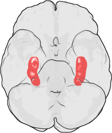
The hippocampus is a part of the forebrain, located in the medial temporal lobe. It belongs to the limbic system and plays major roles in long term memory and spatial navigation. Humans and other mammals have two hippocampi, one in each side of the brain. In rodents, where it has been studied most extensively, the hippocampus is shaped something like a banana. In humans, it has a curved and convoluted shape that reminded early anatomists of a seahorse. The name, in fact, derives from the Greek word for seahorse (Greek: hippos = horse, campos = sea).
In Alzheimer's disease, the hippocampus is one of the first regions of the brain to suffer damage; memory problems and disorientation appear among the first symptoms. Damage to the hippocampus can also result from oxygen starvation (anoxia), encephalitis or mesial temporal lobe epilepsy. People with extensive hippocampal damage may experience amnesia, that is, inability to form or retain new memories.
Functions of the hippocampus
Perhaps the earliest idea was that the hippocampus is involved in olfaction: this seems to have been suggested mainly by its location in the brain, next to the olfactory cortex. There continues to be some interest in hippocampal olfactory responses, but almost nobody now believes that the primary function of the hippocampus is olfactory.
Over the years, three main ideas of hippocampal function have dominated the literature: inhibition, memory, and space. The behavioral inhibition theory (caricatured by O'Keefe and Nadel as "step on the brakes!") was very popular up to the 1960s. It derived much of its force from two observations: first, animals with hippocampal damage tend to be hyperactive; second, animals with hippocampal damage often have difficulty learning to inhibit responses that they have previously been taught. Jeffrey Gray developed this line of thought into a full-fledged theory of the role of the hippocampus in anxiety[1]. The inhibition theory is not, however, very popular at present.
The second important line of thought relates the hippocampus to memory. Although it had precursors, this idea derived its main force from a very well-known report by Scoville and Milner[2] of the results of surgical destruction of the hippocampus (in an attempt to relieve epileptic seizures), in a patient known as H.M. The unexpected outcome was severe amnesia: H.M. was unable to consciously remember events that occurred after his surgery or for several years before it. This case occasioned such enormous interest that H.M. is now said to be the most intensively studied medical case in history. In the ensuing years, other patients with similar levels of hippocampal damage and amnesia (caused by accident or disease) have been studied as well, and literally thousands of experiments have studied the physiology of neural plasticity in the hippocampus. There is now almost universal agreement that the hippocampus plays some sort of important role in memory; however, the precise nature of this role remains widely debated[3][4].

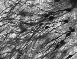
Golgi-stained neurons in the rodent hippocampus.
Role in memory
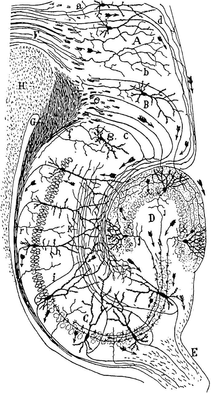
Drawing of the neural circuitry of the rodent hippocampus. S. Ramón y Cajal, 1911.
Psychologists and neuroscientists dispute the precise role of the hippocampus, but generally agree that it has an essential role in the formation of new memories about personally experienced events (episodic or autobiographical memory). Some researchers prefer to consider the hippocampus as part of a larger medial temporal lobe memory system responsible for general declarative memory (memories which can be explicitly verbalized—these would include, for example, memory for facts in addition to episodic memory).
- Main article: Role of the hippocampus in memory
Some evidence implicates the hippocampus in storing and processing spatial information. Studies in rats have shown that neurons in the hippocampus have spatial firing fields. These cells are called place cells. Some cells fire when the animal finds itself in a particular location, regardless of direction of travel, while most are at least partially sensitive to head direction and direction of travel.
- Main article: Role of the hippocampus in spatial memory and navigation
Hippocampus and depression
- Hippocampal volume loss,perhaps reflecting the effects of excessive glucocorticoids on
neurogenesis has been identified in groups of depressed people in comparison with controls.
- Main article: Depression and the hippocampus
History
The anatomist Giulio Cesare Aranzi (circa 1564) first used the term hippocampus to describe the cerebral organ because of its visual resemblance to a seahorse. This organ was initially connected with the sense of smell, rather than with its known function in memory acquisition. The Russian Vladimir Bekhterev noted the role of the hippocampus in memory around 1900, based on observations of a patient with profound memory disturbances. However, for many years, the conventional view of the hippocampus was that, like the rest of the limbic system, it was responsible for emotion.
The importance of the hippocampus in memory was brought to the attention of researchers by patient HM. HM suffered from a number of anterograde and temporally-graded retrograde memory impairments (such impairments are the subject of the movie Memento) following the bilateral removal of various medial-temporal lobe structures (including bilateral ablation of his hippocampi) to relieve frequent epileptic seizures. Of particular importance is that HM was still able to learn procedural tasks (which are associated with the striatum) and had an above-average IQ. HM demonstrated a striking single-dissociation between intelligence and declarative memory. The relative size of the hippocampal formation in relation with the total volume of the brain is often conserved in most of the mammalian species. Nevertheless, it has been found that these areas are relatively hypotrophic in cetaceans.
Anatomy
- Main article: Hippocampus anatomy
Human hippocampus.
Nissl-stained coronal section of the brain of a macaque monkey, showing hippocampus (circled). Source: brainmaps.org
Anatomically, the hippocampus is an elaboration of the edge of the cortex. It can be distinguished as a zone where the cortex narrows into a single layer of very densely packed neurons, which curls into a tight S shape. The structures that line the edge of the cortex make up the so-called limbic system (Latin limbus = border): these include the hippocampus, cingulate cortex, olfactory cortex, and amygdala. Paul MacLean once suggested, as part of his triune brain theory, that the limbic structures comprise the neural basis of emotion. Most neuroscientists no longer believe that the concept of a unified "limbic system" is valid, though.
The hippocampus, as a whole, ends up looks something like a curved tube, which has been analogized variously to a seahorse, or a ram's horn (Cornu Ammonis), or a banana. This general layout holds across the full range of mammalian species, from hedgehog to human, although the details vary. In the rat, the two hippocampi look astonishingly like a pair of bananas, joined at the stem. In human or monkey brains, the portion of the hippocampus down at the bottom, near the base of the temporal lobe, is much broader than the part at the top. One of the consequences of this complex geometry is that cross-sections through the hippocampus can show a bewildering variety of shapes, depending on the angle and location of the cut.
The strongest connections of the hippocampus are with the entorhinal cortex (EC), which lies next to it in the temporal lobe. The superficial layers of the EC provide the most numerous inputs to the hippocampus, and the deep layers of the EC receive the most numerous outputs. The EC, in turn, is strongly, and reciprocally, connected with many other parts of the cortex. The hippocampus also receives a very important projection from the medial septal area. Destruction of the septal area abolishes the hippocampal theta rhythm, and severely impairs certain types of memory. (So-called "date rape" drugs are thought to exert their amnestic effects at least partly by antagonizing the cholinergic projection from the medial septum to the hippocampus.)
Physiology
Examples of rat hippocampal EEG and CA1 neural activity in the theta (awake/behaving) and LIA (slow-wave sleep) modes. Each plot show 20 seconds of data, with a hippocampal EEG trace at the top, spike rasters from 40 simultaneously recorded CA1 pyramidal cells in the middle (each raster line represents a different cell), and a plot of running speed at the bottom. The top plot represents a time period during which the rat was actively searching for scattered food pellets. For the bottom plot, the rat was asleep.
The hippocampus shows two major "modes" of activity, each associated with a distinct pattern of EEG waves and neural population activity. These modes are named after the EEG patterns associated with them: theta and large irregular activity (LIA). Here are some of their main characteristics in the rat, the animal that has been most extensively studied:[5]
The theta mode appears during states of active, alert behavior (especially locomotion), and also during REM (dreaming) sleep. In the theta mode, the EEG is dominated by large regular waves with a frequency range of 6-9 Hz, and the main groups of hippocampal neurons (pyramidal cells and granule cells) show sparse population activity, which means that in any short time interval, the great majority of cells are silent, while the small remaining fraction fire at relatively high rates, up to 50 spikes in one second for the most active of them. An active cell typically stays active for from half a second to a few seconds. As the rat behaves, the active cells fall silent and new cells become active, but the overall percentage of active cells remains more or less constant. In many situations, cell activity is determined largely by the spatial location of the animal, but other behavioral variables also clearly influence it.
The LIA mode appears during slow-wave (non-dreaming) sleep, and also during states of waking immobility, such as resting or eating. In the LIA mode, the EEG is dominated by sharp waves, which are randomly-timed large deflections of the EEG signal lasting for 200-300 msec. These sharp waves also determine the population neural activity patterns. Between them, pyramidal cells and granule cells are very quiet (but not silent). During a sharp wave, as many as 5-10% of the population may emit action potentials during a period of 50 msec; many of these cells emit not one but a burst of spikes.
These two hippocapampal activity modes can be seen in primates as well as rats, with the important exception that it has been difficult to see robust theta rhythmicity in the primate hippocampus. There are, however, qualitatively similar sharp waves, and similar state-dependent changes in neural population activity.[6].
The theta rhythm
Because of its densely packed neural layers, the hippocampus generates some of the largest EEG signals of any brain structure. In some situations the EEG is dominated by regular waves, often continuing for many seconds. This EEG pattern is known as the theta rhythm. It was one of the earliest EEG phenomena to be discovered: the first description came from Jung and Kornmuller, in 1938. It was not until 1954, however, with the publication by Green and Arduini of a long and thorough study of theta rhythm in rabbits, cats, and monkeys, that interest really took off.[7] Perhaps largely because they related the theta rhythm to arousal, which was the hot topic of the day, their paper provoked a flood of followup studies, resulting in the publication of literally hundreds of studies of the physiology and pharmacology of theta during the 1950s and 1960s. In spite of this rather daunting body of work, many questions remained unanswered, especially the question of function. Even at present this most critical of questions has not yet been convincingly answered.
Theta rhythmicity is very obvious in rabbits and rodents, and also clearly present in cats and dogs. Whether theta can be seen in primates is a vexing question. Green and Arduini reported only very short bursts of rather irregular rhythmicity in monkeys, and most later studies have seen little more. However, variations in methodology have made it difficult to draw strong conclusions.[8]
In rats (the animals that have been by far the most extensively studied), theta is seen mainly in two conditions: first, when an animal is walking or in some other way actively interacting with its surroundings; second, during REM sleep.[9] The frequency increases as a function of running speed, starting at about 6.5 Hz on the low end, and increasing to about 9 Hz on the high end, although higher frequencies are sometimes seen for dramatic movements such as jumps across wide gaps. In other, larger, species of animals, theta frequencies are generally a bit lower. The behavioral dependency also seems to vary by species: in cats and rabbits, theta is often observed during states of motionless alertness. This has been reported for rats as well, but only when they are severely frightened.[10]
Theta is not just confined to the hippocampus. In rats, it can be observed in many parts of the brain, including nearly all that interact strongly with the hippocampus. The pacemaker for the rhythm is thought to lie within the medial septal area: this area projects to all of the regions that show theta rhythmicity, and destruction of it eliminates theta throughout the brain. (There may be one exception, a small area in the hypothalamus called the supramamillary nucleus, which seems to be capable of sustaining theta independently of the septum in some situations.[11])
The function of theta, presuming it has one, has not yet been convincingly explained, although numerous theories have been proposed.[12] The most popular trend has been to relate it to learning and memory. It is well established that lesions of the medial septum---the central node of the theta system---cause severe disruptions of memory. However, the medium septum is more than just the controller of theta, it is also the main source of cholinergic projections to the hippocampus. It has not been established that septal lesions exert their effects specifically by eliminating theta.
Sharp waves
During sleep, or during waking states when an animal is resting or otherwise not engaged with its surroundings, the hippocampal EEG shows a pattern of irregular slow waves, somewhat larger in amplitude than theta waves. This pattern is occasionally interrupted by large surges called sharp waves. These events are associated with bursts of spike activity, lasting 50-100 msec, in pyramidal cells of CA3 and CA1. They are also associated with short-lasting high-frequency EEG oscillations called "ripples". Ripples, with frequencies in the range 150-200 Hz in rats, can usually be detected only by electrodes located either inside, or very close to, the CA1 cell body layer. In contrast, electrodes located anywhere inside the hippocampus, or even in neighboring brain structures, will often pick up sharp waves as large slow EEG deflections, lasting 200-400 msec.
In rats, sharp waves are most robust during sleep, when they occur at an average rate around 1 per second, but in a very irregular temporal pattern. Sharp waves also occur during inactive waking states, but they are less frequent then and usually smaller. Sharp waves have also been observed in the human temporal lobe and monkey hippocampus. In monkeys, sharp waves are quite robust, but do not occur nearly as frequently as in rats.
One of the most interesting aspects of sharp waves is that they appear to be associated with memory. Wilson and McNaughton 1994, and numerous later studies, reported that when hippocampal place cells have overlapping spatial firing fields (and therefore often fire in near-simultaneity), they tend to show correlated activity during sleep following the behavioral session. This enhancement of correlation, commonly known as reactivation, has been found to be confined mainly to sharp waves. It has been proposed that sharp waves are, in fact, reactivations of neural activity patterns that were memorized during behavior, driven by strengthening of synaptic connections within the hippocampus. This idea forms a key component of the "two-stage memory" theory, advocated by Buzsaki and others, which proposes that memories are stored within the hippocampus during behavior, and then later transferred to the neocortex during sleep: sharp waves are suggested to drive Hebbian synaptic changes in the neocortical targets of hippocampal output pathways.
Evolution
The hippocampus has a generally similar appearance across the range of mammal species, from basal ones such as the hedgehog to the most "advanced" ones such as humans[13]. The hippocampal-size-to-body-size ratio broadly increases, being about twice as large for primates as for the hedgehog. It does not, however, increase at anywhere close to the rate of the neocortex-to-body-size ratio. Thus, the hippocampus takes up a much larger volume of the cortical mantle in rodents than in primates.
There is also a general relationship between the size of the hippocampus and spatial memory: when comparisons are made between similar species, ones that have a greater capacity for spatial memory tend to have larger hippocampal volumes.[14]. This relationship also extends to sex differences: in species where males and females show strong differences in spatial memory ability, they also tend to show corresponding differences in hippocampal volume[15]
Non-mammalian species do not have a brain structure that looks like the mammalian hippocampus, but they have one that is considered homologous to it. The hippocampus, as pointed out above, is essentially the medial edge of the cortex. Only mammals have a fully developed cortex, but the structure it evolved from, called the pallium, is present in all vertebrates, even the most primitive ones such as the lamprey or hagfish[16]. The pallium is usually divided into three zones: medial, lateral, and dorsal. The medial pallium forms the precursor of the hippocampus. It does not resemble the hippocampus visually, because the layers are not warped into an S shape or enwrapped by the dentate gyrus, but the homology in indicated by strong chemical and functional affinities. There is now evidence that these hippocampal-like stuctures are involved in spatial cognition in birds, reptiles, and fish.[17]
In birds, the correspondence is sufficiently well established that most anatomists refer to the medial pallial zone as the "avian hippocampus".[18] Numerous species of birds have strong spatial skills, particularly those that cache food. There is evidence that food-caching birds have a larger hippocampus than other types of birds, and that damage to the hippocampus causes impairments in spatial memory.[19].
The story for fish is more complex. In teleost fish (which make up the great majority of existing species), the forebrain is weirdly distorted in comparison to other types of vertebrates. Most neuroanatomists believe that the teleost forebrain is essentially everted, like a sock turned inside-out, so that structures that lie in the interior, next to the ventricles, for most vertebrates, are found on the outside in teleost fish, and vice versa.[20] One of the consequences of this is that the medial pallium ("hippocampal" zone) of a typical vertebrate is thought to correspond to the lateral pallium of a typical fish. Several types of fish (particularly goldfish) have been show experimentally to have strong spatial memory abilities, even forming "cognitive maps" of the areas they inhabit.[14] There is evidence that damage to the lateral pallium impairs spatial memory.[21] [22] (Long-distance navigation, such as homing by salmon, seems to rely on different mechanisms, however.)
Thus, the role of the hippocampal region in navigation appears to begin far back in vertebrate evolution, predating splits that occurred hundreds of millions of years ago.[23] It is not yet known whether the medial pallium plays a similar role in even more primitive vertebrates, such as sharks and rays, or even lampreys and hagfish. Some types of insects, and molluscs such as the octopus, also have strong spatial learning and navigation abilities, but these appear to work differently from the mammalian spatial system, so there is as yet no good reason to think that they have a common evolutionary origin; nor is there sufficient similarity in brain structure to enable anything resembling a "hippocampus" to be identified in these species.
See also
References & Bibliography
- ↑ Gray and McNaughton, 2000
- ↑ Scoville and Milner, 1957
- ↑ Squire, 1992
- ↑ Eichenbaum and Cohen, 1993
- ↑ Buzsaki, 2006
- ↑ Skaggs et al., 2007
- ↑ Green and Arduini, 1954
- ↑ Cantero et al., 2003
- ↑ Vanderwolf, 1969
- ↑ Sainsbury et al., 1987
- ↑ Kirk and McNaughton, 1991
- ↑ Buzsáki, 2006
- ↑ West, 1990
- ↑ 14.0 14.1 Jacobs, 2003
- ↑ Jacobs et al., 1990
- ↑ Aboitiz et al., 2003
- ↑ Rodríguez et al., 2002
- ↑ Colombo and Broadbent, 2000
- ↑ Shettleworth, 2003
- ↑ Nieuwenhuys, 1982
- ↑ Portavella et al., 2002
- ↑ Vargas et al., 2006
- ↑ Broglio et al., 2005
Key texts
Books
- Aboitiz, F, Morales D, Montiel J (2003). The evolutionary origin of the mammalian isocortex: Towards an integrated developmental and functional approach. Behav. Brain Sciences 26: 535-552.
- Amaral, D; Lavenex P (2006). "Ch 3. Hippocampal Neuroanatomy" Andersen P, Morris R, Amaral D, Bliss T, O'Keefe J The Hippocampus Book, Oxford University Press.
- Broglio, C, Gómez A, Durán E, Ocaña FM, Jiménez-Moya F, Rodríguez F, Salas C (2002). Hallmarks of a common forebrain vertebrate plan: Specialized pallial areas for spatial, temporal and emotional memory in actinopterygian fish. Brain Res. Bull. 57: 397-399.
- Buzsáki, G (2002). Theta oscillations in the hippocampus. Neuron 33: 325-340.
- Buzsáki, G (2006). Rhythms of the Brain.
- Cantero, JL, Atienza M, Stickgold R, Kahana MJ, Madsen JR, Kocsis B (2003). Sleep-dependent theta oscillations in the human hippocampus and neocortex. J. Neurosci. 23: 10897-10903.
- Colombo, M, Broadbent N (2000). Is the avian hippocampus a functional homologue of the mammalian hippocampus?. Neurosci. Biobehav. Rev. 24: 465-484.
- Eichnbaum, H; Cohen NJ (1993). Memory, Amnesia, and the Hippocampal System, MIT Press.
- Ekstrom, AD, Kahana MJ, Caplan JB, Fields TA, Isham EA, Newman EL, Fried I (2003). Cellular networks underlying human spatial navigation. Nature 425: 184-188.
- Gray, JA; McNaughton N (2000). The Neuropsychology of Anxiety: An Enquiry into the Functions of the Septo-Hippocampal System, Oxford University Press.
- Green, JD, Arduini AA (1954). Hippocampal electrical activity in arousal. J. Neurophysiol. 17: 533-557.
- Jacobs, LF, Gaulin SJ, Sherry DF, Hoffman GE (1990). Evolution of spatial cognition: sex-specific patterns of spatial behavior predict hippocampal size. PNAS 87: 6349-6352.
- Jacobs, LF (2003). The Evolution of the Cognitive Map. Brain Behav. Evol. 62: 128-139.
- Kirk, IJ, McNaughton N (1991). Supramammillary cell firing and hippocampal rhythmical slow activity. Neuroreport 11: 723-725.
- Maguire, EA, Gadian DG, Johnsrude IS, Good CD, Ashburner J, Frackowiak RS, Frith CD (2000). Navigation-related structural change in the hippocampi of taxi drivers. PNAS 97: 4398-4403.
- McNaughton, BL, Battaglia FP, Jensen O, Moser EI, Moser MB (2006). Path integration and the neural basis of the 'cognitive map'. Nat. Rev. Neurosci. 7: 663-678.
- Moser, EI, Kropf E, Moser M-B (2008). Place Cells, Grid Cells, and the Brain's Spatial Representation System. Ann. Rev. Neurosci. 31.
- Nieuwenhuys, R (1982). An Overview of the Organization of the Brain of Actinopterygian Fishes. Am. Zool. 22: 287-310.
- O'Kane, G, Kensinger EA, Corkin S (2004). Evidence for semantic learning in profound amnesia: An investigation with patient H.M.. Hippocampus 14: 417-425.
- O'Keefe, J; Nadel L (1978). The Hippocampus as a Cognitive Map, Oxford University Press.
- Portavella, M, Vargas JP, Torres B, Salas C (2002). The effects of telencephalic pallial lesions on spatial, temporal, and emotional learning in goldfish. Brain Res. Bull. 57: 397-399.
- Rodríguez, F, Lópeza JC, Vargasa JP, Broglioa C, Gómeza Y, Salas C (2002). Spatial memory and hippocampal pallium through vertebrate evolution: insights from reptiles and teleost fish. Brain Res. Bull. 57: 499-503.
- Sainsbury, RS, Heynen A, Montoya CP (1987). Behavioral correlates of hippocampal type 2 theta in the rat. Physiol. Behav. 39: 513-519.
- Scoville, WB, Milner B (1957). Loss of Recent Memory After Bilateral Hippocampal Lesions. J. Neurol. Neurosurg. Psych. 20: 11-21.
- Shettleworth, SJ (2003). Memory and Hippocampal Specialization in Food-Storing Birds: Challenges for Research on Comparative Cognition. Brain Behav. Evol. 62: 108-116.
- Skaggs, WE, McNaughton BL, Wilson MA, Barnes CA (1996). Theta phase precession in hippocampal neuronal populations and the compression of temporal sequences. Hippocampus 6: 149-176.
- Squire, LR (1992). Memory and the hippocampus: a synthesis from findings with rats, monkeys, and humans. Psych. Rev. 99: 195-231.
- Squire, LR; Schacter DL (2002). The Neuropsychology of Memory, Guilford Press.
- Vanderwolf, CH (1969). Hippocampal electrical activity and voluntary movement in the rat. EEG & Clin. Neurophysiol. 26: 407-418.
- Vargas, JP, Bingman VP, Portavella M, López JC (2006). Telencephalon and geometric space in goldfish. Eur. J. Neurosci. 24: 2870-2878.
- West, MJ (1990). Stereological studies of the hippocampus: a comparison of the hippocampal subdivisions of diverse species including hedgehogs, laboratory rodents, wild mice and men.. Prog. Brain Res. 83: 13-36.
Papers
- Amaral DG and Cowan WM. 1980. Subcortical afferents to the hippocampal formation in the monkey. Journal of Comparative Neurology. Feb 15; 189(4):573-91.
- Duvernoy, H. (2005) The Human Hippocampus, 3rd ed. Berlin: Springer-Verlag.
Additional material
Books
Papers
External links
- http://www.psycheducation.org/emotion/hippocampus.htm Great brain tours!!
- BrainMaps at UCDavis hippocampus
- Artificial Hippocampus
- BrainInfo at the University of Washington hier-164
- John O'Keefe & Lynn Nadel (1978) The Hippocampus as a Cognitive Map , Oxford University Press. Full Text
Additional images
")
")
")
.jpg "Hippocampus (brain).jpg (62 KB)")
![Hippocampal areas in a Nissl-stained coronal section of the rat brain. DG: Dentate gyrus.]]](/wiki/File:HippocampalRegions.jpg "HippocampalRegions.jpg (43 KB)")
| Human brain: Limbic system | |
| Amygdala - Cingulate gyrus - Fornicate gyrus - Hippocampus - Hypothalamus - Mammillary body - Nucleus accumbens - Orbitofrontal cortex - Parahippocampal gyrus |
| This page uses Creative Commons Licensed content from Wikipedia (view authors). |