Assessment |
Biopsychology |
Comparative |
Cognitive |
Developmental |
Language |
Individual differences |
Personality |
Philosophy |
Social |
Methods |
Statistics |
Clinical |
Educational |
Industrial |
Professional items |
World psychology |
Biological: Behavioural genetics · Evolutionary psychology · Neuroanatomy · Neurochemistry · Neuroendocrinology · Neuroscience · Psychoneuroimmunology · Physiological Psychology · Psychopharmacology (Index, Outline)
Functional magnetic resonance imaging (fMRI) is the use of MRI to measure the haemodynamic response related to neural activity in the brain or spinal cord of humans or other animals. It is one of the most recently developed forms of neuroimaging.
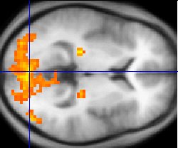
fMRI data (yellow) overlaid on an average of the brain anatomies of multiple humans (gray)
Background
It has been known for over 100 years (Roy and Sherrington 1890) that changes in blood flow and blood oxygenation in the brain (collectively known as hemodynamics) are closely linked to neural activity. When nerve cells are active they consume oxygen carried by hemoglobin in red blood cells from local capillaries. The local response to this oxygen utilisation is an increase in blood flow to regions of increased neural activity, occurring after a delay of approximately 1-5 seconds. This hemodynamic response rises to a peak over 4-5 seconds, before falling back to baseline (and typically undershooting slightly). This leads to local changes in the relative concentration of oxyhemoglobin and deoxyhemoglobin and changes in local cerebral blood volume in addition to this change in local cerebral blood flow.
Hemoglobin is diamagnetic when oxygenated but paramagnetic when deoxygenated. The magnetic resonance (MR) signal of blood is therefore slightly different depending on the level of oxygenation. These differential signals can be detected using an appropriate MR pulse sequence as Blood Oxygenation Level Dependent (BOLD) contrast. Higher BOLD signal intensities arise from decreases in the concentration of deoxygenated hemoglobin since the blood magnetic susceptibility now more closely matches the tissue magnetic susceptibility. By collecting data in an MRI scanner with parameters sensitive to changes in magnetic susceptibility one can assess changes in BOLD contrast. These changes can be either positive or negative depending upon the relative changes in both cerebral blood flow (CBF) and oxygen consumption. Increases in CBF that outstrip changes in oxygen consumption will lead to increased BOLD signal, conversely decreases in CBF that outstrip changes in oxygen consumption will cause decreased BOLD signal intensity.
Neural correlates of BOLD
The precise relationship between neural signals and BOLD is under active research. In general, changes in BOLD signal are well correlated with changes in blood flow. Numerous studies during the past several decades have identified a coupling between blood flow and metabolic rate; that is, the blood supply is tightly regulated in space and time to provide the nutrients for brain metabolism. However, neuroscientists have been seeking a more direct relationship between the blood supply and the neural inputs/outputs that can be related to observable electrical activity and circuit models of brain function.
While current data indicate that local field potentials, an index of integrated electrical activity, form a better correlation with blood flow than the spiking action potentials that are most directly associated with neural communication, no simple measure of electrical activity to date has provided an adequate correlation with metabolism and the blood supply across a wide dynamic range. Presumably, this reflects the complex nature of metabolic processes, which form a superset with regards to electrical activity. Some recent results have suggested that the increase in cerebral blood flow (CBF) following neural activity is not causally related to the metabolic demands of the brain region, but rather is driven by the presence of neurotransmitter, especially glutamate.
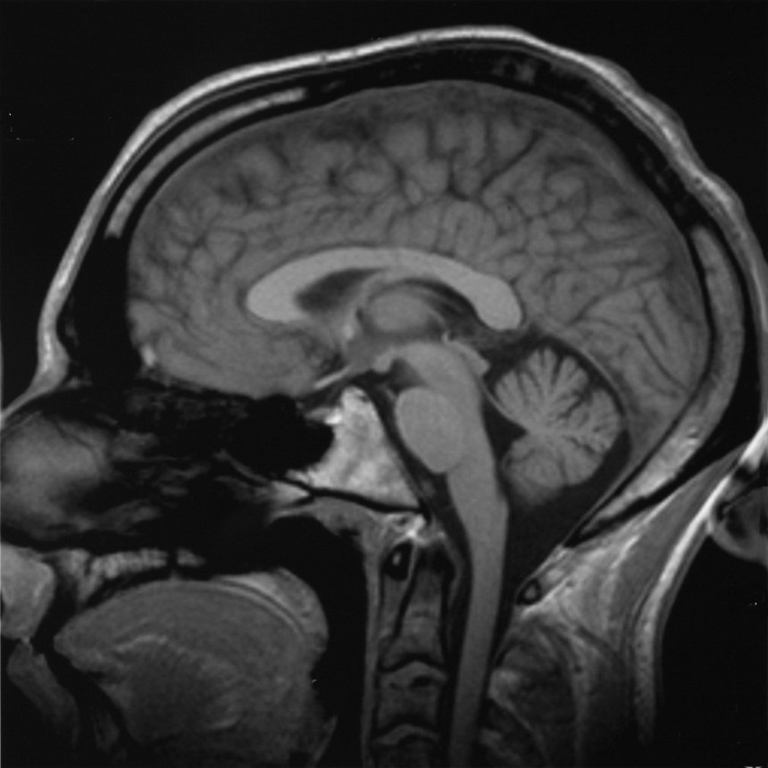
A sagittal slice of a Structural MRI scan of a human head. The nose is to the left.Click here to view an animated sequence of slices.
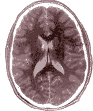
A slice of an MRI scan of the brain. The forehead is at the top and the back of the head is at the bottom. Click here to view an animation of the scan from top to bottom.
Technique
BOLD effects are measured using rapid volumetric acquisition of images with contrast weighed by T2 or T2* (see MRI). Such images can be acquired with moderately good spatial and temporal resolution; images are usually taken every 1-4 seconds, and the voxels in the resulting image typically represent cubes of tissue about 2-4 millimeters on each side in humans. Recent technical advancements, such as the use of high magnetic fields and advanced "multichannel" RF reception, have advanced spatial resolution to the millimeter scale. Although responses to stimuli presented as close together as 1/10 second can be distinguished from one another, the full time course of a BOLD response to a briefly presented stimulus lasts several seconds.
fMRI draws from many disciplines
To use fMRI effectively, an investigator must have a firm grasp of the relevant principles from all of these fields:
- Physics: Researchers should have a reasonable understanding of the tools they are using in their own studies.
- Electrophysiology: Familiarity with neuronal behavior at the electrophysiological level can help investigators design a useful fMRI study.
- Psychology: Many fMRI studies benefit from psychophysical paradigms which allow quantitative measurement of the effect being studied.
- Statistics: Correct application of statistics is essential to "tease out" observations and avoid false-positive results.
- Neuroanatomy: The fMRI signals can be put into the context of previous knowledge only with an understanding of the neuroanatomy. Ultimately, the goal of all functional imaging experiments is to explain human cognition and behavior in terms of physical (anatomical) mechanisms.
Is fMRI worthwhile?
Since its inception, fMRI has been strongly criticised, both as a research technique and in the way its results have been interpreted.
Criticisms leveled at fMRI
- The BOLD signal is only an indirect measure of neural activity, and is therefore susceptible to influence by non-neural changes in the body.
- Different brain areas may have different hemodynamic responses, which would not be accurately reflected by the general linear model often used to filter fMRI time signals.
- fMRI has often been used to ask "where" activations take place in the brain. This has led to the charge that it is simply a modern-day phrenology. Most scientists prefer models which explain "how" psychological mechanisms function. The counter-argument to this criticism is that knowing "where" a cognitive function is located is vitally important. Neuropsychology, invasive manipulation of brain function and functional imaging each give us different windows of understanding into what each brain region does.
- fMRI has often been used to show activation localized to specific regions, and do not reflect the distributed nature of processing in neural networks. Several recent multivariate statistical techniques work around this issue by characterizing interactions between "active" regions found via traditional univariate techniques. Such techniques might prove useful in the future.
- For a non-invasive scan, fMRI has moderately good spatial resolution. However, the temporal response of the blood supply, which is the basis of fMRI, is poor relative to the electrical signals that define neuronal communication. Therefore, some research groups are working around this issue by combining fMRI with data collection techniques such as electroencephalography (EEG) or magnetoencephalography (MEG), which have much higher temporal resolution but rather poorer spatial resolution.
- Many theoretical models used to explain fMRI signals are so poorly specified that they are not falsifiable (a central tenet from the scientific method). Hence, some argue, fMRI is not really a "science". The counter-argument is that a fMRI study can provide evidence to falsify a prior theory if it is well-designed. Also, well-specified mathematical and computational models of the neural processes underlying fMRI can make theories more concrete, allowing them to make predictions that can be verified or falsified by fMRI.
General counterargument
Like any other technique, fMRI is as worthwhile as the design of the experiment using it. Many investigators have used fMRI ineffectively because they were not familiar with all aspects of the technique, or because they received their academic training in disciplines characterized by less rigor than some other branches of neuroscience. Ineffective use of the technique is a problem for the field, but it is not a consequence of the technique itself.
Advantages of fMRI
- It can record brain signals noninvasively.
- It can record brain signals in humans.
- It can record brain signals on a spatial resolution of 1 millimeter. Depending on the type of time course information required, its temporal resolution ranges from 30 seconds to 1/10 of a second.
Scanning in practice

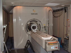
Berkeley's 4T fMRI scanner.
Subjects participating in a fMRI experiment are asked to lie still and are usually restrained with soft pads to prevent small motions from disturbing measurements. Some labs also employ bite bars to reduce motion, although these are unpopular as they can cause some discomfort to subjects. It is possible to correct for some amount of head movement with post-processing of the data, but large transient motion can render these attempts futile. Generally motion in excess of 3 millimeters will result in unusable data. The issue of motion is present for all populations, but most notably within populations that are not physically or emotionally equipped for even short MRI sessions (e.g., those with Alzheimer's Disease or schizophrenia, or young children). In these populations, various positive and negative reinforcement strategies can be employed in an attempt to attenuate motion artifacts, but in general the solution lies in designing a compatible paradigm with these populations.
An fMRI experiment usually lasts 1-2 hours. Depending on the purpose of study, subjects may view movies, hear sounds, smell odors, do cognitive tasks such as memorizing or imagination, or press a few buttons. Researchers are required to give detailed instructions and descriptions of the experiment plan to each subject, who must sign a consent form before the experiment.
Safety is a very important issue in all experiments involving MRI. Potential subjects must ensure that they are able to enter the fMRI environment. Due to the nature of the fMRI scanner, there is an extremely strong magnetic field surrounding the fMRI scanner (at least 1.5 teslas, usually stronger). Potential subjects must be thoroughly examined for any ferromagnetic objects (e.g. watches, glasses, hair pins, pacemakers, bone plates and screws, etc.) before entering the scanning environment.
To this date, fMRI has neither proven therapeutic value nor known damage to the human body. Because fMRI brings no direct benefits to the human subject, cash payment is often used as incentives for researchers to recruit a group of subjects. Typically, payment rates vary from approximately $10 to $40 per hour, depending on the nature of the study.
Related techniques
Aside from fMRI, there are other related ways to probe brain activity using magnetic resonance properties:
Contrast MR
An injected contrast agent such as an iron oxide that has been coated by a sugar or starch (to hide from the body's defense system), causes a local disturbance in the magnetic field that is measurable by the MRI scanner. The signals associated with these kinds of contrast agents are proportional to the cerebral blood volume. While this semi-invasive method presents a considerable disadvantage in terms of studying brain function in normal subjects, it enables far greater detection sensitivity than BOLD signal, which may increase the viability of fMRI in clinical populations. Other methods of investigating blood volume that do not require an injection are a subject of current research, although no alternative technique in theory can match the high sensitivity provided by injection of contrast agent.
Arterial spin labeling
By magnetic labeling the proximal blood supply using "arterial spin labeling" ASL, the associated signal is proportional to the cerebral blood flow, or perfusion. This method provides more quantitative physiological information than BOLD signal, but has less sensitivity for detecting task-induced changes in local brain function.
Magnetic resonance spectroscopic imaging
Magnetic resonance spectroscopic imaging (MRS) is another, NMR-based process for assessing function within the living brain. MRS takes advantage of the fact that protons (hydrogen atoms) residing in differing chemical environments depending upon the molecule they inhabit (H2O vs. protein, for example) possess slightly different resonant properties. For a given volume of brain (typically > 1 cubic cm), the distribution of these H resonances can be displayed as a spectrum.
The area under the peak for each resonance provides a quantitative measure of the relative abundance of that compound. The largest peak is composed of H2O. However, there are also discernible peaks for choline, creatine, n-acetylaspartate (NAA) and lactate. Fortuitously, NAA is mostly inactive within the neuron, serving as a precursor to glutamate and as storage for acetyl groups (to be used in fatty acid synthesis)—but its relative levels are a reasonable approximation of neuronal integrity and functional status. Brain diseases (schizophrenia, stroke, certain tumors, multiple sclerosis) can be characterized by the regional alteration in NAA levels when compared to healthy subjects. Creatine is used a relative control value since its levels remain fairly constant, while choline and lactate levels have been used to evaluate brain tumors.
Diffusion tensor imaging
Diffusion tensor imaging (DTI) is a related use of MR to measure anatomical connectivity between areas. Although it is not strictly a functional imaging technique because it does not measure dynamic changes in brain function, the measures of inter-area connectivity it provides are complementary to images of cortical function provided by BOLD fMRI. As protons are directed along certain axes in the brain (e.g., water flowing down a neuronal axon within a bundle of nerve fibers in cerebral white matter), this directionality can be measured. Connectivity between brain regions may be inferable from diffusion images. Illnesses that disrupt the normal organization or integrity of cerebral white matter (such as multiple sclerosis) have a quantitative impact on DTI measures.
Approaches to fMRI data analysis
External links
- The Functional Imaging Laboratory at University College London
- The Centre for Functional Magnetic Resonance Imaging of the Brain at Oxford University
- About fMRI from Functional MRI Research Center, Columbia University
- Introduction to FMRI from the Oxford Centre for Functional Magnetic Resonance Imaging of the Brain, Oxford University
- FMRLAB Toolbox for fMRI data analysis
de:Funktionelle Magnetresonanztomografie is:Starfræn segulómmyndun pt:Ressonância magnética zh:功能磁共振成像
| This page uses Creative Commons Licensed content from Wikipedia (view authors). |