Assessment |
Biopsychology |
Comparative |
Cognitive |
Developmental |
Language |
Individual differences |
Personality |
Philosophy |
Social |
Methods |
Statistics |
Clinical |
Educational |
Industrial |
Professional items |
World psychology |
Biological: Behavioural genetics · Evolutionary psychology · Neuroanatomy · Neurochemistry · Neuroendocrinology · Neuroscience · Psychoneuroimmunology · Physiological Psychology · Psychopharmacology (Index, Outline)
The vestibular apparatus or vestibular system, or balance system, is the sensory system that provides the dominant input about our movement and orientation in space (see equilibrioception). Together with the cochlea, the auditory organ, it is situated in the vestibulum in the inner ear (Figure 1). As our movements consist of rotations and translations, the vestibular system comprises two components: the semicircular canals, which indicate rotational movements; and the Otoliths, which indicate linear translations. The vestibular system sends signals primarily to the neural structures that control our eye movements, and to the muscle that keep us upright. The projections to the former provide the anatomical basis of the vestibulo-ocular reflex, which is required for clear vision; and the projections to the muscles that control our posture are necessary to keep us upright.
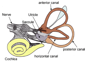
Figure 1 Human Labyrinth, from the left ear. It contains i) the cochlea (yellow), which is the peripheral organ of our auditory system; ii) the semicircular canals (brown), which transduce rotational movements; and iii) the otoliths (in the blue/purple pouches), which transduce linear accelerations. The light blue pouch is the endolymphatic sac, and contains only fluid.
Semicircular canals[]
Our world has three spatial dimensions. Accordingly, our vestibular system contains three semicircular canals in each labyrinth. They are approximately orthogonal to each other, and are called
- horizontal (or lateral)
- anterior (or superior)
- posterior (or inferior)
Push-pull systems[]
The canals are cleverly arranged in such a way that each canal on the left side has an almost parallel counterpart on the right side. Each of these three pairs works in a push-pull fashion: when one canal is stimulated, its corresponding partner on the other side is inhibited, and vice versa.

Figure 2: Push-pull system of the semicircular canals, for a horizontal head movement to the right.
This push-pull system allows us to sense all directions of rotation: while the right horizontal canal gets stimulated during head rotations to the right (Fig 2), the left horizontal canal gets stimulated (and thus predominantly signals) by head rotations to the left.
Vestibulo-ocular reflex (VOR)[]
The vestibular system needs to be fast: if we want clear vision, head movements need to be compensated almost immediately. Otherwise our vision corresponds to a photograph taken with a shaky hand. To achieve clear vision, signals from the semicircular canals are sent as directly as possible to the eye muscles. This direct connection involves only three neurons, and is correspondingly called Three-neuron-arc (Fig 3). Using these direct connections, eye movements lag the head movements by less than 10 ms, one of the fastest reflexes in the human body. The automatic generation of eye movements from movements of the head is called vestibulo-ocular reflex, or short VOR.
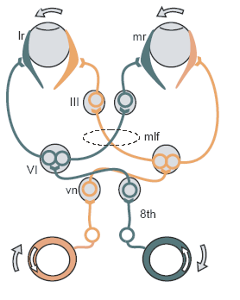
Figure 3 Three-neuron arc, during a head movement to the right. 8th cranial nerve, from the peripheral vestibular sensors to vn, the vestibular nuclei in the brainstem. VI abducens nucleus. The medial longitudinal fascicle (mlf) projects from the abducens nucleus to III, the oculomotor nucleus. The left lateral rectus muscle lr and the right medial rectus muscle mr get contracted, turning the eyes to the left. The green objects are excited, the orange ones inhibited.
This reflex, combined with the push-pull principle described above, forms the physiological basis of the Rapid head impulse test or Halmagyi-Curthoys-test: when the function of your right balance system is reduced by a disease or by an accident, quick head movements to the right cannot be sensed properly any more. As a consequence, no compensatory eye movements are generated, and the patient cannot fixate a point in space during this rapid head movement. Another way of testing the VOR response is to attempt to induce nystagmus (compensatory eye movements in the absence of head motion) by pouring cold or warm water into the ear.
Mechanics[]
The mechanics of the semicircular canals can be described by a damped oscillator. If we designate the deflection of the cupula with , and the head velocity with , the cupula deflection is approximately


α is a proportionality factor, and s corresponds to the frequency. For humans, the time constants T1 and T2 are approximately 3 ms and 5 s, respectively. As a result, for typical head movements, which cover the frequency range of 0.1 Hz and 10 Hz, the deflection of the cupula is approximately proportional to the head-velocity (!). This is very useful, since the velocity of the eyes must be opposite to the velocity of the head in order to have clear vision.
Central Processing[]
Signals from the vestibular system also project to the Cerebellum (where they are used to keep the VOR working, a task usually referred to as Learning or Adaptation) and to different areas in the cortex. The projections to the cortex are spread out over different areas, and their implications are currently not clearly understood.
Otoliths[]
While the semicircular canals respond to rotations, the otoliths sense linear accelerations. We have two on each side, one called Utricle, the other Saccule. Figure 4C shows a cross section through an otolith: the otoconia crystals in the Otoconia Layer (Fig. 4, top layer) rest on a viscous gel layer, and are heavier than their surroundings. Therefore they get displaced during linear acceleration, which in turn deflects the Hair cells (Fig. 4, bottom layer) and thus produces a sensory signal. Most of the utricular signals elicit eye movements, while the majority of the saccular signals projects to muscles that control our posture. While the interpretation of the rotation signals from the semicircular canals is straightforward, the interpretation of otolith signals is more difficult: since gravity is equivalent to a constant linear acceleration, we somehow have to distinguish otolith signals that are caused by linear movements from such that are caused by gravity. We can do that quite well, but the neural mechanisms underlying this separation are not yet fully understood.
Pathologies[]
Diseases of the vestibular system can take different forms, and usually induce vertigo and instability, often accompanied by nausea. The most common ones are Vestibular neuritis, a related condition called Labyrinthitis, and BPPV. In addition, the function of the vestibular system can be affected by tumors on the cochleo-vestibular nerve, an infarct in the brain stem or in cortical regions related to the processing of vestibular signals, and cerebellar atrophy. Less severe, but often also with large consequences, is vertigo caused by the intake of large amounts of alcohol.
BPPV[]
BPPV, which is short for Benign Paroxysmal Positional Vertigo, is probably caused by pieces that have broken off from the Otoliths, and have slipped into one of the semicircular canals. In most cases it is the posterior canal that is affected. In certain head positions, these particles push on the cupula of the canal affected, which leads to dizziness and vertigo. This problem occurs rather frequently, often after hits to the head or after long bed rest. The tell-tale sign of BPPV are vertigo attacks which repeatably appear when the head is brought into a specific orientation. In most cases BPPV can be eliminated (for the patient in an almost miraculous way) by lying down, bringing the head in the right orientation, and sitting up quickly.
External links[]
- SensesWeb, which has been created by Tutis Vilis, contains fantastic animations - at a high level(!) - off all sensory systems, as well as the corresponding PDF-Files, and additional further links.
- Vestibular Primer (by David Dickman, Ph.D.) A very good, up-to-date introduction to the vestibular system.
References[]
- The Vestibular System
Stephen M. Highstein, Richard R. Fay, Arthur N. Popper (eds) Springer-Verlag (2004) ISBN 0-387-98314-7 Comment: A book for experts, summarizing the state of the art in our understanding of the balance system
- Vertigo : Its Multisensory Syndromes
Thomas Brandt Springer-Verlag (2003) ISBN 0-387-40500-3 Comment: For clinicians, and other professionals working with dizzy patients.
- Driver Drowsiness: Is something missing?
J. Christopher Brill, Peter A. Hancock, Richard D. Gilson University of Central Florida (2003)
Comment: Research on driver or motion-induced sleepiness aka 'sopite syndrome' links it to the vestibular labrynths.
| This page uses Creative Commons Licensed content from Wikipedia (view authors). |