Assessment |
Biopsychology |
Comparative |
Cognitive |
Developmental |
Language |
Individual differences |
Personality |
Philosophy |
Social |
Methods |
Statistics |
Clinical |
Educational |
Industrial |
Professional items |
World psychology |
Biological: Behavioural genetics · Evolutionary psychology · Neuroanatomy · Neurochemistry · Neuroendocrinology · Neuroscience · Psychoneuroimmunology · Physiological Psychology · Psychopharmacology (Index, Outline)
Immunohistochemistry or IHC refers to the process of localizing proteins in cells of a tissue section exploiting the principle of antibodies binding specifically to antigens in biological tissues. Immunohistochemical staining is widely used in the diagnosis and treatment of cancer. Specific molecular markers are characteristic of particular cancer types. IHC is also widely used in basic research to understand the distribution and localization of biomarkers in different parts of a tissue.
Visualising an antibody-antigen interaction can be accomplished in a number of ways. In the most common instance, an antibody is conjugated to an enzyme, such as peroxidase, that can catalyse a colour-producing reaction (see immunoperoxidase staining). Alternatively, the antibody can also be tagged to a fluorophore, such as FITC, rhodamine, or Texas Red, (see immunofluorescence). The latter method is of great use in confocal laser scanning microscopy, which is highly sensitive and can also be used to visualise the interactions between multiple proteins.
Antibody types[]
The antibodies used for specific detection can be polyclonal or monoclonal. Monoclonal antibodies are generally considered to exhibit greater specificity. Polyclonal antibodies are made by injecting animals with peptide antigens, and then after a secondary immune response is stimulated, isolating antibodies from whole serum. Thus, polyclonal antibodies are a heterogeneous mix of antibodies that recognize several epitopes
Direct vs indirect IHC[]
There are two strategies used for the immmunohistochemical detection of antigens in tissue, the direct method and the indirect method.
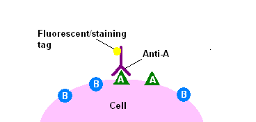
The direct method of immunohistochemical staining uses one labelled antibody, which binds directly to the antigen being stained for.
The direct method is a one-step staining method, and involves a labeled antibody (e.g. FITC conjugated antiserum) reacting directly with the antigen in tissue sections. This technique utilizes only one antibody and the procedure is therefore simple and rapid. However, it can suffer problems with sensitivity due to little signal amplification and is in less common use than indirect methods.
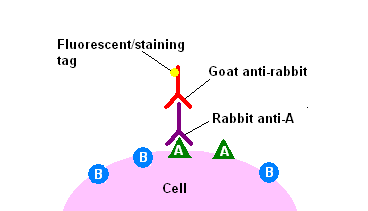
The indirect method of immunohistochemical staining uses one antibody against the antigen being probed for, and a second, labelled, antibody against the first.
The indirect method involves an unlabeled primary antibody (first layer) which reacts with tissue antigen, and a labeled secondary antibody (second layer) which reacts with the primary antibody. (The secondary antibody must be against the IgG of the animal species in which the primary antibody has been raised.) This method is more sensitive due to signal amplification through several secondary antibody reactions with different antigenic sites on the primary antibody. The second layer antibody can be labeled with a fluorescent dye or an enzyme.
Examples of diagnostic IHC markers[]
- Carcinoembryonic antigen (CEA): used for identification of Adenocarcinomas. Not specific for site.
- CD15 and CD30 : used for Hodgkin's disease
- Alpha fetoprotein: for yolk sac tumors and hepatocellular carcinoma
- CD117: for gastrointestinal stromal tumors (GIST)
- Prostate specific antigen (PSA): for prostate cancer
- estrogens and progesterone staining for tumour identification
- Identification of B-cell lymphomas using CD20
Immunotherapy[]
- Main article: Monoclonal antibody therapy
Many proteins shown to be highly upregulated in pathological states by immunohistochemistry are porential targets for therapies utilising monoclonal antibodies.
For example, Her-2/neu (also known as Erb-B2) is highly expressed in a variety of cancer cell types. As such, antibodies against Her-2/neu have been FDA approved for clinical treatment of cancer under the drug name Herceptin.
See also[]
- Methods in Neurochemistry
References & Bibliography[]
Key texts[]
Books[]
Papers[]
Additional material[]
Books[]
Papers[]
External links[]
- Immunohistochemistry Protocol
- Official publication of the society of Applied Immunohistochemistry
- Immunohistochemical Staining Protocols
- Immunohistochemistry Image Gallery
- Immunohistochemistry Protocol Database
- Online Information Center for Immunohistochemistry
- Antibody Search Engine and Antibody Review
- Yale Core Tissue Microarray Facility
- Immunohistochemistry Protocols, Buffers and Troubleshooting
- HistoWiki entry for Immunohistochemistry
- Answers.com Topic for Immunohistochemistry
- http://www.ncbi.nlm.nih.gov/entrez/query.fcgi?cmd=Retrieve&db=PubMed&list_uids=9129199&dopt=Abstract
- de:Antikörperfärbung
- fr:Immunohistochimie
- he:אימונוהיסטוכימיה
- pl:Immunohistochemia
- sv:Immunohistokemi
| This page uses Creative Commons Licensed content from Wikipedia (view authors). |