| Brain: Falx cerebri | ||
|---|---|---|
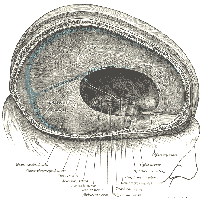 | ||
| Dura mater and its processes exposed by removing part of the right half of the skull and the brain. | ||
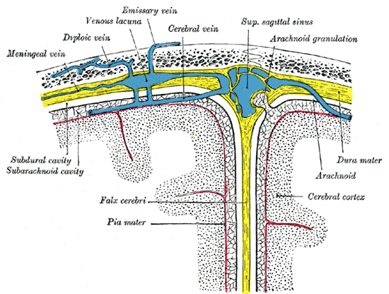 | ||
| Diagrammatic representation of a section across the top of the skull, showing the membranes of the brain, etc. (Falx cerebri is yellow line running down center.) | ||
| Latin | ' | |
| Gray's | subject #193 873 | |
| Part of | ||
| Components | ||
| Artery | ||
| Vein | ||
| BrainInfo/UW | ancil-258 | |
| MeSH | [1] | |
The falx cerebri (Latin: "scythe of the brain") is an extension of the protective dura mater that projects into the longitudinal fissure that separates the two cerebral hemispheres.
Details from Gray's anatomy[]
The falx cerebri, so named from its sickle-like form, is a strong, arched process which descends vertically in the longitudinal fissure between the cerebral hemispheres.
It is narrow in front, where it is attached to the crista galli of the ethmoid; and broad behind, where it is connected with the upper surface of the tentorium cerebelli.
Its upper margin is convex, and attached to the inner surface of the skull in the middle line, as far back as the internal occipital protuberance; it contains the superior sagittal sinus. Its lower margin is free and concave, and contains the inferior sagittal sinus.
External links[]
This article was originally based on an entry from a public domain edition of Gray's Anatomy. As such, some of the information contained herein may be outdated. Please edit the article if this is the case, and feel free to remove this notice when it is no longer relevant.
| Meninges of the brain and medulla spinalis |
|
Dura mater - Falx cerebri - Tentorium cerebelli - Falx cerebelli - Arachnoid mater - Subarachnoid space - Cistern - Cisterna magna - Median aperture - Cerebrospinal fluid - Arachnoid granulation - Pia mater |
| This page uses Creative Commons Licensed content from Wikipedia (view authors). |