Assessment |
Biopsychology |
Comparative |
Cognitive |
Developmental |
Language |
Individual differences |
Personality |
Philosophy |
Social |
Methods |
Statistics |
Clinical |
Educational |
Industrial |
Professional items |
World psychology |
Biological: Behavioural genetics · Evolutionary psychology · Neuroanatomy · Neurochemistry · Neuroendocrinology · Neuroscience · Psychoneuroimmunology · Physiological Psychology · Psychopharmacology (Index, Outline)
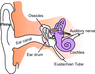
Anatomy of the human ear.
- Main article: Middle ear
The Eustachian tube (or auditory tube) is a tube that links the pharynx to the middle ear. In adults the Eustachian tube is approximately 35 mm long. It is named after the 16th century anatomist Bartolomeo Eustachi. Some modern medical books call this the pharyngotympanic tube.
Functions
Pressure equalization
Normally the Eustachian tube is closed, but it can open to let a small amount of air through to equalize the pressure between the middle ear and the atmosphere. When this happens we hear a small pop, an event familiar to airplane travelers or drivers in mountainous regions. Yawning or swallowing can pull on muscles in the neck, causing the tube to open. Some people are born with the ability to contract just these muscles voluntarily, similar to people who can wiggle their ears. Without this airway, the middle ear would be isolated from the atmosphere, and could be easily damaged by pressure changes.
Mucus drainage
The Eustachian tube also drains mucus from the middle ear. Upper airway infections or allergies can cause the Eustachian tube to become swollen, trapping bacteria and causing ear infections. This swelling can be reduced through the use of pseudoephedrine. Earaches are more common in children because the tube is more horizontal, making the movement of fluid harder.
An alternative method of relieving the pain felt by an earache is to have a physician or chiropractor perform endonasal therapy, which is a useful form of local treatment for catarrh problems. Endonasal therapy is a basic treatment used to initiate the draining and cleansing processes necessary to remove congestion in the Eustachian tubes.
Behind the nose and up above the tonsils is a small indentation called the "fossa of Rosenmuller." In this area the proximal end of the Eustachian tube opens into the throat. The Eustachian tube begins in the middle ear, passing downward, to come to an end in this fossa. Owing to the nature of the surface anatomy and of the draining pathways of the mucus, the fossa of Rosenmuller invariably becomes clogged with this draining fluid in catarrhal conditions. With the passage of time, the material that accumulates in this small cavity solidifies and becomes jelly-like. In this stage, it may clog the opening of the Eustachian tube and even some of the sinus drain tubes. In time the accumulated material becomes harder; both small capillaries and adhesions may form in this mass as time goes on. Because of this material's placement at the end of the Eustachian tube, its persistent pressure can cause catarrhal afflictions of the ear.
Embryologic development
The Eustachian tube is derived from the first pharyngeal pouch, which during embryogenesis forms a recess called the tubotympanic sulcus. The sulcus deepens to meet the first pharyngeal cleft forming the tympanic membrane. The distal part of the tubotympanic sulcus gives rise to the tympanic cavity, while the proximal tubular structure becomes the Eustachian tube.
Muscles
There are four muscles associated with the function of the eustachian tube:
- levator veli palatini
- salpingopharyngeus
- tensor tympani
- tensor veli palatini
References
- Cairns, W. (1998). The patulous Eustachian tube syndrome: Palliative Medicine Vol 12(1) Jan 1998, 59-60.
- Jeter, I. K. (1976). Waveform patterns of reflex and voluntary contraction of the middle ear muscles: Journal of Auditory Research Vol 16(3) Jul 1976, 183-191.
- Karwautz, A., Hafferl, A., Ungar, D., & Sailer, H. (1999). Patulous Eustachian tube in a case of adolescent anorexia nervosa: International Journal of Eating Disorders Vol 25(3) Apr 1999, 353-355.
- Oghan, F., Harputluoglu, U., Guclu, E., Guvey, A., Turan, N., & Ozturk, O. (2007). Permanent t-tube insertion in two patients with Hurler's syndrome: International Journal of Audiology Vol 46(2) Feb 2007, 94-96.
- Petrova, P., Freeman, S., & Sohmer, H. (2007). Mechanism and rate of middle ear fluid absorption: Audiology & Neurotology Vol 12(3) Apr 2007, 155-159.
- Riedel, C. L., Wiley, T. L., & Block, M. G. (1987). Tympanometric measures of Eustachian tube function: Journal of Speech & Hearing Research Vol 30(2) Jun 1987, 207-214.
- Venkatesh Aithal, H. (1982). Eustachian tube function in geriatric subjects: Journal of the All-India Institute of Speech & Hearing Vol 13 1982, 181-191.
- Weaver, R. M., Arbon, R. A., Watkins, P. L., & Olsen, R. G. (1976). Comparisons among two different electro-acoustic impedance measures and otoscopy by an ENT specialist in identifying middle ear anomalies in mental retardates: Journal of Auditory Research Vol 16(4) 1976, 239-246.
Sensory system: Auditory and Vestibular systems (TA A15.3, GA 10.1029) | |||||||||||||||
|---|---|---|---|---|---|---|---|---|---|---|---|---|---|---|---|
| Outer ear |
Pinna (Helix, Antihelix, Tragus, Antitragus, Incisura anterior auris, Earlobe) • Ear canal • Auricular muscles | ||||||||||||||
| Middle ear |
| ||||||||||||||
| Inner ear/ (membranous labyrinth, bony labyrinth) |
| ||||||||||||||
| {| class="navbox collapsible nowraplinks" style="margin:auto; " | |||||||||||||||
| |||||||||||||||
|}
<!-Interlinks->
de:Eustachi-Röhre es:Trompa de Eustaquio fr:Trompe d'Eustache lt:Ausies trimitas nl:Buis van Eustachius pt:Trompa de Eustáquio sv:Örontrumpet
| This page uses Creative Commons Licensed content from Wikipedia (view authors). |