Assessment |
Biopsychology |
Comparative |
Cognitive |
Developmental |
Language |
Individual differences |
Personality |
Philosophy |
Social |
Methods |
Statistics |
Clinical |
Educational |
Industrial |
Professional items |
World psychology |
Biological: Behavioural genetics · Evolutionary psychology · Neuroanatomy · Neurochemistry · Neuroendocrinology · Neuroscience · Psychoneuroimmunology · Physiological Psychology · Psychopharmacology (Index, Outline)
| Brain: Cochlear nuclei | ||
|---|---|---|
| Dissection of brain-stem. Dorsal view. ("Cochlear nucleus" is labeled on left, fifth from the bottom.) | ||
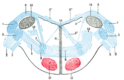
| Terminal nuclei of the cochlear nerve, with their upper connections. (Schematic.) The vestibular nerve with its terminal nuclei and their efferent fibers have been suppressed. On the other hand, in order not to obscure the trapezoid body, the efferent fibers of the terminal nuclei on the right side have been resected in a considerable portion of their extent. The trapezoid body, therefore, shows only one-half of its fibers, viz., those which come from the left. 1. Vestibular nerve, divided at its entrance into the medulla oblongata. 2. Cochlear nerve. 3. Accessory nucleus of acoustic nerve. 4. Tuberculum acusticum. 5. Efferent fibers of accessory nucleus. 6. Efferent fibers of tuberculum acusticum, forming the striae medullares, with 6’, their direct bundle going to the superior olivary nucleus of the same side; 6’’, their decussating bundles going to the superior olivary nucleus of the opposite side. 7. Superior olivary nucleus. 8. Trapezoid body. 9. Trapezoid nucleus. 10. Central acoustic tract (lateral lemniscus). 11. Raphé. 12. Cerebrospinal fasciculus. 13. Fourth ventricle. 14. Inferior peduncle. | ||
| Latin | ' | |
| Gray's | subject #187 788 | |
| Part of | ||
| Components | ||
| Artery | ||
| Vein | ||
| BrainInfo/UW | hier-717 | |
| MeSH | [1] | |
The cochlear nuclei consist of:
- (a) the lateral cochlear nucleus, corresponding to the tuberculum acusticum on the dorso-lateral surface of the inferior peduncle; and
- (b) the ventral or accessory cochlear nucleus, placed between the two divisions of the nerve, on the ventral aspect of the inferior peduncle.
Relationship to auditory system
The cochlear nucleus is the first site of the neuronal processing of the newly converted “digital” data from the inner ear. The information is brought via the cochlear nerve (also called the VIII "8th" nerve) to the aforementioned nucleus, where there is an organization of sound information. The lower frequency axons follow a similar path, innervating the ventral portions of the dorsal cochlear nucleus and the ventrolateral portions of the anteroventral cochlear nucleus. In contrast, the axons from the higher frequency hair cells project to the dorsal portion of the anteroventral cochlear nucleus and the uppermost dorsal portions of the dorsal cochlear nucleus. The mid frequency projections end up in between the two extremes, in this way the frequency spectrum is preserved.
There are four types of cells found in the cochlear nucleus. Stellate cells have a radial, star-like, shape which is where they get their first name. Their second name, chopper cells, is in reference to their ability to fire consistently despite background noise. Chopper cells are also not affected by slight variations in frequency, and in this way encode for the characteristic frequency of the specific neuron, or preceding hair cell. These cells are only found in the ventral half of the cochlear nucleus.
Bushy cells are composed of a single, very short dendrite with numerous small branchings, which cause it to resemble a “bush”. These cells are usually only innervated by a select few axons, which dominate its firing patterns. These efferent axons wrap their branches around the entire soma, choking the bushy cells with their large end bulbs into firing whenever they please. This being said, a single unit recording of an electrically stimulated bushy neuron characteristically produces exactly one action potential. In this way, the bushy cell is thought to respond only to the occurrence of a new sound and to help in its localization. Along with the chopper cells, the bushy cells are only found in the ventral portion of the cochlear nucleus.
Fusiform cells have been known to be excitatory or inhibitory and located solely within the dorsal cochlear nucleus. Whist the bushy cells aide in the location of a sound stimulus on the horizontal axis, fusiform cells locate the sound stimulus on the vertical axis. With the combined power of these two types of cells, an ordinary man can locate where a firecracker explodes without the use of his eyes. These cells are known to reside solely in the dorsal cochlear nucleus.
Whilst the fusiform and bushy cells take care of locating a sound stimulus in the first and second dimensions of space, the octopus cells seem to take care of the fourth dimension (17). Electrical stimuli to the auditory nerve have been shown to evoke a graded post synaptic potential in the octopus cells. These EPSP’s are known to be brief and consistently 1 millisecond, and is not dependent on the stimulus strength, although there is minimum voltages requirement necessary elicit this effect. In this way, the octopus cells have been thought to be active in the process of timing. These cells are located in the posteroventral cochlear nucleus, but make several connections with many of the auditory nerve fibers. This is thought to be the pathway which carries information concerning action potentials (2).
Figure four, seen below, illustrates individual stereotypical behavior for the four major types of cells in the cochlear nucleus. The column labeled the “intrinsic properties” indicates the action potentials generated by the labeled neuron in response to a typical depolarizing stimulus. This column also displays the reaction of the neurons to depolarizing currents. The depolarizing and hyperpolarizing currents are equal and opposite in magnitude, one being 0.4 nA and the other being -0.4 nA. The EPSP column shows the stereotypical post synaptic potential evoked by these neurons when stimulated by an instantaneous short burst or electrical activity. This is in contrast to the left column which illustrates a pulse sustained for 50 milliseconds.
Projections from cochlear nucleus
There are three major projections from the cochlear nucleus. Through the medulla, one projection bifurcates, and shoots to the contralateral the superior olivary complex via the trapezoid body, whilst the other half shoots to the ipsilateral SOC. This projection is called the ventral acoustic stria. Another projection, called the dorsal acoustic stria, rises above the medulla into the pons where it hits the nucleus of the lateral lemniscus along with its kin, the intermediate acoustic stria. The IAS decussates across the medulla, before hitting the contralateral lateral lemniscus. The lateral lemniscus, in turn projects to the contralateral lateral lemniscus, as well as the inferior colliculus. The inferior colliculus receives projections from the superior olivary complex and the contralateral dorsal acoustic stria, as well as the aforementioned lateral lemniscus.
It is here at the inferior colliculus that the simple processing of sound stops for some species. The indirect projections from the ipsilateral cochlear nucleus stop at the inferior colliculus. The contralateral projections are the only ones which continue on to the medial geniculate nucleus of the thalamus, and consequentially the cortex. The auditory cortex is a part of the superior temporal gyrus.
Assessment |
Biopsychology |
Comparative |
Cognitive |
Developmental |
Language |
Individual differences |
Personality |
Philosophy |
Social |
Methods |
Statistics |
Clinical |
Educational |
Industrial |
Professional items |
World psychology |
Biological: Behavioural genetics · Evolutionary psychology · Neuroanatomy · Neurochemistry · Neuroendocrinology · Neuroscience · Psychoneuroimmunology · Physiological Psychology · Psychopharmacology (Index, Outline)
See also
References
2) Kandel, et al Principles of Neuroscience. Fourth ed. pp 591-624. Copyright 2000, by McGraw-Hill Co.
17) Ferragamo MJ. Octopus cells of the mammalian ventral cochlear nucleus sense the rate of depolarization. J Neurophysiol. 2002 May;87(5):2262-70
External links
This article was originally based on an entry from a public domain edition of Gray's Anatomy. As such, some of the information contained herein may be outdated. Please edit the article if this is the case, and feel free to remove this notice when it is no longer relevant.
Sensory system: Auditory and Vestibular systems (TA A15.3, GA 10.1029) | |||||||||||||||
|---|---|---|---|---|---|---|---|---|---|---|---|---|---|---|---|
| Outer ear |
Pinna (Helix, Antihelix, Tragus, Antitragus, Incisura anterior auris, Earlobe) • Ear canal • Auricular muscles | ||||||||||||||
| Middle ear |
| ||||||||||||||
| Inner ear/ (membranous labyrinth, bony labyrinth) |
| ||||||||||||||
| {| class="navbox collapsible nowraplinks" style="margin:auto; " | |||||||||||||||
| |||||||||||||||
|}
| This page uses Creative Commons Licensed content from Wikipedia (view authors). |

