No edit summary |
No edit summary |
||
| Line 17: | Line 17: | ||
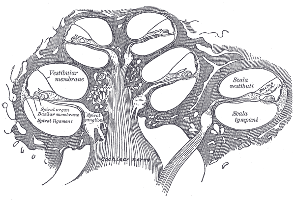
| − | The cochlear nerve is one of two branches of the auditory nerve (the VIII cranial nerve), the other being the vestibular nerve. Auditory nerve fibres provide a direct synaptic connection between the hair cells of the cochlea and the cochlear nucleus. The cochlear nerve fibres originate in the spiral ganglion of the cochlea, which in turn connect to the hair cells. In humans, there are about 30,000 ganglion cells in each cochlea. It was once believed that most of the cochlear nerve fibres were directed to the outer hair cells, but it is now understood that at least 90% of the cochlear ganglion cells terminate on inner hair cells, the rest terminating on the outer hair cells. Each axon innervates only a single hair cell, but each hair cell directs its output to an average of 10 nerve fibres. The transmission between the inner hair cells and the neurons is chemical, using glutamate as a neurotransmitter. |
+ | The cochlear nerve is one of two branches of the [[auditory nerve]] (the VIII cranial nerve), the other being the [[vestibular nerve]]. Auditory nerve fibres provide a direct synaptic connection between the [[hair cells]] of the [[cochlea]] and the cochlear nucleus. The cochlear nerve fibres originate in the spiral ganglion of the cochlea, which in turn connect to the hair cells. In humans, there are about 30,000 ganglion cells in each cochlea. It was once believed that most of the cochlear nerve fibres were directed to the outer hair cells, but it is now understood that at least 90% of the cochlear ganglion cells terminate on inner hair cells, the rest terminating on the outer hair cells. Each axon innervates only a single hair cell, but each hair cell directs its output to an average of 10 nerve fibres. The transmission between the inner hair cells and the neurons is chemical, using [[glutamate]] as a [[neurotransmitter]]. |
| − | The cochlear neurons can be divided into two groups: Type I and Type II. Type I neurons make up 90-95% of the neurons and innervate the inner hair cells. They have a relatively large diameter, and are bipolar and myelinated. Type II cells, which have a relatively small diameter, connect with the outer hair cells, are monopolar and are not myelinated. |
+ | The cochlear neurons can be divided into two groups: Type I and Type II. Type I neurons make up 90-95% of the neurons and innervate the inner hair cells. They have a relatively large diameter, and are bipolar and [[myelinated]]. Type II cells, which have a relatively small diameter, connect with the outer hair cells, are monopolar and are not myelinated. |
| − | The axons from each cochlear nerve terminate in the cochlear nuclear complex which are ipsilaterally located in the medulla of the brainstem. The cochlear nucleus is the first 'relay station' of the auditory nervous system and receives mainly ipsilateral afferent input. The three major components of the cochlear nuclear complex are the dorsal cochlear nucleus (DCN), the anteroventral cochlear nucleus (AVCN) and the posteroventral cochlear nucleus (PVCN; see Fig 1). Each of the three cochlear nuclei are tonotopically organised. The axons from the lower frequency area of the cochlea innervate the ventral portion of the dorsal cochlear nucleus and the ventrolateral portions of the anteroventral cochlear nucleus, while the higher frequency axons project into the dorsal portion of the anteroventral cochlear nucleus and the uppermost dorsal portions of the dorsal cochlear nucleus. The mid frequency projections end up in between the two extremes, in this way the frequency spectrum is preserved. |
+ | The axons from each cochlear nerve terminate in the cochlear nuclear complex which are ipsilaterally located in the [[medulla]] of the [[brainstem]]. The cochlear nucleus is the first 'relay station' of the [[auditory system|auditory nervous system]] and receives mainly ipsilateral afferent input. The three major components of the cochlear nuclear complex are the [[dorsal cochlear nucleus]] (DCN), the [[anteroventral cochlear nucleus]] (AVCN) and the [[posteroventral cochlear nucleus]] (PVCN; see Fig 1). Each of the three cochlear nuclei are [[tonotopically]] organised. The axons from the lower frequency area of the cochlea innervate the [[ventral]] portion of the [[dorsal]] cochlear nucleus and the ventrolateral portions of the anteroventral cochlear nucleus, while the higher frequency axons project into the dorsal portion of the anteroventral cochlear nucleus and the uppermost dorsal portions of the dorsal cochlear nucleus. The mid frequency projections end up in between the two extremes, in this way the frequency spectrum is preserved. |
[[http://upload.wikimedia.org/wikipedia/en/thumb/0/0b/Cochlear_nucleus_innervated_by_a_branching_auditory_nerve_fibre.JPG/800px-Cochlear_nucleus_innervated_by_a_branching_auditory_nerve_fibre.jpg]] |
[[http://upload.wikimedia.org/wikipedia/en/thumb/0/0b/Cochlear_nucleus_innervated_by_a_branching_auditory_nerve_fibre.JPG/800px-Cochlear_nucleus_innervated_by_a_branching_auditory_nerve_fibre.jpg]] |
||
Revision as of 00:26, 8 December 2006
Assessment |
Biopsychology |
Comparative |
Cognitive |
Developmental |
Language |
Individual differences |
Personality |
Philosophy |
Social |
Methods |
Statistics |
Clinical |
Educational |
Industrial |
Professional items |
World psychology |
Biological: Behavioural genetics · Evolutionary psychology · Neuroanatomy · Neurochemistry · Neuroendocrinology · Neuroscience · Psychoneuroimmunology · Physiological Psychology · Psychopharmacology (Index, Outline)
| Nerve: Cochlear nerve | ||
|---|---|---|
 | ||
| Diagrammatic longitudinal section of the cochlea. (Cochlear nerve is in center, shown as striped.) | ||
 | ||
| Part of the cochlear division of the acoustic nerve, highly magnified. | ||
| Latin | n. cochlearis | |
| Gray's | subject #203 906 | |
| Innervates | ||
| From | Vestibulocochlear nerve | |
| To | ||
| MeSH | A08.800.800.120.910.120 | |
The cochlear nerve is one of two branches of the auditory nerve (the VIII cranial nerve), the other being the vestibular nerve. Auditory nerve fibres provide a direct synaptic connection between the hair cells of the cochlea and the cochlear nucleus. The cochlear nerve fibres originate in the spiral ganglion of the cochlea, which in turn connect to the hair cells. In humans, there are about 30,000 ganglion cells in each cochlea. It was once believed that most of the cochlear nerve fibres were directed to the outer hair cells, but it is now understood that at least 90% of the cochlear ganglion cells terminate on inner hair cells, the rest terminating on the outer hair cells. Each axon innervates only a single hair cell, but each hair cell directs its output to an average of 10 nerve fibres. The transmission between the inner hair cells and the neurons is chemical, using glutamate as a neurotransmitter.
The cochlear neurons can be divided into two groups: Type I and Type II. Type I neurons make up 90-95% of the neurons and innervate the inner hair cells. They have a relatively large diameter, and are bipolar and myelinated. Type II cells, which have a relatively small diameter, connect with the outer hair cells, are monopolar and are not myelinated.
The axons from each cochlear nerve terminate in the cochlear nuclear complex which are ipsilaterally located in the medulla of the brainstem. The cochlear nucleus is the first 'relay station' of the auditory nervous system and receives mainly ipsilateral afferent input. The three major components of the cochlear nuclear complex are the dorsal cochlear nucleus (DCN), the anteroventral cochlear nucleus (AVCN) and the posteroventral cochlear nucleus (PVCN; see Fig 1). Each of the three cochlear nuclei are tonotopically organised. The axons from the lower frequency area of the cochlea innervate the ventral portion of the dorsal cochlear nucleus and the ventrolateral portions of the anteroventral cochlear nucleus, while the higher frequency axons project into the dorsal portion of the anteroventral cochlear nucleus and the uppermost dorsal portions of the dorsal cochlear nucleus. The mid frequency projections end up in between the two extremes, in this way the frequency spectrum is preserved.
[[1]]
See also
External links
- Cochlear Structures (Flash animation)
- Penn State
- Illustration
- Illustration
- Gray's
Sensory system: Auditory and Vestibular systems (TA A15.3, GA 10.1029) | |||||||||||||||
|---|---|---|---|---|---|---|---|---|---|---|---|---|---|---|---|
| Outer ear |
Pinna (Helix, Antihelix, Tragus, Antitragus, Incisura anterior auris, Earlobe) • Ear canal • Auricular muscles | ||||||||||||||
| Middle ear |
| ||||||||||||||
| Inner ear/ (membranous labyrinth, bony labyrinth) |
| ||||||||||||||
| {| class="navbox collapsible nowraplinks" style="margin:auto; " | |||||||||||||||
| |||||||||||||||
|}
I-IV: olfactory - optic - oculomotor - trochlear
V: trigeminal: trigeminal ganglion
V1: ophthalmic: lacrimal - frontal (supratrochlear, supraorbital) - nasociliary (long root of ciliary, long ciliary, infratrochlear, posterior ethmoidal, anterior ethmoidal) - ciliary ganglion (short ciliary)
V2: maxillary: middle meningeal - in the pterygopalatine fossa (zygomatic, zygomaticotemporal, zygomaticofacial, sphenopalatine, posterior superior alveolar)
in the infraorbital canal/infraorbital nerve (middle superior alveolar, anterior superior alveolar)
on the face (inferior palpebral, external nasal, superior labial, infraorbital plexus) - pterygopalatine ganglion (deep petrosal, nerve of pterygoid canal)
branches of distribution (palatine, nasopalatine, pharyngeal)
V3: mandibular: nervus spinosus - medial pterygoid - anterior (masseteric, deep temporal, buccal, lateral pterygoid)
posterior (auriculotemporal, lingual, inferior alveolar, mylohyoid, mental) - otic ganglion - submandibular ganglion
VI: abducens
VII: facial: nervus intermedius - geniculate - inside facial canal (greater petrosal, nerve to the stapedius, chorda tympani)
at exit from stylomastoid foramen (posterior auricular, digastric - stylohyoid)
on face (temporal, zygomatic, buccal, mandibular, cervical)
VIII: vestibulocochlear: cochlear (striae medullares, lateral lemniscus) - vestibular
IX: glossopharyngeal: fasciculus solitarius - nucleus ambiguus - ganglia (superior, petrous) - tympanic - carotid sinus
X: vagus: ganglia (jugular, nodose) - Alderman's nerve - in the neck (pharyngeal branch, superior laryngeal ext and int, recurrent laryngeal)
in the thorax (pulmonary branches, esophageal plexus) - in the abdomen (gastric plexuses, celiac plexus, gastric plexus)
XI: accessory XII: hypoglossal
| This page uses Creative Commons Licensed content from Wikipedia (view authors). |