Assessment |
Biopsychology |
Comparative |
Cognitive |
Developmental |
Language |
Individual differences |
Personality |
Philosophy |
Social |
Methods |
Statistics |
Clinical |
Educational |
Industrial |
Professional items |
World psychology |
Language: Linguistics · Semiotics · Speech
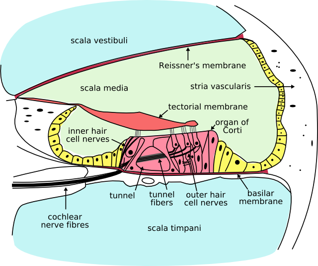
Cross section of the cochlea.
Named after the Latin word for snail shell, the cochlea is a coiled, tapered tube containing the auditory branch of the mammalian inner ear. Its core component is the Organ of Corti, the sensory organ of hearing.
Anatomy
Structures
The cochlea is a spiralled, hollow, conical chamber of bone. Structures include:
- the scala tympani (containing perilymph), which lies superior to the cochlea duct and abuts the oval window.
- the scala vestibuli (containing perilymph), which lies inferior to the scala media and terminates at the round window.
- the scala media (containing endolymph), which is the membraneous cochlea duct containing the organ of Corti.
- the helicotrema is the location where the scala tympani and the scala vestibuli merge
- Reissner's membrane separates the scala vestibuli from the scala media.
- The basilar membrane separates the scala media from the scala tympani.
- The Organ of Corti is a cellular layer sitting on top of the basilar membrane. It is lined with hair cells — sensory cells topped with hair-like structures called stereocilia.
Function
The stapes transmits vibrations to the fenestra ovalis (oval window) on the outside of the cochlea, which vibrates the perilymph in the scala vestibuli.
This in turn vibrates the endolymph in the scala media, thus causing movements of the hair bundles of the hair cells, which are acoustic sensor cells that convert vibration into electrical potentials. The hair cells in the organ of Corti are tuned to certain sound frequencies, being responsive to high frequencies near the oval window and to low frequencies near the apex of the cochlea.
The hair cells are arranged in four rows in the Organ of Corti along the entire length of the cochlear coil. Three rows consist of outer hair cells (OHCs) and one row consists of inner hair cells (IHCs). The inner hair cells provide the main neural output of the cochlea. The outer hair cells, instead, mainly receive neural input from the brain, which influences their motility as part of the cochlea’s mechanical pre-amplifier.
For very low frequencies (below 20Hz), the pressure waves propagate along the complete route of the cochlea - up scala vestibuli, around helicotrema and down scala tympani to the round window. Frequencies this low do not activate the organ of Corti and are below the threshold for hearing. Higher frequencies do not propagate to the helicotrema but are transmitted through the endolymph in the cochlea duct to the perilymph in the scala tympani.
A very strong movement of the endolymph due to very loud noise may cause hair cells to die. This is a common cause of partial hearing loss and is the reason why anyone using firearms or heavy machinery should wear earmuffs or earplugs.
Comparative physiology
The coiled form of cochlea is unique for mammals. In birds and in other non-mammalian vertebrates the compartment containing the sensory cells for hearing is occasionally also called “cochlea”, although it is not coiled up. Instead it forms a blind-ended tube, also called the cochlear duct. This difference apparently evolved in parallel with the differences in frequency range of hearing and in frequency resolution between mammals and non-mammalian vertebrates. Most bird species do not hear above 4–5 kHz, the currently known maximum being ~ 11 kHz in the barn owl. Some marine mammals hear up to 200 kHz. The superior frequency resolution in mammals is due to their unique mechanism of pre-amplification of sound by active cell-body vibrations of outer hair cells. A long coiled compartment, rather than a short and straight one, provides more space for frequency dispersion and is therefore better adapted to the highly derived functions in mammalian hearing.
References
- Vater M, Meng J, Fox RC. Hearing organ evolution and specialization: Early and later mammals. In: GA Manley, AN Popper, RR Fay (Eds). Evolution of the Vertebrate Auditory System, Springer-Verlag, New York 2004, pp 256-288.
See also
External links
Sensory system: Auditory and Vestibular systems (TA A15.3, GA 10.1029) | |||||||||||||||
|---|---|---|---|---|---|---|---|---|---|---|---|---|---|---|---|
| Outer ear |
Pinna (Helix, Antihelix, Tragus, Antitragus, Incisura anterior auris, Earlobe) • Ear canal • Auricular muscles | ||||||||||||||
| Middle ear |
| ||||||||||||||
| Inner ear/ (membranous labyrinth, bony labyrinth) |
| ||||||||||||||
| {| class="navbox collapsible nowraplinks" style="margin:auto; " | |||||||||||||||
| |||||||||||||||
|}
de:Innenohr es:Cóclea nl:Slakkenhuis (oor)
| This page uses Creative Commons Licensed content from Wikipedia (view authors). |