(Revert vandalism) |
No edit summary |
||
| (6 intermediate revisions by 4 users not shown) | |||
| Line 20: | Line 20: | ||
DorlandsSuf = | |
DorlandsSuf = | |
||
}} |
}} |
||
| + | The '''cerebellum''' (Latin for ''little brain'') is a region of the [[brain]] that plays an important role in [[motor control]]. It may also be involved in some [[cognition|cognitive function]]s such as [[attention]] and [[language]], and in regulating [[fear]] and [[pleasure]] responses,<ref>{{cite journal |author=Wolf U, Rapoport MJ, Schweizer TA |title=Evaluating the affective component of the cerebellar cognitive affective syndrome |journal=J. Neuropsychiatry Clin. Neurosci. |volume=21 |pages=245–53 |year=2009 |pmid=19776302 |doi=10.1176/appi.neuropsych.21.3.245 |issue=3}}</ref> but its movement-related functions are the most solidly established. The cerebellum does not initiate movement, but it contributes to [[Motor coordination|coordination]], precision, and accurate timing. It receives input from [[sensory system]]s of the [[spinal cord]] and from other parts of the [[brain]], and integrates these inputs to fine tune motor activity.<ref name="Fine">{{cite journal | author=Fine EJ, Ionita CC, Lohr L | title=The history of the development of the cerebellar examination | journal=Semin Neurol | year=2002 | pages=375–84 | volume=22 | issue=4 | pmid=12539058 | doi = 10.1055/s-2002-36759}}</ref> Because of this fine-tuning function, [[lesion|damage]] to the cerebellum does not cause [[paralysis]], but instead produces disorders in fine movement, [[Equilibrioception|equilibrium]], [[Human positions|posture]], and [[motor learning]].<ref name="Fine"/> |
||
| + | In its anatomy, the cerebellum has the appearance of a separate structure attached to the bottom of the brain, tucked underneath the [[cerebral hemisphere]]s. The surface of the cerebellum is covered with finely spaced parallel grooves, in striking contrast to the broad irregular convolutions of the [[cerebral cortex]]. These parallel grooves conceal the fact that the cerebellum is actually a continuous thin layer of tissue (the cerebellar cortex), tightly folded in the style of an [[accordion]]. Within this thin layer are several types of [[neuron]]s with a highly regular arrangement, the most important being [[Purkinje cell]]s and [[Cerebellum granule cell|granule cell]]s. This complex neural network gives rise to a massive signal-processing capability, but almost all of its output is directed to a set of small [[deep cerebellar nuclei]] lying in the interior of the cerebellum. |
||
| − | {{portalpar|Neuroscience|Neuro logo.png}} |
||
| + | In addition to its direct role in motor control, the cerebellum also is necessary for several types of [[motor learning]], the most notable one being learning to adjust to changes in sensorimotor relationships. Several theoretical models have been developed to explain sensorimotor calibration in terms of [[synaptic plasticity]] within the cerebellum. Most of them derive from early models formulated by [[David Marr (neuroscientist)|David Marr]] and [[James S. Albus|James Albus]], which were motivated by the observation that each cerebellar Purkinje cell receives two dramatically different types of input: On one hand, thousands of inputs from [[parallel fiber]]s, each individually very weak; on the other hand, input from one single [[climbing fiber]], which is, however, so strong that a single climbing fiber action potential will reliably cause a target Purkinje cell to fire a burst of action potentials. The basic concept of the Marr-Albus theory is that the climbing fiber serves as a "teaching signal", which induces a long-lasting change in the strength of synchronously activated parallel fiber inputs. Observations of [[long-term depression]] in parallel fiber inputs have provided support for theories of this type, but their validity remains controversial. |
||
| − | The '''cerebellum''' ([[Latin]]: "little brain") is a region of the [[brain]] that plays an important role in the integration of [[perception|sensory perception]] and [[motoneuron|motor]] control. In order to coordinate motor control, there are many [[neural pathway]]s linking the cerebellum with the [[cerebrum|cerebral]] [[motor cortex]] (which sends information to the [[muscle]]s causing them to move) and the [[spinocerebellar tract]] (which provides [[proprioception|proprioceptive]] feedback on the position of the body in space). The cerebellum integrates these pathways, like a train conductor, using the constant feedback on body position to fine-tune motor movements.<ref name="Fine">{{cite journal | author=Fine EJ, Ionita CC, Lohr L | title=The history of the development of the cerebellar examination | journal=Semin Neurol | year=2002 | pages=375-84 | volume=22 | issue=4 | id=PMID 12539058}}</ref> |
||
Because of this 'updating' function of the cerebellum, [[lesion]]s within it are not so debilitating as to cause [[paralysis]], but rather present as [[feedback]] deficits resulting in disorders in fine movement, [[Equilibrioception|equilibrium]], [[Human position|posture]], and [[motor learning]]. Initial observations by [[physiology|physiologists]] during the 18th century indicated that patients with cerebellar damage show problems with [[motor coordination]] and movement. Research into cerebellar function during the early to mid 19th century was done via lesion and ablation studies in [[animal]]s. Research physiologists noted that such lesions led to animals with strange movements, awkward gait, and muscular weakness. These observations and studies led to the conclusion that the cerebellum was a motor control structure.<ref name="Fine"/> However, modern research shows that the cerebellum has a broader role in a number of key cognitive functions, including [[attention]] and the processing of [[language]], [[music]], and other sensory temporal stimuli.<ref>{{cite book |last=Rapp |first=Brenda |title=The Handbook of Cognitive Neuropsychology: What Deficits Reveal about the Human Mind |year=2001 |publisher=Psychology Press |isbn=1841690449 |pages=481 }}</ref> |
Because of this 'updating' function of the cerebellum, [[lesion]]s within it are not so debilitating as to cause [[paralysis]], but rather present as [[feedback]] deficits resulting in disorders in fine movement, [[Equilibrioception|equilibrium]], [[Human position|posture]], and [[motor learning]]. Initial observations by [[physiology|physiologists]] during the 18th century indicated that patients with cerebellar damage show problems with [[motor coordination]] and movement. Research into cerebellar function during the early to mid 19th century was done via lesion and ablation studies in [[animal]]s. Research physiologists noted that such lesions led to animals with strange movements, awkward gait, and muscular weakness. These observations and studies led to the conclusion that the cerebellum was a motor control structure.<ref name="Fine"/> However, modern research shows that the cerebellum has a broader role in a number of key cognitive functions, including [[attention]] and the processing of [[language]], [[music]], and other sensory temporal stimuli.<ref>{{cite book |last=Rapp |first=Brenda |title=The Handbook of Cognitive Neuropsychology: What Deficits Reveal about the Human Mind |year=2001 |publisher=Psychology Press |isbn=1841690449 |pages=481 }}</ref> |
||
| + | ==Structure== |
||
| − | ==General features== |
||
| + | {{Main|Anatomy of the cerebellum}} |
||
| − | The cerebellum is located in the inferior posterior portion of the head (the [[rhombencephalon|hindbrain]]), directly dorsal to the [[pons]], and inferior to the [[occipital lobe]] (Figs. 1 and 3). Because of its large number of tiny [[granule cell]]s, the cerebellum contains more than 50% of all [[neuron]]s in the brain, but it only takes up 10% of total brain volume.<ref>http://thebrain.mcgill.ca/flash/d/d_06/d_06_cr/d_06_cr_mou/d_06_cr_mou.html</ref> The cerebellum receives nearly 200 million input fibers; in contrast, the [[optic nerve]] is composed of a mere one million fibers. |
||
| + | At the level of large scale anatomy, the cerebellum consists of a tightly folded and crumpled layer of cortex, with white matter underneath, several deep nuclei embedded in the white matter, and a fluid-filled [[Fourth ventricle|ventricle]] at the base. At the microscopic level, each part of the cortex consists of the same small set of neuronal elements, laid out with a highly stereotyped geometry. At an intermediate level, the cerebellum and its auxiliary structures can be decomposed into several hundred or thousand independently functioning modules called "microzones" or "microcompartments". |
||
| − | The cerebellum is divided into two large hemispheres, much like the [[telencephalon|cerebrum]], and contains ten smaller lobules. The [[cytoarchitectonics|cytoarchitecture]] ([[cell (biology)|cellular]] organization) of the cerebellum is highly uniform, with connections organized into a rough, three-dimensional array of perpendicular [[biological neural network|circuit]] elements. This organizational uniformity makes the nerve circuitry relatively easy to study. To envision this "perpendicular array," one might imagine a tree-lined street with wires running straight through the branches of one tree to the next.{{clarifyme}} |
||
| ⚫ | |||
| ⚫ | |||
| + | [[File:Human cerebellum anterior view description.JPG|thumb|right|Anterior view of the human cerebellum, with numbers indicating salient landmarks]] |
||
| − | [[Image:CajalCerebellum.jpg|right|thumb|300px|Figure 2: Drawing of the [[cell (biology)|cells]] in the [[chicken]] cerebellum by [[Santiago Ramón y Cajal|S. Ramón y Cajal]].]] |
||
| + | The cerebellum is located at the bottom of the brain, with the large mass of the [[cerebral cortex]] above it and the portion of the [[brainstem]] called the [[pons]] in front of it.<ref name=Ghez/> It is separated from the overlying cerebrum by a layer of leathery [[dura mater]]; all of its connections with other parts of the brain travel through the pons. Anatomists classify the cerebellum as part of the [[metencephalon]], which also includes the pons; the metencephalon is the upper part of the [[rhombencephalon]] or "hindbrain". Like the cerebral cortex, the cerebellum is divided into two hemispheres; it also contains a narrow midline zone called the ''vermis''. A set of large folds is, by convention, used to divide the overall structure into 10 smaller "lobules". Because of its large number of tiny [[granule cell]]s, the cerebellum contains more [[neuron]]s than the rest of the brain put together, but it takes up only 10% of total brain volume.<ref name=SOB/> The number of neurons in the cerebellum is related to the number of neurons in the [[neocortex]]. There are about 3.6 times as many neurons in the cerebellum as in neocortex, a number that is conserved across many different mammalian species.<ref>{{cite journal |author=Herculano-Houzel S |year=2010 |title=Coordinated scaling of cortical and cerebellar numbers of neurons |journal=Front. Neuroanat. |volume=4:12 |doi=10.3389/fnana.2010.00012}}</ref> |
||
| + | [[File:Gray704.png|thumb|left|Vertical cross-section of the human cerebellum, showing folding pattern of the cortex, and interior structures]] |
||
| − | During the early stages of [[embryogenesis|embryonic development]], the brain starts to form in three distinct segments: the [[prosencephalon]], [[mesencephalon]], and [[rhombencephalon]]. The rhombencephalon is the most caudal (toward the tail) segment of the embryonic brain; it is from this segment that the cerebellum develops. Along the embryonic rhombencephalic segment develop eight swellings, called [[rhombomere]]s. The cerebellum arises from two rhombomeres located in the [[alar plate]] of the [[neural tube]], a structure that eventually forms the brain and spinal cord. The specific rhombomeres from which the cerebellum forms are rhombomere 1 (Rh.1) caudally (near the tail) and the "isthmus" rostrally (near the front).<!-- |
||
| + | The unusual surface appearance of the cerebellum conceals the fact that most of its volume is made up of a very tightly folded layer of [[gray matter]], the cerebellar cortex. It has been estimated that, if the human cerebellar cortex were completely unfolded, it would give rise to a layer of neural tissue about 1 meter long and averaging 5 centimeters wide — a total surface area of about 500 square cm, packed within a volume of dimensions 6 cm × 5 cm × 10 cm.<ref name=SOB/> Underneath the gray matter of the cortex lies [[white matter]], made up largely of [[myelin]]ated nerve fibers running to and from the cortex. Embedded within the white matter — which is sometimes called the ''[[arbor vitae (anatomy)|arbor vitae]]'' (Tree of Life) because of its branched, tree-like appearance in cross-section — are four [[deep cerebellar nuclei]], composed of gray matter.<ref name=Ghez/> |
||
| − | --><ref name="Muller">{{cite journal | author=Muller F, O'Rahilly R | title=The human brain at stages 21–23, with particular reference to the cerebral cortical plate and to the development of the cerebellum | journal=Anat Embryol (Berl) | year=1990 | pages=375–400 | volume=182 | issue=4 | id=PMID 2252222}}</ref> |
||
| + | ====Subdivisions==== |
||
| − | Two primary regions are thought to give rise to the neurons that make up the cerebellum. The first region is the ventricular zone in the roof of the [[fourth ventricle]]. This area produces [[Purkinje cell]]s and deep cerebellar [[nucleus (neuroanatomy)|nuclear]] neurons. These cells are the primary output neurons of the cerebellar cortex and cerebellum. The second germinal zone (cellular birthplace) is known as the external granular layer. This layer of cells—found on the exterior the cerebellum—produces the granule neurons. Once born, the granule neurons migrate from this exterior layer to form an inner layer known as the internal granule layer. The external granular layer ceases to exist in the mature cerebellum, leaving only granule cells in the internal granule layer. The cerebellar [[white matter]] may be a third germinal zone in the cerebellum; however, its function as a germinal zone is controversial. |
||
| + | Based on surface appearance, three lobes can be distinguished in the cerebellum, called the [[flocculonodular lobe]], [[Anterior lobe of cerebellum|anterior lobe]] (above the primary fissure), and [[Posterior lobe of cerebellum|posterior lobe]] (below the primary fissure). These lobes divide the cerebellum from rostral to caudal (in humans, top to bottom). In terms of function, however, there is a more important distinction along the medial-to-lateral dimension. Leaving out the flocculonodular part, which has distinct connections and functions, the cerebellum can be parsed functionally into a medial sector called the spinocerebellum and a larger lateral sector called the cerebrocerebellum.<ref name=Ghez/> A narrow strip of protruding tissue along the midline is called the [[cerebellar vermis|vermis]] (Latin for "worm").<ref name=Ghez/> |
||
| ⚫ | |||
| − | The cerebellum is of [[archipallium|archipalliar]] [[phylogeny|phylogenetic]] origin. The [[pallium (anatomy)|pallium]] is a term for gray matter that forms the cortex. The archipallium is the one of the most [[evolution]]arily primitive brain regions. The circuits in the cerebellar cortex look similar across all [[class (biology)|class]]es of [[vertebrate]]s, including [[fish]], [[reptiles]], [[birds]], and [[mammals]] (e.g., Fig. 2). This has been taken as evidence that the cerebellum performs functions important to all vertebrate [[species]]. |
||
| ⚫ | | [[File:CerebellumRegions.jpg|thumb|center|325px|Cerebellum and surrounding regions; sagittal view of one hemisphere. A: [[Midbrain]]. B: [[Pons]]. C: [[Medulla oblongata|Medulla]]. D: [[Spinal cord]]. E: [[Fourth ventricle]]. F: [[Arbor vitae (anatomy)|''Arbor vitae'']]. G: [[Cerebellar tonsils|Tonsil]]. H: Anterior lobe. I: Posterior lobe.]] |
||
| + | | [[File:CerebellumDiv.png|thumb|center|380px|Schematic representation of the major anatomical subdivisions of the cerebellum. Superior view of an "unrolled" cerebellum, placing the vermis in one plane.]] |
||
| ⚫ | |||
| + | The smallest region, the flocculonodular lobe, is often called the [[Anatomy of the cerebellum#Phylogenetic and functional divisions|vestibulocerebellum]]. It is the oldest part in evolutionary terms (archicerebellum) and participates mainly in balance and spatial orientation; its primary connections are with the [[vestibular nuclei]], although it also receives visual and other sensory input. Damage to it causes disturbances of balance and [[gait]].<ref name=Ghez/> |
||
| ⚫ | |||
| − | The cerebellum contains similar [[gray matter|gray]] and white matter divisions as the [[cerebrum]]. Embedded within the white matter—which is known as the ''[[arbor vitae (anatomy)|arbor vitae]]'' (Tree of Life) in the cerebellum due to its branched, [[tree]]like appearance—are four deep cerebellar nuclei. Three gross phylogenetic segments are largely grouped by general function. The three cortical layers contain various cellular types that often create various feedback and feedforward loops. [[Oxygen]]ated [[blood]] is supplied by three [[artery|arterial]] branches off the [[basilar artery|basilar]] and [[vertebral artery|vertebral arteries]]. |
||
| + | The medial zone of the anterior and posterior lobes constitutes the [[Anatomy of the cerebellum#Phylogenetic and functional divisions|spinocerebellum]], also known as paleocerebellum. This sector of the cerebellum functions mainly to fine-tune body and limb movements. It receives [[proprioceptive|proprioception]] input from the dorsal columns of the [[spinal cord]] (including the [[spinocerebellar tract]]) and from the [[trigeminal nerve]], as well as from visual and [[auditory system|auditory]] systems. It sends fibres to deep cerebellar nuclei that, in turn, project to both the cerebral cortex and the brain stem, thus providing modulation of descending motor systems.<ref name=Ghez/> |
||
| − | ===Divisions=== |
||
| − | The cerebellum can be divided according to three different criteria: gross anatomical, phyologenetical, and functional. |
||
| + | The lateral zone, which in humans is by far the largest part, constitutes the [[Anatomy of the cerebellum#Phylogenetic and functional divisions|cerebrocerebellum]], also known as neocerebellum. It receives input exclusively from the cerebral cortex (especially the [[parietal lobe]]) via the [[pontine nuclei]] (forming cortico-ponto-cerebellar pathways), and sends output mainly to the ventrolateral [[thalamus]] (in turn connected to motor areas of the [[premotor cortex]] and [[primary motor area]] of the cerebral cortex) and to the [[red nucleus]].<ref name=Ghez/> There is disagreement about the best way to describe the functions of the lateral cerebellum: It is thought to be involved in planning movement that is about to occur,<ref>{{cite book |author=Kingsley RE |title=Concise Text of Neuroscience |edition=2nd |publisher=Lippincott Williams and Wilkins |year=2000 |isbn=0-683-30460-7}}</ref> in evaluating sensory information for action,<ref name=Ghez/> and in a number of purely cognitive functions as well, such as determining the verb which best fits with a certain noun (as in 'sit' for 'chair').<ref>{{cite journal |title=Cerebellar contributions to cognitive functions: a progress report after two decades of research |journal=Cerebellum |volume=6 |pages=159–62 |year=2007 |pmid=17786810 |doi=10.1080/14734220701496448 |author=Timmann D, Daum I |issue=3}}</ref><ref>{{cite journal|last=Lenhoff|first=Howard|coauthors=Paul Wang|title=Williams Syndrome and the Brain|journal=Scientific American|date=December 1997|page=72|bibcode=1997SciAm.277f..68L|last2=Wang|last3=Greenberg|last4=Bellugi|volume=277|doi=10.1038/scientificamerican1297-68|issue=6}}</ref> |
||
| − | ====Gross anatomical divisions==== |
||
| − | On gross inspection, three lobes can be distinguished in the cerebellum: the '''flocculonodular lobe''', the '''anterior lobe''' (rostral to the "primary fissure"), and the '''posterior lobe''' (dorsal to the "primary fissure"). The latter two can be further divided in a midline '''[[cerebellar vermis]]''' and lateral '''cerebellar hemispheres'''. |
||
| + | ===Cellular components=== |
||
| ⚫ | |||
| ⚫ | [[File:CerebCircuit.png|thumb|left|275px|Microcircuitry of the cerebellum. (+): excitatory; (-): inhibitory; MF: [[Mossy fiber (cerebellum)|Mossy fiber]]; DCN: [[Deep cerebellar nuclei]]; IO: [[Inferior olivary nucleus|Inferior olive]]; CF: [[Climbing fiber]]; GC: [[granule cell (cerebellum)|Granule cell]]; PF: [[Parallel fiber]]; PC: [[Purkinje cell]]; GgC: [[Golgi cell]]; SC: [[Stellate cell]]; BC: [[Basket cell]]]] |
||
| ⚫ | | [[ |
||
| + | [[File:Gray706.png|thumb|250px|right|Transverse section of a cerebellar [[folium (brain)|folium]], showing principal cell types and connections.]] |
||
| ⚫ | |||
| + | Two types of neuron play dominant roles in the cerebellar circuit: [[Purkinje cell]]s and [[granule cell (cerebellum)|granule cell]]s. Three types of axons also play dominant roles: mossy fibers and climbing fibers (which enter the cerebellum from outside), and parallel fibers (which are the axons of granule cells). There are two main pathways through the cerebellar circuit, originating from [[mossy fiber (cerebellum)|mossy fiber]]s and [[climbing fiber]]s, both eventually terminating in the deep cerebellar nuclei. |
||
| − | ====Phylogenetic and functional divisions==== |
||
| − | The cerebellum can also be divided in three parts based on both [[phylogenetics|phylogenetic]] criteria (the evolutionary age of each part) and on functional criteria (the incoming and outgoing connections each part has and the role played in normal cerebellar function). From the phylogenetically oldest to the newest, the three parts are: |
||
| + | Mossy fibers project directly to the deep nuclei, but also give rise to the pathway: mossy fiber → granule cells → parallel fibers → Purkinje cells → deep nuclei. Climbing fibers project to Purkinje cells and also send collaterals directly to the deep nuclei.<ref name=SOB/> The mossy fiber and climbing fiber inputs each carry fiber-specific information; the cerebellum also receives [[dopamine]]rgic, [[serotonin|serotonergic]], [[norepinephrine|noradrenergic]], and [[acetylcholine|cholinergic]] inputs that presumably perform global modulation.<ref>{{cite journal |title=Cerebellar aminergic neuromodulation: towards a functional understanding |journal=Brain Res. Brain Res. Rev. |volume=44 |pages=103–116 |pmid=15003388 |author=Schweighofer N, Doya K, Kuroda S |doi=10.1016/j.brainresrev.2003.10.004 |year=2004 |issue=2–3}}</ref> |
||
| − | {| class="wikitable" |
||
| − | | '''Functional denomination''' (''phylogenetic denomination)'' || '''Anatomical parts''' || '''Role''' |
||
| − | |- |
||
| − | | '''Vestibulocerebellum''' ''(Archicerebellum)'' || Flocculonodular lobe (and immediately adjacent vermis) || The vestibulocerebellum regulates balance and eye movements. It receives [[vestibular system|vestibular]] input from both the [[semicircular canals]] and from the [[vestibular nuclei]], and sends fibres back to the medial and lateral vestibular nuclei. It also receives [[visual system|visual]] input from the [[superior colliculi]] and from the [[visual cortex]] (the latter via the [[pontine nuclei]], forming a cortico-ponto-cerebellar pathway). Lesions of the vestibulocerebellum cause disturbances of balance and [[gait]]. |
||
| − | |- |
||
| − | | '''Spinocerebellum''' ''(Paleocerebellum)'' || Vermis and intermediate parts of the hemispheres ("paravermis") || The spinocerebellum regulates body and limb movements. It receives [[proprioceptive|proprioception]] input from the dorsal columns of the [[spinal cord]] (including the [[spinocerebellar tract]]) as well as from the [[trigeminal nerve]], as well as from visual and [[auditory system|auditory]] systems. It sends fibres to deep cerebellar nuclei which in turn project to both the cerebral cortex and the brain stem, thus providing modulation of descending motor systems. The spinocerebellum contains sensory maps as it receives data on the position of various body parts in space: in particular, the vermis receives fibres from the trunk and proximal portions of limbs, while the intermediate parts of the hemispheres receive fibres from the distal portions of limbs. The spinocerebellum is able to elaborate proprioceptive input in order to anticipate the future position of a body part during the course of a movement, in a "feed forward" manner. |
||
| − | |- |
||
| − | | '''Cerebrocerebellum''' ''(Neocerebellum)'' || Lateral parts of the hemispheres || The neocerebellum is involved in planning movement and evaluating sensory information for action. It receives input exclusively from the cerebral cortex (especially the [[parietal lobe]]) via the pontine nuclei (forming cortico-ponto-cerebellar pathways), and sends fibres mainly to the ventrolateral [[thalamus]] (in turn connected to motor areas of the [[premotor cortex]] and [[primary motor area]] of the cerebral cortex) and to the [[red nucleus]] (in turn connected to the [[inferior olivary nucleus]], which links back to the cerebellar hemispheres). The neocerebellum is involved in planning movement that is about to occur<ref>{{cite book|last=Kingsley |first=R. E.|authorlink=|title=Consise Text of Neuroscience|edition=2nd edition|publisher=Lippincott Williams and Wilkins|location=|year=2000|isbn=0-683-30460-7|series=}}</ref> and has purely cognitive functions as well. |
||
| − | |} |
||
| + | The cerebellar cortex is divided into three layers. At the bottom lies the thick granular layer, densely packed with granule cells, along with interneurons, mainly [[Golgi cell]]s but also including [[Lugaro cells]] and [[unipolar brush cells]]. In the middle lies the Purkinje layer, a narrow zone that contains only the cell bodies of Purkinje cells. At the top lies the molecular layer, which contains the flattened dendritic trees of Purkinje cells, along with the huge array of parallel fibers penetrating the Purkinje cell dendritic trees at right angles. This outermost layer of the cerebellar cortex also contains two types of inhibitory [[interneuron]]s, [[stellate cell]]s, and [[basket cell]]s. Both stellate and basket cells form [[Gamma-aminobutyric acid|GABAergic]] synapses onto Purkinje cell dendrites.<ref name=SOB/> |
||
| − | Much of what is understood about the functions of the cerebellum stems from careful documentation of the effects of focal lesions in human patients who have suffered from injury or disease or through animal lesion research. |
||
| − | === |
+ | ====Purkinje cells==== |
| + | [[File:Purkinje cell by Cajal.png|thumb|150px|right|Drawing of a Purkinje cell from cat cerebellum]] |
||
| + | [[Purkinje cell]]s are among the most distinctive neurons in the brain, and also among the earliest types to be recognized — they were first described by the Czech anatomist [[Jan Evangelista Purkyně]] in 1837. They are distinguished by the shape of the dendritic tree: The dendrites branch very profusely, but are severely flattened in a plane perpendicular to the cerebellar folds. Thus, the dendrites of a Purkinje cell form a dense planar net, through which parallel fibers pass at right angles.<ref name=SOB>{{cite book |title=The Synaptic Organization of the Brain |editor=Shepherd GM |chapter=Ch. 7 ''Cerebellum'' |year=2004 |publisher=Oxford University Press |location=New York |isbn=0-19-515955-1 |author=Llinas RR, Walton KD, Lang EJ}}</ref> The dendrites are covered with [[dendritic spine]]s, each of which receives synaptic input from a parallel fiber. Purkinje cells receive more synaptic inputs than any other type of cell in the brain — estimates of the number of spines on a single human Purkinje cell run as high as 200,000.<ref name=SOB/> The large, spherical cell bodies of Purkinje cells are packed into a narrow layer (one cell thick) of the cerebellar cortex, called the ''Purkinje layer''. After emitting collaterals that innervate nearby parts of the cortex, their axons travel into the [[deep cerebellar nuclei]], where they make on the order of 1,000 contacts each with several types of nuclear cells, all within a small domain. Purkinje cells use [[GABA]] as their neurotransmitter, and therefore exert inhibitory effects on their targets.<ref name=SOB/> |
||
| + | |||
| + | Purkinje cells form the heart of the cerebellar circuit, and their large size and distinctive activity patterns have made it relatively easy to study their response patterns in behaving animals using [[extracellular field potential|extracellular]] recording techniques. Purkinje cells normally emit action potentials at a high rate even in the absence of synaptic input. In awake, behaving animals, mean rates averaging around 40 Hz are typical. The spike trains show a mixture of what are called simple and complex spikes. A simple spike is a single action potential followed by a refractory period of about 10 ms; a complex spike is a stereotyped sequence of action potentials with very short inter-spike intervals and declining amplitudes.{{Citation needed|date=June 2010}} Physiological studies have shown that complex spikes (which occur at baseline rates around 1 Hz and never at rates much higher than 10 Hz) are reliably associated with climbing fiber activation, while simple spikes are produced by a combination of baseline activity and parallel fiber input. Complex spikes are often followed by a pause of several hundred milliseconds during which simple spike activity is suppressed.<ref name=Simpson>{{cite journal |title=On climbing fiber signals and their consequence(s) |author=Simpson JI, Wylie DR, De Zeeuw CI |journal=Behav. Brain Sci. |volume=19 |year=1996 |pages=384–398 |doi=10.1017/S0140525X00081486 |issue=3}}</ref> |
||
| + | |||
| ⚫ | |||
| + | Cerebellar [[granule cell (cerebellum)|granule cell]]s, in contrast to Purkinje cells, are among the smallest neurons in the brain. They are also easily the most numerous neurons in the brain: In humans, estimates of their total number average around 50 billion, which means that about 3/4 of the brain's neurons are cerebellar granule cells.<ref name=SOB/> Their cell bodies are packed into a thick layer at the bottom of the cerebellar cortex. A granule cell emits only four to five dendrites, each of which ends in an enlargement called a ''dendritic claw''.<ref name=SOB/> These enlargements are sites of excitatory input from mossy fibers and inhibitory input from [[Golgi cell]]s. |
||
| + | |||
| + | [[File:Parallel-fibers.png|left|thumb|Granule cells, parallel fibers, and Purkinje cells with flattened dendritic trees]] |
||
| + | The thin, unmyelinated axons of granule cells rise vertically to the upper (molecular) layer of the cortex, where they split in two, with each branch traveling horizontally to form a [[parallel fiber]]; the splitting of the vertical branch into two horizontal branches gives rise to a distinctive "T" shape. A parallel fiber runs for an average of 3 mm in each direction from the split, for a total length of about 6 mm (about 1/10 of the total width of the cortical layer).<ref name=SOB/> As they run along, the parallel fibers pass through the dendritic trees of Purkinje cells, contacting one of every 3–5 that they pass, making a total of 80–100 synaptic connections with Purkinje cell dendritic spines.<ref name=SOB/> Granule cells use [[glutamate]] as their neurotransmitter, and therefore exert excitatory effects on their targets. |
||
| + | |||
| + | Granule cells receive all of their input from mossy fibers, but outnumber them 200 to 1 (in humans). Thus, the information in the granule cell population activity state is the same as the information in the mossy fibers, but recoded in a much more expansive way. Because granule cells are so small and so densely packed, it has been very difficult to record their spike activity in behaving animals, so there is little data to use as a basis of theorizing. The most popular concept of their function was proposed by [[David Marr (neuroscientist)|David Marr]], who suggested that they could encode combinations of mossy fiber inputs. The idea is that with each granule cell receiving input from only 4–5 mossy fibers, a granule cell would not respond if only a single one of its inputs were active, but would respond if more than one were active. This combinatorial coding scheme would potentially allow the cerebellum to make much finer distinctions between input patterns than the mossy fibers alone would permit.<ref name=Marr/> |
||
| + | |||
| + | ====Mossy fibers==== |
||
| + | [[Mossy fiber (cerebellum)|Mossy fiber]]s enter the granular layer from their points of origin, many arising from the pontine nuclei, others from the spinal cord, vestibular nuclei, etc. In the human cerebellum, the total number of mossy fibers has been estimated at about 200 million.<ref name=SOB/> These fibers form excitatory synapses with the granule cells and the cells of the deep cerebellar nuclei. Within the granular layer, a mossy fiber generates a series of enlargements called ''rosettes''. The contacts between mossy fibers and granule cell dendrites take place within structures called [[glomerulus (cerebellum)|glomeruli]]. Each glomerulus has a mossy fiber rosette at its center, and up to 20 granule cell dendritic claws contacting it. Terminals from [[Golgi cell]]s infiltrate the structure and make inhibitory synapses onto the granule cell dendrites. The entire assemblage is surrounded by a sheath of glial cells.<ref name=SOB/> Each mossy fiber sends collateral branches to several cerebellar folia, generating a total of 20–30 rosettes; thus a single mossy fiber makes contact with an estimated 400–600 granule cells.<ref name=SOB/> |
||
| + | |||
| + | ====Climbing fibers==== |
||
| ⚫ | Purkinje cells also receive input from the [[inferior olivary nucleus]] (IO) on the contralateral side of the brainstem, via [[climbing fiber]]s. Although the IO lies in the medulla oblongata, and receives input from the spinal cord, brainstem, and cerebral cortex, its output goes entirely to the cerebellum. A climbing fiber gives off collaterals to the deep cerebellar nuclei before entering the cerebellar cortex, where it splits into about 10 terminal branches, each of which innervates a single Purkinje cell.<ref name=SOB/> In striking contrast to the 100,000-plus inputs from parallel fibers, each Purkinje cell receives input from exactly one climbing fiber; but this single fiber "climbs" the dendrites of the Purkinje cell, winding around them and making a total of up to 300 synapses as it goes.<ref name=SOB/> The net input is so strong that a single [[action potential]] from a climbing fiber is capable of producing an extended complex spike in the Purkinje cell: a burst of several spikes in a row, with diminishing amplitude, followed by a pause during which activity is suppressed. The climbing fiber synapses cover the cell body and proximal dendrites; this zone is devoid of parallel fiber inputs.<ref name=SOB/> |
||
| + | |||
| + | Climbing fibers fire at low rates, but a single climbing fiber action potential induces a burst of several action potentials in a target Purkinje cell (a complex spike). The contrast between parallel fiber and climbing fiber inputs to Purkinje cells (over 100,000 of one type versus exactly one of the other type) is perhaps the most provocative feature of cerebellar anatomy, and has motivated much of the theorizing. In fact, the function of climbing fibers is the most controversial topic concerning the cerebellum. There are two schools of thought, one following Marr and Albus in holding that climbing fiber input serves primarily as a teaching signal, the other holding that its function is to shape cerebellar output directly. Both views have been defended in great length in numerous publications. In the words of one review, "In trying to synthesize the various hypotheses on the function of the climbing fibers, one has the sense of looking at a drawing by Escher. Each point of view seems to account for a certain collection of findings, but when one attempts to put the different views together, a coherent picture of what the climbing fibers are doing does not appear. For the majority of researchers, the climbing fibers signal errors in motor performance, either in the usual manner of discharge frequency modulation or as a single announcement of an 'unexpected event'. For other investigators, the message lies in the degree of ensemble synchrony and rhythmicity among a population of climbing fibers."<ref name=Simpson/> |
||
| + | |||
| + | ====Deep nuclei==== |
||
{{Main|Deep cerebellar nuclei}} |
{{Main|Deep cerebellar nuclei}} |
||
| + | [[File:Gray707.png|thumb|right|Cross-section of human cerebellum, showing the dentate nucleus, as well as the pons and inferior olivary nucleus]] |
||
| − | The four deep cerebellar [[Nucleus (neuroanatomy)|nuclei]] are in the center of the cerebellum, embedded in the white matter. These nuclei receive [[Inhibitory postsynaptic potential|inhibitory]] ([[GABA]]ergic) inputs from Purkinje cells in the cerebellar cortex and [[Excitatory postsynaptic potential|excitatory]] ([[glutamate]]rgic) inputs from [[mossy fiber]] pathways. Most output fibers of the cerebellum originate from these nuclei. One exception is that fibers from the flocculonodular lobe synapse directly on [[vestibular nuclei]] without first passing through the deep cerebellar nuclei. The vestibular nuclei in the [[brainstem]] are analogous structures to the deep nuclei, since they receive both mossy fiber and Purkinje cell inputs. |
||
| + | The deep nuclei of the cerebellum are clusters of gray matter lying within the white matter at the core of the cerebellum. They are, with the minor exception of the nearby [[vestibular nuclei]], the sole sources of output from the cerebellum. These nuclei receive collateral projections from mossy fibers and climbing fibers, as well as inhibitory input from the Purkinje cells of the cerebellar cortex. The three nuclei (dentate, interpositus, and fastigial) each communicate with different parts of the brain and cerebellar cortex. The fastigial and interpositus nuclei belong to the spinocerebellum. The dentate nucleus, which in mammals is much larger than the others, is formed as a thin, convoluted layer of gray matter, and communicates exclusively with the lateral parts of the cerebellar cortex. The flocculonodular lobe is the only part of the cerebellar cortex that does not project to the deep nuclei — its output goes to the vestibular nuclei instead.<ref name=SOB/> |
||
| + | The majority of neurons in the deep nuclei have large cell bodies and spherical dendritic trees with a radius of about 400 μm, and use [[glutamate]] as their neurotransmitter. These cells project to a variety of targets outside the cerebellum. Intermixed with them is a lesser number of small cells, which use [[GABA]] as neurotransmitter and project exclusively to the [[inferior olivary nucleus]], the source of [[climbing fiber]]s. Thus, the nucleo-olivary projection provides an inhibitory feedback to match the excitatory projection of climbing fibers to the nuclei. There is evidence that each small cluster of nuclear cells projects to the same cluster of olivary cells that send climbing fibers to it; there is strong and matching topography in both directions.<ref name=SOB/> |
||
| − | From lateral to medial, the four deep cerebellar nuclei are the [[dentate nucleus|dentate]], [[Nucleus_emboliformis|emboliform]], [[Globose nucleus|globose]], and [[fastigial nucleus|fastigial]]. An easy [[mnemonic]] device to remember these names and positions relative to their position from the midline is the phrase "'''D'''on't '''E'''at '''G'''reasy '''F'''ood", where each letter indicates the lateral to medial location in the cerebellar white matter. Some animals do not have distinct emboliform and globose nuclei, instead having a single, fused nucleus interpositus (interposed nucleus). In animals with distinct emboliform and globose nuclei, the term ''interposed nucleus'' is often used to refer collectively to these two nuclei. |
||
| + | When a Purkinje cell axon enters one of the deep nuclei, it branches to make contact with both large and small nuclear cells, but the total number of cells contacted is only about 35 (in cats). On the converse, a single deep nuclear cell receives input from approximately 860 Purkinje cells (again in cats).<ref name=SOB/> |
||
| − | In general, each pair of deep nuclei is associated with a corresponding region of cerebellar surface anatomy. The dentate nuclei are deep within the lateral hemispheres, the interposed nuclei are located in the paravermal (intermediate) zone, and the fastigial nuclei are in the vermis. These structural relationships are generally maintained in the neuronal connections between the nuclei and associated cerebellar cortex, with the dentate nucleus receiving most of its connections from the lateral hemispheres, the interposed nuclei receiving inputs mostly from the paravermis, and the fastigial nucleus receiving primarily afferents from the vermis. |
||
| − | === |
+ | ===Compartmentalization=== |
| + | [[File:microzone.svg|thumb|250px|right|Schematic illustration of the structure of zones and microzones in the cerebellar cortex]] |
||
| ⚫ | [[ |
||
| + | From the viewpoint of gross anatomy, the cerebellar cortex appears to be a homogeneous sheet of tissue, and, from the viewpoint of microanatomy, all parts of this sheet appear to have the same internal structure. There are, however, a number of respects in which the structure of the cerebellum is compartmentalized. There are large compartments that are generally known as ''zones''; these can be decomposed into smaller compartments known as ''microzones''.<ref name=AppsGarwicz/> |
||
| + | The first indications of compartmental structure came from studies of the receptive fields of cells in various parts of the cerebellum cortex.<ref name=AppsGarwicz/> Each body part maps to specific points in the cerebellum, but there are numerous repetitions of the basic map, forming an arrangement that has been called "fractured somatotopy".<ref>{{cite journal |author=Manni E, Petrosini L |title=A century of cerebellar somatotopy: a debated representation |journal=Nat. Rev. Neurosci. |volume=5 |pages=241–9 |year=2004 |pmid=14976523 |doi=10.1038/nrn1347 |url= |issue=3}}</ref> A clearer indication of compartmentalization is obtained by [[immunostain]]ing the cerebellum for certain types of protein. The best-known of these markers are called "zebrins", because staining for them gives rise to a complex pattern reminiscent of the stripes on a zebra. The stripes generated by zebrins and other compartmentalization markers are oriented perpendicular to the cerebellar folds — that is, they are narrow in the mediolateral direction, but much more extended in the longitudinal direction. Different markers generate different sets of stripes, and the widths and lengths vary as a function of location, but they all have the same general shape.<ref name=AppsGarwicz/> |
||
| − | [[Image:L7cerebellum.png|thumb|300px|right|Figure 6: [[Confocal laser scanning microscopy|Confocal]] [[micrograph]] from [[mouse]] cerebellum expressing green-fluorescent protein in [[Purkinje cells]].]] |
||
| + | Oscarsson in the late 1970s proposed that these cortical zones can be partitioned into smaller units called microzones.<ref>{{cite journal |author=Oscarsson O |title=Functional units of the cerebellum-sagittal zones and microzones |journal=Trends Neurosci. |year=1979 |volume=2 |pages=143–145 | doi = 10.1016/0166-2236(79)90057-2 }}</ref> A microzone is defined as a group of Purkinje cells all having the same somatotopic receptive field. Microzones were found to contain on the order of 1000 Purkinje cells each, arranged in a long, narrow strip, oriented perpendicular to the cortical folds.<ref name=AppsGarwicz/> Thus, as the adjoining diagram illustrates, Purkinje cell dendrites are flattened in the same direction as the microzones extend, while [[parallel fiber]]s cross them at right angles. |
||
| − | There are three layers to the cerebellar cortex; from outer to inner layer, these are the molecular, Purkinje, and granular layers. The function of the cerebellar cortex is essentially to modulate information flowing through the deep nuclei. The microcircuitry of the cerebellum is schematized in Figure 5. [[Mossy fiber|Mossy]] and [[climbing fiber]]s carry sensorimotor information into the deep nuclei, which in turn pass it on to various premotor areas, thus regulating the [[Gain#Electronics|gain]] and timing of motor actions. Mossy and climbing fibers also feed this information into the cerebellar cortex, which performs various computations, resulting in the regulation of Purkinje cell firing. Purkinje neurons feed back into the deep nuclei via a potent inhibitory [[synapse]]. This synapse regulates the extent to which mossy and climbing fibers activate the deep nuclei, and thus control the ultimate effect of the cerebellum on motor function. The synaptic strength of almost every synapse in the cerebellar cortex has been shown to undergo [[synaptic plasticity]]. This allows the circuitry of the cerebellar cortex to continuously adjust and fine-tune the output of the cerebellum, forming the basis of some types of motor learning and coordination. Each layer in the cerebellar cortex contains the various cell types that comprise this circuitry. |
||
| + | It is not only receptive fields that define the microzone structure: The [[climbing fiber]] input from the [[inferior olivary nucleus]] is equally important. The branches of a climbing fiber (usually numbering about 10) usually innervate Purkinje cells belonging to the same microzone. Moreover, olivary neurons that send climbing fibers to the same microzone tend to be coupled by [[gap junction]]s, which synchronize their activity, causing Purkinje cells within a microzone to show correlated complex spike activity on a millisecond time scale.<ref name=AppsGarwicz/> Also, the Purkinje cells belonging to a microzone all send their axons to the same small cluster of output cells within the [[deep cerebellar nuclei]].<ref name=AppsGarwicz/> Finally, the axons of [[basket cell]]s are much longer in the longitudinal direction than in the mediolateral direction, causing them to be confined largely to a single microzone.<ref name=AppsGarwicz/> The consequence of all this structure is that cellular interactions within a microzone are much stronger than interactions between different microzones. |
||
| ⚫ | |||
| − | The innermost layer contains the cell bodies of two types of cells: the numerous and tiny [[granule cell]]s, and the larger [[Golgi cell]]s. Mossy fibers enter the granular layer from their main point of origin, the pontine nuclei. These fibers form excitatory synapses with the granule cells and the cells of the deep cerebellar nuclei. The granule cells send their T-shaped axons—known as [[parallel fiber]]s—up into the superficial molecular layer, where they form hundreds of thousands of synapses with Purkinje cell [[dendrite]]s. The human cerebellum contains on the order of 60 to 80 billion granule cells, making this single cell type by far the most numerous neuron in the brain (roughly 70% of all neurons in the brain and spinal cord, combined). Golgi cells provide inhibitory feedback to granule cells, forming a synapse with them and projecting an axon into the molecular layer. |
||
| + | In 2005, Richard Apps and Martin Garwicz summarized evidence that microzones themselves form part of a larger entity they call a multizonal microcomplex. Such a microcomplex includes several spatially separated cortical microzones, all of which project to the same group of deep cerebellar neurons, plus a group of coupled olivary neurons that project to all of the included microzones as well as to the deep nuclear area.<ref name=AppsGarwicz>{{cite journal |author=Apps R, Garwicz M |year=2005 |title=Anatomical and physiological foundations of cerebellar information processing |journal=Nat. Rev. Neurosci. |volume=6 |pages=297–311 |pmid=15803161 |doi=10.1038/nrn1646 |issue=4}}</ref> |
||
| − | ====Purkinje layer==== |
||
| − | The middle layer contains only one type of cell body—that of the large [[Purkinje cell]]. Purkinje cells are the primary integrative neurons of the cerebellar cortex and provide its sole output. Purkinje cell dendrites are large arbors with hundreds of spiny branches reaching up into the molecular layer (Fig. 6). These dendritic arbors are flat—nearly all of them lie in planes—with neighboring Purkinje arbors in parallel planes. Each parallel fiber from the granule cells runs [[orthogonality|orthogonally]] through these arbors, like a wire passing through many layers. Purkinje neurons are GABAergic—meaning they have inhibitory synapses—with the neurons of the deep cerebellar and vestibular nuclei in the brainstem. Each Purkinje cell receives excitatory input from 100,000 to 200,000 parallel fibers. Parallel fibers are said to be responsible for the simple (all or nothing, [[amplitude]] invariant) spiking of the Purkinje cell. |
||
| ⚫ | |||
| ⚫ | Purkinje cells also receive input from the [[inferior olivary nucleus]] via [[climbing fiber]]s. |
||
| + | The strongest clues to the function of the cerebellum have come from examining the consequences of damage to it. Animals and humans with cerebellar dysfunction show, above all, problems with motor control, on the side of the body [[ipsilateral]] to the damaged cerebellum. They continue to be able to generate motor activity, but it loses precision, producing erratic, uncoordinated, or incorrectly timed movements. A standard test of cerebellar function is to reach with the tip of the finger for a target at arm's length: A healthy person will move the fingertip in a rapid straight trajectory, whereas a person with cerebellar damage will reach slowly and erratically, with many mid-course corrections. Deficits in non-motor functions are more difficult to detect. Thus, the general conclusion reached decades ago is that the basic function of the cerebellum is not to initiate movements, or to decide which movements to execute, but rather to calibrate the detailed form of a movement.<ref name=Ghez/> |
||
| + | Prior to the 1990s, the function of the cerebellum was almost universally believed to be purely motor-related, but newer findings have brought that view strongly into question. Functional imaging studies have shown cerebellar activation in relation to language, attention, and mental imagery; correlation studies have shown interactions between the cerebellum and non-motoric areas of the cerebral cortex; and a variety of non-motor symptoms have been recognized in people with damage that appears to be confined to the cerebellum.<ref>{{cite book |last=Rapp |first=Brenda |title=The Handbook of Cognitive Neuropsychology: What Deficits Reveal about the Human Mind |year=2001 |publisher=Psychology Press |isbn=1-84169-044-9 |page=481 }}</ref><ref name=Doya>{{cite journal |title=Complementary roles of basal ganglia and cerebellum in learning and motor control |author=Doya K |journal=Curr. Op. Neurobiology |year=2000 |volume=10 |pages=732–739 |pmid=11240282 |doi=10.1016/S0959-4388(00)00153-7 |issue=6}}</ref> In particular, the [[Cerebellar Cognitive Affective Syndrome]] has been described in adults <ref>Schmahmann, J. D., & Sherman, J. C. "The cerebellar cognitive affective syndrome", Brain, 121(Pt 4), 561–579, 1998.</ref> and children.<ref>Levisohn, L., Cronin-Golomb, A., & Schmahmann, J. D. "Neuropsychological consequences of cerebellar tumour resection in children: cerebellar cognitive affective syndrome in a paediatric population", Brain, 123(Pt 5), 1041–1050, 2000.</ref> |
||
| − | ====Molecular layer==== |
||
| − | This outermost layer of the cerebellar cortex contains two types of inhibitory [[interneuron]]s: the [[stellate cell|stellate]] and [[basket cell]]s. It also contains the dendritic arbors of Purkinje neurons and parallel fiber tracts from the granule cells. Both stellate and basket cells form GABAergic synapses onto Purkinje cell dendrites. |
||
| + | Kenji Doya has argued that the function of the cerebellum is best understood not in terms of what behaviors it is involved in but rather in terms of what neural computations it performs; the cerebellum consists of a large number of more or less independent modules, all with the same geometrically regular internal structure, and therefore all, it is presumed, performing the same computation. If the input and output connections of a module are with motor areas (as many are), then the module will be involved in motor behavior; but, if the connections are with areas involved in non-motor cognition, the module will show other types of behavioral correlates. The cerebellum, Doya proposes, is best understood as a device for [[supervised learning]], in contrast to the [[basal ganglia]], which perform [[reinforcement learning]], and the [[cerebral cortex]], which performs [[unsupervised learning]].<ref name=Doya/> |
||
| − | ===Peduncles=== |
||
| − | Similarly, the cerebellum follows the trend of "threes", with three major input and output peduncles (fiber bundles). These are the superior (brachium conjunctivum), middle (brachium pontis), and inferior (restiform body) cerebellar peduncles. |
||
| + | ===Principles=== |
||
| − | {| class="wikitable" |
||
| + | The comparative simplicity and regularity of the cerebellar anatomy led to an early hope that it might imply a similar simplicity of computational function, as expressed in one of the first books on cerebellar electrophysiology, ''The Cerebellum as a Neuronal Machine'' by [[John C. Eccles]], [[Masao Ito]], and [[Janos Szentágothai]].<ref>{{cite book|title=The Cerebellum as a Neuronal Machine |publisher=Springer-Verlag |year=1967 |author=Eccles JC, Ito M, Szentágothai J}}</ref> Although a full understanding of cerebellar function has remained elusive, at least four principles have been identified as important: (1) feedforward processing, (2) divergence and convergence, (3) modularity, and (4) plasticity. |
||
| − | | '''Peduncle''' || '''Description''' |
||
| − | |- |
||
| − | | [[Superior cerebellar peduncles|Superior]] || While there are some afferent fibers from the [[anterior spinocerebellar tract]] that are conveyed to the anterior cerebellar lobe via this peduncle, most of the fibers are efferents. Thus, the superior cerebellar peduncle is the major output pathway of the cerebellum. Most of the efferent fibers originate within the [[dentate nucleus]] which in turn project to various [[midbrain]] structures including the [[red nucleus]], the ventral lateral/ventral anterior nucleus of the [[thalamus]], and the [[medulla]]. The [[dentatorubrothalamocortical]] (dentate nucleus > [[red nucleus]] > [[thalamus]] > [[premotor cortex]]) and [[cerebellothalamocortical]] (cerebellum > thalamus > premotor cortex) pathways are two major pathways that pass through this peduncle and are important in motor planning. |
||
| − | |- |
||
| − | | [[Middle cerebellar peduncles|Middle]] || This is composed entirely of afferent fibers originating within the [[pontine nuclei]] as part of the massive [[corticopontocerebellar tract]] (cerebral cortex > pons > cerebellum). These fibers descend from the sensory and motor areas of the cerebral [[neopallium|neocortex]] and make the middle cerebellar peduncle the largest of the three cerebellar peduncles. |
||
| − | |- |
||
| − | | [[Inferior cerebellar peduncle|Inferior]] || This carries many types of input and output fibers that are mainly concerned with integrating [[Proprioception|proprioceptive]] sensory input with motor [[Vestibular system|vestibular functions]] such as balance and posture maintenance. Proprioceptive information from the body is carried to the cerebellum via the dorsal [[spinocerebellar tract]]. This tract passes through the inferior cerebellar peduncle and synapses within the paleocerebellum. Vestibular information projects onto the archicerebellum.<BR>The [[climbing fiber]]s of the [[Inferior olivary nucleus|inferior olive]] run through the inferior cerebellar peduncle.<BR>This peduncle also carries information directly from the [[Purkinje cells]] out to the [[vestibular nuclei]] in the dorsal brainstem located at the junction between the [[pons]] and [[medulla]]. |
||
| − | |} |
||
| + | 1. '''Feedforward processing''': The cerebellum differs from most other parts of the brain (especially the cerebral cortex) in that the signal processing is almost entirely feedforward - that is, signals move unidirectionally through the system from input to output, with very little recurrent internal transmission. The small amount of recurrence that does exist consists of mutual inhibition; there are no mutually excitatory circuits. This feedforward mode of operation means that the cerebellum, in contrast to the cerebral cortex, cannot generate self-sustaining patterns of neural activity. Signals enter the circuit, are processed by each stage in sequential order, and then leave. As Eccles, Ito, and Szentágothai wrote, "This elimination in the design of all possibility of reverberatory chains of neuronal excitation is undoubtedly a great advantage in the performance of the cerebellum as a computer, because what the rest of the nervous system requires from the cerebellum is presumably not some output expressing the operation of complex reverberatory circuits in the cerebellum but rather a quick and clear response to the input of any particular set of information."<ref>''The Cerebellum as a Neuronal Machine'', p. 311</ref> |
||
| − | There are three sources of input to the cerebellum, in two categories consisting of mossy and climbing fibers, respectively. Mossy fibers can originate from the pontine nuclei, which are clusters of neurons located in the pons that carry information from the contralateral cerebral cortex. They may also arise within the spinocerebellar tract whose origin is located in the [[Anatomical position|ipsilateral]] [[spinal cord]]. Most of the output from the cerebellum initially synapses onto the deep cerebellar nuclei before exiting via the three peduncles. The most notable exception is the direct inhibition of the vestibular nuclei by Purkinje cells. |
||
| + | 2. '''Divergence and convergence''': In the human cerebellum, information from 200 million [[mossy fiber (cerebellum)|mossy fiber]] inputs is expanded to 40 billion [[granule cell (cerebellum)|granule cell]]s, whose [[parallel fiber]] outputs then converge onto 15 million [[Purkinje cell]]s.<ref name=SOB/> Because of the way that they are lined up longitudinally, the 1000 or so Purkinje cells belonging to a microzone may receive input from as many as 100 million parallel fibers, and focus their own output down to a group of less than 50 [[deep cerebellar nuclei|deep nuclear]] cells.<ref name=AppsGarwicz/> Thus, the cerebellar network receives a modest number of inputs, processes them very extensively through its rigorously structured internal network, and sends out the results via a very limited number of output cells. |
||
| − | ===Blood supply=== |
||
| − | [[Image:CerebellumArteries.jpg|right|thumb|300px|Figure 7: The three major arteries of the cerebellum: the SCA, AICA, and PICA.]] |
||
| − | Three arteries supply blood to the cerebellum (Fig. 7): the [[superior cerebellar artery]] (SCA), [[anterior inferior cerebellar artery]] (AICA), and [[posterior inferior cerebellar artery]] (PICA). |
||
| + | 3. '''Modularity''': The cerebellar system is functionally divided into more or less independent modules, which probably number in the hundreds to thousands. All modules have a similar internal structure, but different inputs and outputs. A module (a multizonal microcompartment in the terminology of Apps and Garwicz) consists of a small cluster of neurons in the inferior olivary nucleus, a set of long narrow strips of Purkinje cells in the cerebellar cortex (microzones), and a small cluster of neurons in one of the deep cerebellar nuclei. Different modules share input from mossy fibers and parallel fibers, but in other respects they appear to function independently — the output of one module does not appear to significantly influence the activity of other modules.<ref name=AppsGarwicz/> |
||
| − | ====Superior cerebellar artery==== |
||
| − | The SCA branches off the lateral portion of the basilar artery, just inferior to its bifurcation into the posterior cerebral artery. Here it wraps posteriorly around the pons (to which it also supplies blood) before reaching the cerebellum. The SCA supplies blood to most of the cerebellar cortex, the cerebellar nuclei, and the middle and superior cerebellar peduncles. |
||
| + | 4. '''Plasticity''': The synapses between parallel fibers and Purkinje cells, and the synapses between mossy fibers and deep nuclear cells, are both susceptible to modification of their strength. In a single cerebellar module, input from as many as a billion parallel fibers converges onto a group of less than 50 deep nuclear cells, and the influence of each parallel fiber on those nuclear cells is adjustable. This arrangement gives tremendous flexibility for fine-tuning the relationship between cerebellar inputs and outputs.<ref name=Boyden>{{cite journal |author=Boyden ES, Katoh A, Raymond JL |title=Cerebellum-dependent learning: the role of multiple plasticity mechanisms |journal=Annu. Rev. Neurosci. |volume=27 |issue= |pages=581–609 |year=2004 |pmid=15217344 |doi=10.1146/annurev.neuro.27.070203.144238}}</ref> |
||
| − | ====Anterior inferior cerebellar artery==== |
||
| − | The AICA branches off the lateral portion of the basilar artery, just superior to the junction of the vertebral arteries. From its origin, it branches along the inferior portion of the pons at the cerebellopontine angle before reaching the cerebellum. This artery supplies blood to the anterior portion of the inferior cerebellum, and to the [[facial nerve|facial]] (CN VII) and [[vestibulocochlear nerve]]s (CN VIII). |
||
| + | ===Learning=== |
||
| − | Obstruction of the AICA can cause [[paresis]], [[paralysis]], and loss of sensation in the face; it can also cause [[hearing impairment]]. Moreover, it could cause an infarct of the cerebellopontine angle. This could lead to [[hyperacusia]] (dysfunction of the stapedius muscle, innervated by [[CN VII]]) and [[vertigo (medical)|vertigo]] (wrong interpretation from the vestibular semi-circular canal's [[endolymph]] acceleration caused by alteration of [[CN VIII]]). |
||
| + | There is considerable evidence that the cerebellum plays an essential role in some types of motor learning. The tasks where the cerebellum most clearly comes into play are those in which it is necessary to make fine adjustments to the way an action is performed. There has, however, been much dispute about whether learning takes place within the cerebellum itself, or whether it merely serves to provide signals that promote learning in other brain structures.<ref name="Boyden">{{cite journal | author=Boyden ES, Katoh A, Raymond JL | title=Cerebellum-dependent learning: the role of multiple plasticity mechanisms | journal=Annu Rev Neurosci | year=2004 | pages=581–609 | volume=27 | pmid=15217344 | doi = 10.1146/annurev.neuro.27.070203.144238}}</ref> Most theories that assign learning to the circuitry of the cerebellum are derived from early ideas of [[David Marr (neuroscientist)|David Marr]] and [[James S. Albus|James Albus]], who postulated that [[climbing fiber]]s provide a teaching signal that induces synaptic modification in [[parallel fiber]]—[[Purkinje cell]] synapses.<ref name=Houk1996/> Marr assumed that climbing fiber input would cause synchronously activated parallel fiber inputs to be strengthened. Most later cerebellar-learning models, however, have followed Albus in assuming that climbing fiber activity would be an ''error'' signal, and would cause synchronously activated parallel fiber inputs to be weakened. Some of these later models, such as the ''Adaptive Filter'' model of Fujita<ref>{{cite journal |author=Fujita M |title=Adaptive filter model of the cerebellum |journal=Biol Cybern |volume=45 |pages=195–206 |year=1982 |pmid=7171642 |doi=10.1007/BF00336192 |issue=3}}</ref> made attempts to understand cerebellar function in terms of [[optimal control theory]]. |
||
| + | The idea that climbing fiber activity functions as an error signal has been examined in many experimental studies, with some supporting it but others casting doubt.<ref name=Simpson/> In a pioneering study by Gilbert and Thach from 1977, Purkinje cells from monkeys learning a reaching task showed increased complex spike activity — which is known to reliably indicate activity of the cell's climbing fiber input — during periods when performance was poor.<ref>{{cite journal |title=Purkinje cell activity during motor learning |journal=Brain Res. |volume=128 |pages=309–28 |year=1977 |pmid=194656 |author=Gilbert PF, Thach WT |doi=10.1016/0006-8993(77)90997-0 |issue=2}}</ref> Several studies of motor learning in cats observed complex spike activity when there was a mismatch between an intended movement and the movement that was actually executed. Studies of the [[vestibulo-ocular reflex]] (which stabilizes the visual image on the retina when the head turns) found that climbing fiber activity indicated "retinal slip", although not in a very straightforward way.<ref name=Simpson/> |
||
| − | ====Posterior inferior cerebellar artery==== |
||
| − | The PICA branches off the lateral portion of the vertebral arteries just inferior to their junction with the basilar artery. Before reaching the inferior surface of the cerebellum, the PICA sends branches into the medulla, supplying blood to several [[cranial nerve]] nuclei. In the cerebellum, the PICA supplies blood to the posterior inferior portion of the cerebellum, the inferior cerebellar peduncle, the [[nucleus ambiguus]], the [[vagus nerve|vagus]] motor nucleus, the spinal [[trigeminal nerve|trigeminal]] nucleus, the [[solitary nucleus]], and the [[vestibulocochlear nerve|vestibulocochlear]] nuclei. |
||
| + | One of the most extensively studied cerebellar learning tasks is the [[eyeblink conditioning]] paradigm, in which a neutral conditioned stimulus such as a tone or a light is repeatedly paired with an unconditioned stimulus, such as an air puff, that elicits a blink response. After such repeated presentations of the CS and US, the CS will eventually elicit a blink before the US, a conditioned response or CR. Experiments showed that lesions localized either to a specific part of the interpositus nucleus (one of the [[deep cerebellar nuclei]]) or to a few specific points in the cerebellar cortex would abolish learning of a correctly timed blink response. If cerebellar outputs are pharmacologically inactivated while leaving the inputs and intracellular circuits intact, learning takes place even while the animal fails to show any response, whereas, if intracerebellar circuits are disrupted, no learning takes place — these facts taken together make a strong case that the learning, indeed, occurs inside the cerebellum.<ref>{{cite journal |author=Christian KM, Thompson RF |title=Neural substrates of eyeblink conditioning: acquisition and retention |journal=Learn. Mem. |volume=10 |pages=427–55 |year=2003 |pmid=14657256 |doi=10.1101/lm.59603 |issue=6}}</ref> |
||
| ⚫ | |||
| + | ===Theories and computational models=== |
||
| − | Functionally, the [[climbing fiber]] and the [[mossy fiber]]-granule cell-parallel fiber pathways are the two main types of afferents to the cerebellum as a whole and to the Purkinje cells in particular ( <ref> Ramon y Cajal. R. (1904). "La Textura del Sistema Nervioso del Hombre y los Vertebrados". Madrid: Moya.</ref>, <ref> Eccles J.C Ito, M and Szentagothai J. (1967). "The cerebellum as a neuronal machine". Springer Verlag </ref>. These afferent systems differ dramatically in their connectivity. The Purkinje cell and its climbing fiber afferent have a one-to-one relationship and the overall projection is organized to produce synchronous activation of specific groupings of Purkinje cells in a rostrocaudal orientation. The relationship between the Purkinje cell and the mossy fiber-parallel fiber system can be characterized as many-to-many. With the directionality being mediolateral orientation within the molecular layer i.e. at right angles to the Purkinje cell dendrites which are isoplanar . |
||
| + | [[File:Model of Cerebellar Perceptron.jpg|thumb|right|Model of a cerebellar perceptron, as formulated by [[James Albus]] ]] |
||
| + | The large base of knowledge about the anatomical structure and behavioral functions of the cerebellum have made it a fertile ground for theorizing — there are perhaps more theories of the function of the cerebellum than of any other part of the brain. The most basic distinction among them is between "learning theories" and "performance theories" — that is, theories that make use of [[synaptic plasticity]] within the cerebellum to account for its role in learning, versus theories that account for aspects of ongoing behavior on the basis of cerebellar signal processing. Several theories of both types have been formulated as [[mathematical model]]s and simulated using computers.<ref name=Houk1996>{{cite journal |title=Models of the cerebellum and motor learning |journal=Behav. Brain Sci. |volume=19 |pages=368–383 |year=1996 |author=Houk JC, Buckingham JT, Barto AG |doi=10.1017/S0140525X00081474 |issue=3}}</ref> |
||
| + | Perhaps the earliest "performance" theory was the "delay line" hypothesis of Valentino Braitenberg. The original theory put forth by Braitenberg and Atwood in 1958 proposed that slow propagation of signals along parallel fibers imposes predictable delays that allow the cerebellum to detect time relationships within a certain window.<ref>{{cite journal |year=1958 |title=Morphological observations on the cerebellar cortex |journal=J. Comp. Neurol. |volume=109 |pages=1–33 |pmid=13563670 |author=Braitenberg V, Atwood RP |doi=10.1002/cne.901090102 |issue=1}}</ref> Experimental data did not support the original form of the theory, but Braitenberg continued to argue for modified versions.<ref>{{cite journal |journal=Behav. Brain Sciences |title=The detection and generation of sequences as a key to cerebellar function: Experiments and theory |year=1997 |volume=20 |pages=229–277 |pmid=10096998 |author=Braitenberg V, Heck D, Sultan F |issue=2}}</ref> The hypothesis that the cerebellum functions essentially as a timing system has also been advocated by Richard Ivry.<ref>{{cite journal |title=The cerebellum and event timing |journal=Ann. N. Y. Acad. Sci. |volume=978 |year=2002 |pages=302–307 |pmid=12582062 |author=Ivry RB, Spencer RM, Zelaznik HN, Diedrichsen J |doi=10.1111/j.1749-6632.2002.tb07576.x}}</ref> Another influential "performance" theory is the [[Tensor Network Theory]] of Pellionisz and Llinás, which provided an advanced mathematical formulation of the idea that the fundamental computation performed by the cerebellum is to transform sensory into motor coordinates.<ref>{{cite journal | author=Pellionisz A, Llinas R | title=Space-time representation in the brain. The cerebellum as a predictive space-time metric tensor | journal=Neuroscience | year=1982 | pages=2949–70 | volume=7 | pmid=7162624 | doi=10.1016/0306-4522(82)90224-X | issue=12}}</ref> |
||
| − | The climbing fiber system. |
||
| + | Theories in the "learning" category almost all derive from early publications by [[David Marr (neuroscientist)|David Marr]]<ref name=Marr>{{cite journal|author=[[David Marr (neuroscientist)|Marr D]]|year=1969|title=A theory of cerebellar cortex |journal=J. Physiol. |volume=202 |pages=437–70 |pmid= 5784296 |pmc=1351491|issue=2}}</ref> and [[James S. Albus|James Albus]].<ref name=Albus>{{cite journal |title=A theory of cerebellar function |author=Albus JS |journal=Math. Biosciences |year=1971 |volume=10 |issue=1–2 |pages=25–61 |doi=10.1016/0025-5564(71)90051-4}}</ref> Marr's 1969 paper proposed that the cerebellum is a device for learning to associate elemental movements encoded by climbing fibers with mossy fiber inputs that encode the sensory context. Albus proposed that a cerebellar Purkinje cell functions as a [[perceptron]], a neurally inspired abstract learning device. The most basic difference between the Marr and Albus theories is that Marr assumed that climbing fiber activity would cause parallel fiber synapses to be strengthened, whereas Albus proposed that they would be weakened. Albus also formulated his version as a software algorithm he called a CMAC (Cerebellar Model Articulation Controller), which has been tested in a number of applications.<ref>{{cite journal |title=CMAC: Reconsidering an old neural network |author=Horváth G |journal=Intelligent Control Systems and Signal Processing |year=2003 |url=http://www.mit.bme.hu/~horvath/papers/CMAC_reconsidered.pdf |format=PDF|accessdate=2009-12-24}}</ref> |
||
| − | Originates from the contralateral inferior olive. As a result of the electrical coupling between inferior olivary neurons, their dynamic decoupling via return inhibition from the cerebellar nuclei <ref> Llinás, R., Baker, R. and Sotelo, C. (1974). "Electrotonic coupling between neurons in cat inferior olive". J. Neurophysiol 37: 560-571.</ref> and the topography of the olivocerebellar projection, this system generates synchronous (on a millisecond time scale) complex spike activation of Purkinje cells, in rostrocaudally oriented bands. These activity bands are about 250 µm wide in the mediolateral direction but can be several millimeters long in the rostrocaudal direction and extend down the walls of the cerebellar folia and across several lobules <ref>Sugihara, I., Lang, E.J. and Llinás, R. (1993). "Uniform olivocerebellar conduction time underlies Purkinje cell complex spike synchronicity in the rat cerebellum". J. Physiol. Lond. 470: 243-271.</ref>. The moment–to–moment synchrony distribution of motor control is dynamically modulated by the inferior olive with the major role of the olivary afferents being to determine the pattern of "effective" electronic coupling between olivary neurons and thereby the distribution of synchronous complex spike activity across the cerebellar cortex. Changes in synchrony patterns are associated with movements made by animals performing a motor task <ref> Welsh, J.P., Lang, E.J., Sugihara, I. and Llinás, R. (1995). "Dynamic organization of motor control within the olivocerebellar system". Nature 374: 453-457</ref>. <ref> Lang, E.J. (2001). "Organization of olivocerebellar activity in the absence of excitatory glutamatergic input". J. Neurosci. 21: 1663-1675 </ref>. Indeed. The olivocerebellar system can be considered an electrically malleable substrate from which unique motor synergies can be sculpted. |
||
| + | ==Pathology== |
||
| − | The Mossy Fiber-Parallel fiber system |
||
| + | {{Main|Ataxia}} |
||
| − | |||
| − | In contrast to the punctate nature of cerebellar activation by the olivocerebellar system, the mossy fiber-parallel fiber system provides a continuous and very delicate regulation of the excitability of the [[cerebellar nuclei]], brought about by the tonic activation of simple spikes in Purkinje cells, which ultimately generates the fine control of movement known as motor coordination. The fact that the mossy fibers inform the cerebellar cortex of both ascending and descending messages to and from the motor centers in the spinal cord and brainstem gives us an idea of the ultimate role of the mossy fiber system: it informs the cortex of the place and rate of movement of limbs and puts the motor intentions generated by the brain into the context of the status of the body at the time the movement is to be executed. Moreover, through its effects on the inhibitory GABAergic cerebellar nuclear cells, which project back to the inferior olive, it helps shape the pattern of coupling among olivary cells and hence the synchrony distribution in the upcoming olivocerebellar discharge. |
||
| + | [[File:Thomas-fig68,69-p127.png|thumb|right|Altered walking gait of a woman with cerebellar disease]] |
||
| − | The cerebellar Nuclei |
||
| + | The most salient symptoms of cerebellar dysfunction are motor-related — the specific symptoms depend on which part of the cerebellum is involved and how it is disrupted. Damage to the flocculonodular lobe (the [[vestibular]] part) may show up as a loss of equilibrium and, in particular, an altered walking gait, with a wide stance that indicates difficulty in balancing.<ref name=Ghez /> Damage to the lateral zone, or the cerebrocerebellum, results in problems with skilled voluntary and planned movements. This can cause errors in the force, direction, speed and amplitude of movements. Some manifestations include [[hypotonia]] (decreased muscle tone), [[dysarthria]] (problems with speech articulation), [[dysmetria]] (problems judging distances or ranges of movement), [[dysdiadochokinesia]] (inability to perform rapid alternating movements), impaired check reflex or rebound phenomenon, and tremors (involuntary movement caused by alternating contractions of opposing muscle groups).<ref>{{cite book |author=Schmitz TJ |year=2007 |chapter=Examination of Coordination |editors=O'Sullivan SB, Schmitz TJ |title=Physical rehabilitation |pages=193–225 |location=Philadelphia |publisher=F.A. Davis}}</ref> Damage to the midline portion may disrupt whole-body movements, whereas damage localized more laterally is more likely to disrupt fine movements of the hands or limbs. Damage to the upper part of the cerebellum tends to cause gait impairments and other problems with leg coordination; damage to the lower part is more likely to cause uncoordinated or poorly aimed movements of the arms and hands, as well as difficulties in speed.<ref name=Ghez>{{cite book |title=Principles of Neural Science, 2nd edition |chapter=The cerebellum |editors=Kandel ER, Schwartz JH |location=New York |publisher=Elsevier |year=1985 |pages=502–522 |author=Ghez C, Fahn S}}</ref> This complex of motor symptoms is called "ataxia". To identify cerebellar problems, the [[neurological examination]] includes assessment of gait (a broad-based gait being indicative of ataxia), finger-pointing tests and assessment of posture.<ref name="Fine"/> If cerebellar dysfunction is indicated, a [[magnetic resonance imaging]] scan can be used to obtain a detailed picture of any structural alterations that may exist.<ref>{{cite journal |author=Gilman S |title=Imaging the brain. Second of two parts |journal=N. Engl. J. Med. |volume=338 |issue=13 |pages=889–96 |year=1998 |pmid=9516225 |doi=10.1056/NEJM199803263381307}}</ref> |
||
| + | [[File:Thomas-fig67-p124.png|thumb|left|The lower trace shows an attempt by a patient with cerebellar disease to reproduce the upper trace.]] |
||
| − | The Purkinje cells are the only output of the cerebellar cortex and are inhibitory in nature <ref> Ito, M., Yoshida, M. and Obata, K. (1964). "Monosynaptic inhibition of the intracerebellar nuclei induced from the cerebellar cortex". Experientia 20: 575-576.</ref> Their axons contact the cerebellar and Deiters vestibular nucleus as their only target. The activity of the cerebellar nuclei is regulated in three ways: (1) by excitatory input from collaterals of the cerebellar afferent systems, (2) by inhibitory inputs from Purkinje cells activated over the mossy fiber pathways, and (3) by inputs from Purkinje cells activated by the climbing fiber system |
||
| + | The list of medical problems that can produce cerebellar damage is long: it includes [[stroke]]; hemorrhage; tumors; alcoholism; physical trauma such as gunshot wounds; and chronic degenerative conditions such as [[olivopontocerebellar atrophy]].<ref name=Ghez/> Some forms of migraine headache may also produce temporary dysfunction of the cerebellum, of variable severity.<ref>{{cite journal |author=Vincent M, Hadjikhani N |title=The cerebellum and migraine |journal=Headache |volume=47 |pages=820–33 |year=2007 |pmid=17578530 |doi=10.1111/j.1526-4610.2006.00715.x |issue=6}}</ref> |
||
| + | ===Aging=== |
||
| − | Overall Cerebellar Function |
||
| + | The human cerebellum changes with age. These changes may differ from those of other parts of the brain, for example the [[gene expression]] pattern in the human cerebellum shows less age-related alteration than in the [[cerebral cortex]].<ref>{{Cite journal| title = Aging and Gene Expression in the Primate Brain| journal = [[PLoS Biology]]| volume = 3 | page = e274| year = 2005| doi = 10.1371/journal.pbio.0030274| pmid = 16048372 |pmc=1181540 |author = Fraser HB, Khaitovich P, Plotkin JB, [[Svante Pääbo|Pääbo S]], Eisen MB| issue = 9}}</ref> |
||
| + | Some studies have reported reductions in numbers of cells or volume of tissue, but the amount of data relating to this question is not very large.<ref>{{Cite journal| title = Aging of the human cerebellum: A stereological study| journal = J. Comp. Neurol.| volume = 466| year = 2003| pages = 356–365| doi = 10.1002/cne.10884| pmid = 14556293| author = Andersen BB, Gunderson HJG, Pakkenberg B| issue = 3}}</ref><ref>{{Cite journal |author = Raz N, Gunning-Dixon F, Head D, Williamson A, Acker JD |title = Age and sex differences in the cerebellum and the ventral pons: A prospective MR study of healthy adults |journal = Am. J. Neuroradiol. |volume = 22 |pages = 1161–1167 |year = 2001 |url = http://www.ajnr.org/cgi/reprint/22/6/1161.pdf |pmid = 11415913 |format=PDF |issue = 6}}</ref> |
||
| + | ===Hemorrhage=== |
||
| − | The output of the cerebellum (the cerebellar nuclei axons) proceed to generate the background activity that serves to set the overall tone and posture that gives the motor cortex the ability to execute movements on the basis of intention (the strategy of movement). In this context the cerebellum provides the tactics of the multiple muscle activation required to support such define movements. And so, while the motor brain determines where to move (executive imperative) the cerebellum implements its proper timing and modulates the force given to every motor command, as the coordination of movement is a non-continuous function. <ref> Llinas R. (1991) " The noncontinuous nature of movement execution. In: Motor Control: Concepts and Issues, eds. D.R. Humphrey and H.J. Freund, John Wiley & Sons Ltd., Dahlem Konferenzen pp 223-242" </ref> |
||
| + | Cerebellar hemorrhage is differentiated from other intracranial bleeding in that there is no hemiparesis. Symptoms can include occipitial headaches, ataxia, [[nystagmus]], gaze palsy, facial weakness and vomiting. The most important risk factor is hypertension.<ref>{{cite web|last=Huff, MD|first=J Stephen|title=Cerebellar Hemorrhage|url=http://emedicine.medscape.com/article/1163554|publisher=Medscape|accessdate=9 September 2012}}</ref> |
||
| + | Early diagnosis is important since emergent decompression may be life-saving.<ref>{{cite journal|last=Towfighi, MD|first=Amytis|coauthors=Jonathan Rosand, MD, MSc|title=Stroke Clinical Updates|journal=National Stroke Association|year=2005|month=Mar-Apr|volume=XV|issue=2|pages=1–5|url=http://www.stroke.org/site/DocServer/SCU_-_Mar-April_2005.pdf|accessdate=9 September 2012}}</ref> |
||
| − | == |
+ | ===Tumors=== |
| − | {{ |
+ | {{Main|Brain tumors}} |
| + | The cerebellum is occasionally afflicted by tumors. In adults, metastatic tumors are very common. |
||
| + | Tumors that commonly arise in the cerebellum include [[pilocytic astrocytoma]]s, [[medulloblastoma]]s (especially in children), [[ependymoma]]s, and [[hemangioblastoma]]s (often in the context of [[von Hippel-Lindau syndrome]]). |
||
| − | ''Ataxia'' is a complex of symptoms, generally involving a lack of coordination, that is often found in disease processes affecting the cerebellum. To identify cerebellar problems, the [[neurological examination]] includes assessment of gait (a broad-based gait being indicative of ataxia), finger-pointing tests and assessment of posture.<ref name="Fine"/> Structural abnormalities of the cerebellum (hemorrhage, infarction, neoplasm, degeneration) may be identified on cross-sectional imaging. [[Magnetic resonance imaging]] is the modality of choice, as [[computed tomography]] is insufficiently sensitive for detecting structural abnormalities of the cerebellum.<ref>{{cite journal |author=Gilman S |title=Imaging the brain. Second of two parts |journal=N. Engl. J. Med. |volume=338 |issue=13 |pages=889-96 |year=1998 |pmid=9516225 |doi=}}</ref> |
||
| ⚫ | |||
| − | ==Theories about cerebellar function== |
||
| + | [[File:Porbeagle shark brain.png|thumb|right|Cross-section of the brain of a porbeagle shark, with the cerebellum highlighted]] |
||
| − | Two main theories address the function of the cerebellum, both dealing with motor coordination. One claims that the cerebellum functions as a regulator of the "timing of movements". This has emerged from studies of patients whose timed movements are disrupted.<!-- |
||
| + | The circuits in the cerebellum are similar across all [[class (biology)|class]]es of [[vertebrate]]s, including fish, reptiles, birds, and mammals.<ref name=Bell/> There is also an analogous brain structure in [[cephalopod]]s with well-developed brains, such as [[octopus]]es.<ref>{{cite journal|author=Woodhams PL|year=1977|title= The ultrastructure of a cerebellar analogue in octopus|journal= J Comp Neurol|volume=174|issue=2|pages=329–45 |doi=10.1002/cne.901740209|pmid=864041}}</ref> This has been taken as evidence that the cerebellum performs functions important to all animal [[species]] with a brain. |
||
| − | --><ref name="Ivry">{{cite journal | author=Ivry RB, Keele SW, Diener HC | title=Dissociation of the lateral and medial cerebellum in movement timing and movement execution | journal=Exp Brain Res | year=1988 | pages=167-80 | volume=73 | issue=1 | id=PMID 3208855}}</ref> |
||
| + | There is considerable variation in the size and shape of the cerebellum in different vertebrate species. In [[amphibian]]s, [[lamprey]]s, and [[hagfish]], the cerebellum is little developed; in the latter two groups, it is barely distinguishable from the brain-stem. Although the spinocerebellum is present in these groups, the primary structures are small paired nuclei corresponding to the vestibulocerebellum.<ref name=VB/> The cerebellum is a bit larger in reptiles, considerably larger in birds, and larger yet in mammals. The large paired and convoluted lobes found in humans are typical of mammals, but the cerebellum is, in general, a single median lobe in other groups, and is either smooth or only slightly grooved. In mammals, the neocerebellum is the major part of the cerebellum by mass, but, in other vertebrates, it is typically the spinocerebellum.<ref name=VB>{{cite book |year=1977 |title=The Vertebrate Body |publisher=Holt-Saunders International |location= Philadelphia|page= 531|isbn=0-03-910284-X |author=Romer AS, Parsons TS}}</ref> |
||
| − | The second, "Tensor Network Theory" provides a mathematical model of transformation of sensory (covariant) space-time coordinates into motor (contravariant) coordinates by cerebellar neuronal networks.<!-- |
||
| − | --><ref name="Neuroscience1980-Pellionisz">{{cite journal | author =Pellionisz, A., Llinás, R. | year =1980 | month = | title =Tensorial Approach To The Geometry Of Brain Function: Cerebellar Coordination Via A Metric Tensor | journal = Neuroscience | volume =5 | issue = | pages = 1125—-1136 | id = | url= http://usa-siliconvalley.com/inst/pellionisz/80_metric/80_metric.html}}</ref><!-- |
||
| − | --><ref name="Neuroscience1985-Pellionisz">{{cite journal | author = Pellionisz, A., Llinás, R. | year =1985 | month = | title= Tensor Network Theory Of The Metaorganization Of Functional Geometries In The Central Nervous System | journal = Neuroscience | volume =16 | issue =2 | pages = 245–273| url = http://usa-siliconvalley.com/inst/pellionisz/85_metaorganization/85_metaorganization.html }}</ref> |
||
| + | The cerebellum of [[cartilaginous fish|cartilaginous]] and [[bony fish]]es is extraordinarily large and complex. In at least one important respect, it differs in internal structure from the mammalian cerebellum: The fish cerebellum does not contain discrete [[deep cerebellar nuclei]]. Instead, the primary targets of Purkinje cells are a distinct type of cell distributed across the cerebellar cortex, a type not seen in mammals. In [[mormyridae|mormyrid fish]] (a family of weakly electrosensitive freshwater fish), the cerebellum is considerably larger than the rest of the brain put together. The largest part of it is a special structure called the ''valvula'', which has an unusually regular architecture and receives much of its input from the electrosensory system.<ref>{{cite journal |author=Shi Z, Zhang Y, Meek J, Qiao J, Han VZ |title=The neuronal organization of a unique cerebellar specialization: the valvula cerebelli of a mormyrid fish |journal=J. Comp. Neurol. |volume=509 |pages=449–73 |year=2008 |pmid=18537139 |doi=10.1002/cne.21735 |url= |issue=5}}</ref> |
||
| − | Like many controversies in the physical sciences, there is evidence supporting each of the above hypotheses. Studies of motor learning in the [[vestibulo-ocular reflex]] and [[eyeblink conditioning]] demonstrate that the timing and [[amplitude]] of learned movements are encoded by the cerebellum.<!-- |
||
| − | --><ref name="Boyden"> {{cite journal | author=Boyden ES, Katoh A, Raymond JL | title=Cerebellum-dependent learning: the role of multiple plasticity mechanisms | journal=Annu Rev Neurosci | year=2004 | pages=581–609 | volume=27 | id=PMID 15217344}}</ref> |
||
| − | Many [[synaptic plasticity]] mechanisms have been found throughout the cerebellum. The Marr-Albus model mostly attributes motor learning to a single plasticity mechanism: the [[long-term depression]] of parallel fiber synapses. The Tensor Network Theory of sensorimotor transformations by the cerebellum has also been experimentally supported.<!-- |
||
| − | --><ref name="Neuroscience1982-Pellionisz">{{cite journal | author=Pellionisz A, Llinas R | title=Space-time representation in the brain. the cerebellum as a predictive space-time metric tensor | journal=Neuroscience | year=1982 | pages=2949–70 | volume=7 | issue=12 | id=PMID 7162624}}</ref><!-- |
||
| − | --><ref name="Neuroscience1986-Gielen">{{cite journal | author=Gielen CC, van Zuylen EJ | title=Coordination of arm muscles during flexion and supination: application of the tensor analysis approach | journal=Neuroscience | year=1986 | pages=527-39 | volume=17 | issue=3 | id=PMID 3703248}}</ref> |
||
| + | The hallmark of the mammalian cerebellum is an expansion of the lateral lobes, whose main interactions are with the neocortex. As monkeys evolved into great apes, the expansion of the lateral lobes continued, in tandem with the expansion of the frontal lobes of the neocortex. In ancestral hominids, and in [[homo sapiens]] until the middle [[Pleistocene]] period, the cerebellum continued to expand, but the frontal lobes expanded more rapidly. The most recent period of human evolution, however, may actually have been associated with an increase in the relative size of the cerebellum, as the neocortex reduced its size somewhat while the cerebellum expanded.<ref>{{cite journal |title=Reciprocal evolution of the cerebellum and neocortex in fossil humans |author=Weaver AH |journal=Proc. Natl. Acad. Sci. USA. |year=2005 |volume=102 |pages=3576–3580 |pmid=15731345 |pmc=553338 |doi=10.1073/pnas.0500692102 |issue=10}}</ref> |
||
| − | With the advent of more sophisticated [[neuroimaging]] techniques such as [[positron emission tomography]] (PET),<!-- |
||
| − | --><ref name="Obayashi">{{cite journal | author=Obayashi S | title=Possible mechanism for transfer of motor skill learning: implication of the cerebellum | journal=Cerebellum | year=2004 | pages=204-11 | volume=3 | issue=4 | id=PMID 15686098}}</ref> |
||
| − | and fMRI,<!-- |
||
| − | --><ref name="Kim">{{cite journal | author=Kim SG, Ugurbil K, Strick PL | title=Activation of a cerebellar output nucleus during cognitive processing | journal=Science | year=1994 | pages=949-51 | volume=265 | issue=5174 | id=PMID 8052851}}</ref> |
||
| − | numerous diverse functions are now at least partially attributed to the cerebellum. What was once thought to be primarily a motor/sensory integration region is now proving to be involved in many diverse cognitive functions. Paradoxically, despite the importance of this region and the heterogeneous role it plays in motor and sensory functions, people who have lost their entire cerebellum through disease, injury, or surgery can live reasonably normal lives.{{Fact|date=October 2007}} |
||
| + | ===Cerebellum-like structures=== |
||
| − | ==Cerebellar modeling== |
||
| + | Many vertebrate species have brain areas that resemble the cerebellum in terms of cellular architecture and neurochemistry.<ref name=Bell>{{cite journal |author=Bell CC, Han V, Sawtell NB |title=Cerebellum-like structures and their implications for cerebellar function |journal=Annu. Rev. Neurosci. |volume=31 |issue= |pages=1–24 |year=2008 |pmid=18275284 |doi=10.1146/annurev.neuro.30.051606.094225}}</ref> The only one found in mammals is the [[dorsal cochlear nucleus]] (DCN), one of the two primary sensory nuclei that receive input directly from the [[auditory nerve]]. The DCN is a layered structure, with the bottom layer containing granule cells similar to those of the cerebellum, giving rise to [[parallel fiber]]s that rise to the superficial layer and travel across it horizontally. The superficial layer contains a set of GABAergic neurons called [[cartwheel cells]] that resemble Purkinje cells anatomically and chemically — they receive parallel fiber input, but do not have any inputs that resemble [[climbing fiber]]s. The output neurons of the DCN are called [[fusiform cell]]s. They are glutamatergic, but also resemble Purkinje cells in some respects — they have spiny, flattened superficial dendritic trees that receive parallel fiber input, but they also have basal dendrites that receive input from auditory nerve fibers, which travel across the DCN in a direction at right angles to the parallel fibers. The DCN is most highly developed in rodents and other small animals, and is considerably reduced in primates. Its function is not well understood; the most popular speculations relate it to spatial hearing in one way or another.<ref>{{cite journal |author=Roberts PD, Portfors CV |title=Design principles of sensory processing in cerebellum-like structures. Early stage processing of electrosensory and auditory objects |journal=Biol Cybern |volume=98 |pages=491–507 |year=2008 |pmid=18491162 |doi=10.1007/s00422-008-0217-1 |url= |issue=6}}</ref> |
||
| − | As mentioned in the preceding section, there have been many attempts to [[Mathematical model|model]] the cerebellar function.<!-- |
||
| + | |||
| − | --><ref name="BrianTheory-Marr">{{cite book | last =Pellionisz | first =A | editor=Palm G., & Aertsen, A. | year =1986 | title =Brain Theory | chapter = David Marr's Theory of the Cerebellar Cortex: A Model in Brain Theory for the 'Galilean Combination of Simplification, Unification and Mathematization' | chapterurl =http://www.usa-siliconvalley.com/inst/pellionisz/marr/marr.html | pages =253–257 | publisher =Springer Verlag}}</ref> |
||
| + | Most species of fish and amphibians possess a [[lateral line]] system that senses pressure waves in water. One of the brain areas that receives primary input from the lateral line organ, the medial octavolateral nucleus, has a cerebellum-like structure, with granule cells and parallel fibers. In electrosensitive fish, the input from the electrosensory system goes to the dorsal octavolateral nucleus, which also has a cerebellum-like structure. In [[actinopterygii|ray-finned fishes]] (by far the largest group), the [[superior colliculus|optic tectum]] has a layer — the marginal layer — that is cerebellum-like.<ref name=Bell/> |
||
| − | The insights provided by the models have also led to extrapolations in the domains of [[artificial intelligence]] methodologies, especially [[neural networks]]. Some of the notable achievements have been ''Cerebellatron '',<!-- |
||
| + | |||
| − | --><ref name="Lecturenotes-Hines">{{cite web | author = Dr Evor L. Hines | url = http://www.eng.warwick.ac.uk/eng/staff/elh/ise/session06/ise0629.html | title = Intelligent Systems Engineering - Major Characteristics of Expert Systems and Artificial Neural Networks | work = Intelligent Systems Engineering (ES3770) Lecture Notes | publisher School of Engineering, Warwick University = | accessdate=2006-03-28}}</ref> |
||
| + | All of these cerebellum-like structures appear to be primarily sensory-related rather than motor-related. All of them have granule cells that give rise to parallel fibers that connect to Purkinje-like neurons with [[synaptic plasticity|modifiable synapse]]s, but none have climbing fibers comparable to those of the cerebellum — instead they receive direct input from peripheral sensory organs. None has a demonstrated function, but the most influential speculation is that they serve to transform sensory inputs in some sophisticated way, perhaps to compensate for changes in body posture.<ref name=Bell/> In fact, James Bower and others have argued, partly on the basis of these structures and partly on the basis of cerebellar studies, that the cerebellum itself is fundamentally a sensory structure, and that it contributes to motor control by moving the body in a way that controls the resulting sensory signals.<ref>{{cite journal |author=Bower JM |title=Is the cerebellum sensory for motor's sake, or motor for sensory's sake: the view from the whiskers of a rat? |journal=Prog Brain Res. |year=1997 |volume=114 |pages=463–96 |pmid=9193161 |doi=10.1016/S0079-6123(08)63381-6 |series=Progress in Brain Research |isbn=978-0-444-82313-7}}</ref> |
||
| − | ''Cerebellar Model Associative Memory'' or ''CMAC'' networks,<!-- |
||
| + | |||
| − | --><!-- [http://athene.riv.csu.edu.au/~dcornfor/Papers/Cornforth_ANNES2001_25.pdf] as of 2006-03-28 bad link --> |
||
| + | ==History== |
||
| − | and ''SpikeFORCE'' for robotic movement control,<!-- |
||
| + | [[File:1543,AndreasVesalius'Fabrica,BaseOfTheBrain.jpg|thumb|right|Base of the human brain, as drawn by [[Andreas Vesalius]] in 1543]] |
||
| − | --><ref name="SpikeFORCE">{{cite web | author = | authorlink = | coauthors = | year = | url = http://www.neuro-it.net/Activities/Alicante2003/spikeforce | title = SpikeFORCE: Real-time Spiking Networks for Robot Control | format = PDF | work = | publisher = | accessdate =2006-03-28 }}</ref> |
||
| + | The distinctive appearance of the cerebellum caused even the earliest anatomists to recognize it. Aristotle and Galen, however, did not consider it truly part of the brain: They called it the ''parencephalon'' ("same-as-brain"), as opposed to the ''encephalon'' or brain proper. Galen was the first to give an extensive description, noting that the cerebellar tissue seemed more solid than the rest of the brain, he speculated that its function is to strengthen the motor nerves.<ref>{{cite book|title=The Human Brain and Spinal Cord| edition= 2nd |chapter=Ch. 11: Cerebellum| publisher=Norman Publishing |year=1996 |isbn=0-930405-25-0| author=Clarke E, O'Malley CD}}</ref> |
||
| − | and "Tensor Network Theory".<!-- |
||
| + | |||
| − | --><ref name="EncNeuro-Pellionisz">{{cite book | last = Pellionisz | first = András J. | editor = George Adelman | chapter = Tensor Network Theory of the Central Nervous System | chapterurl = http://usa-siliconvalley.com/inst/pellionisz/encyclopaedia/encyclopaedia.html | pages = 1196–1198 | title =Encyclopedia of Neuroscience | edition=II | publisher =Birkhauser}}</ref> |
||
| + | Further significant developments did not come until the Renaissance. [[Andreas Vesalius|Vesalius]] discussed the cerebellum briefly, and the anatomy was described more thoroughly by [[Thomas Willis]] in 1664. More anatomical work was done during the 18th century, but it was not until early in the 19th century that the first insights into the function of the cerebellum were obtained. [[Luigi Rolando]] in 1809 established the key insight that damage to the cerebellum results in motor disturbances. [[Jean Pierre Flourens]] in the first half of the 19th century carried out detailed experimental work, which revealed that animals with cerebellar damage can still move, but with a loss of coordination (strange movements, awkward gait, and muscular weakness), and that recovery after the lesion can be nearly complete unless the lesion is very extensive.<ref name=Ito2002>{{cite journal |journal=Ann. N. Y. Acad. Sci. |author = Ito M |title= Historical review of the significance of the cerebellum and the role of Purkinje cells in motor learning |year=2002 |volume=978 |pages=273–288 |doi=10.1111/j.1749-6632.2002.tb07574.x |pmid=12582060}}</ref> By the dawn of the 20th century, it was widely accepted that the primary function of the cerebellum relates to motor control; the first half of the 20th century produced several detailed descriptions of the clinical symptoms associated with cerebellar disease in humans.<ref name="Fine"/> |
||
==Additional images== |
==Additional images== |
||
| Line 200: | Line 203: | ||
*[[Brain]] |
*[[Brain]] |
||
*[[Central nervous system]] |
*[[Central nervous system]] |
||
| + | *[[Cerebellar Model Articulation Controller]] |
||
| + | *[[cerebellar peduncle (disambiguation)|Cerebellar peduncles]] |
||
| + | *[[Cerebellar theory of dyslexia]] |
||
| + | *[[Cerebellar stroke syndrome]] |
||
*[[List of regions in the human brain | Regions in the human brain]] |
*[[List of regions in the human brain | Regions in the human brain]] |
||
| + | *[[Paraneoplastic cerebellar degeneration]] |
||
==References== |
==References== |
||
| Line 214: | Line 222: | ||
*Miall R.C. (2001) Cerebellum. in Encyclopaedia of Neurological Science, Academic Press: in press.[http://prism.bham.ac.uk/pdf_files/Miall_2002_Encycl_Neurol_Sci.pdf Full Text] |
*Miall R.C. (2001) Cerebellum. in Encyclopaedia of Neurological Science, Academic Press: in press.[http://prism.bham.ac.uk/pdf_files/Miall_2002_Encycl_Neurol_Sci.pdf Full Text] |
||
*Miall R.C. & Wolpert D.M. (1995) The cerebellum as a predictive model of the motor system: A Smith Predictor hypothesis. in Neural Control of Movement. W.R. Ferrell & U. Proske (eds.), Plenum Press, New York, pp. 215-223. [http://prism.bham.ac.uk/pdf_files/Miall_Wolpert_1995.pdf Full text] |
*Miall R.C. & Wolpert D.M. (1995) The cerebellum as a predictive model of the motor system: A Smith Predictor hypothesis. in Neural Control of Movement. W.R. Ferrell & U. Proske (eds.), Plenum Press, New York, pp. 215-223. [http://prism.bham.ac.uk/pdf_files/Miall_Wolpert_1995.pdf Full text] |
||
| + | * {{cite book |title=The cerebellum and its disorders |publisher=Cambridge University Press |year=2002 |isbn=978-0-521-77156-6 |author=Manto M-U, Pandolfo M}} |
||
| + | * {{cite journal |title=The Cerebellum (journal) |url=http://www.springer.com/biomed/neuroscience/journal/12311 |issn=1473-4222 |publisher=Springer New York |editor=Manto M}} |
||
| + | |||
==Papers== |
==Papers== |
||
| Line 226: | Line 237: | ||
*[http://biology.about.com/library/organs/brain/blcerebellumimage.htm Cerebellum images] |
*[http://biology.about.com/library/organs/brain/blcerebellumimage.htm Cerebellum images] |
||
*[http://brainmaps.org/index.php?q=cerebellum Images of histological sections of the macaque monkey cerebellum] |
*[http://brainmaps.org/index.php?q=cerebellum Images of histological sections of the macaque monkey cerebellum] |
||
| + | * [http://ccdb.ucsd.edu/sand/main?stype=lite&keyword=cerebellum&Submit=Go&event=display&start=1 Cerebellum - Cell Centered Database] |
||
| + | * {{BrainMaps|cerebellum}} |
||
{{Rhombencephalon}} |
{{Rhombencephalon}} |
||
Latest revision as of 11:08, 3 July 2013
Assessment |
Biopsychology |
Comparative |
Cognitive |
Developmental |
Language |
Individual differences |
Personality |
Philosophy |
Social |
Methods |
Statistics |
Clinical |
Educational |
Industrial |
Professional items |
World psychology |
Biological: Behavioural genetics · Evolutionary psychology · Neuroanatomy · Neurochemistry · Neuroendocrinology · Neuroscience · Psychoneuroimmunology · Physiological Psychology · Psychopharmacology (Index, Outline)
| Brain: Cerebellum | ||
|---|---|---|
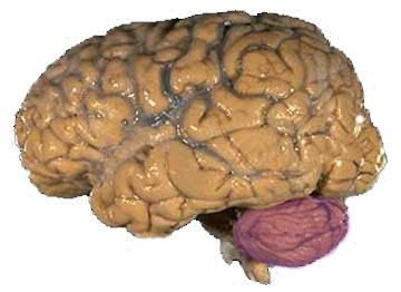 | ||
| Figure 1a: A human brain, with the cerebellum in purple. | ||
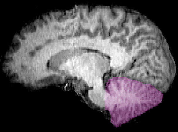 | ||
| Figure 1b: MRI image showing a mid-sagittal view of the human brain, with the cerebellum in purple. | ||
| Latin | ' | |
| Gray's | subject # | |
| Part of | Brain | |
| Components | ||
| Artery | SCA, AICA, PICA | |
| Vein | superior, inferior | |
| BrainInfo/UW | - | |
| MeSH | A08.186.211.132.810.428.200 | |
The cerebellum (Latin for little brain) is a region of the brain that plays an important role in motor control. It may also be involved in some cognitive functions such as attention and language, and in regulating fear and pleasure responses,[1] but its movement-related functions are the most solidly established. The cerebellum does not initiate movement, but it contributes to coordination, precision, and accurate timing. It receives input from sensory systems of the spinal cord and from other parts of the brain, and integrates these inputs to fine tune motor activity.[2] Because of this fine-tuning function, damage to the cerebellum does not cause paralysis, but instead produces disorders in fine movement, equilibrium, posture, and motor learning.[2]
In its anatomy, the cerebellum has the appearance of a separate structure attached to the bottom of the brain, tucked underneath the cerebral hemispheres. The surface of the cerebellum is covered with finely spaced parallel grooves, in striking contrast to the broad irregular convolutions of the cerebral cortex. These parallel grooves conceal the fact that the cerebellum is actually a continuous thin layer of tissue (the cerebellar cortex), tightly folded in the style of an accordion. Within this thin layer are several types of neurons with a highly regular arrangement, the most important being Purkinje cells and granule cells. This complex neural network gives rise to a massive signal-processing capability, but almost all of its output is directed to a set of small deep cerebellar nuclei lying in the interior of the cerebellum.
In addition to its direct role in motor control, the cerebellum also is necessary for several types of motor learning, the most notable one being learning to adjust to changes in sensorimotor relationships. Several theoretical models have been developed to explain sensorimotor calibration in terms of synaptic plasticity within the cerebellum. Most of them derive from early models formulated by David Marr and James Albus, which were motivated by the observation that each cerebellar Purkinje cell receives two dramatically different types of input: On one hand, thousands of inputs from parallel fibers, each individually very weak; on the other hand, input from one single climbing fiber, which is, however, so strong that a single climbing fiber action potential will reliably cause a target Purkinje cell to fire a burst of action potentials. The basic concept of the Marr-Albus theory is that the climbing fiber serves as a "teaching signal", which induces a long-lasting change in the strength of synchronously activated parallel fiber inputs. Observations of long-term depression in parallel fiber inputs have provided support for theories of this type, but their validity remains controversial.
Because of this 'updating' function of the cerebellum, lesions within it are not so debilitating as to cause paralysis, but rather present as feedback deficits resulting in disorders in fine movement, equilibrium, posture, and motor learning. Initial observations by physiologists during the 18th century indicated that patients with cerebellar damage show problems with motor coordination and movement. Research into cerebellar function during the early to mid 19th century was done via lesion and ablation studies in animals. Research physiologists noted that such lesions led to animals with strange movements, awkward gait, and muscular weakness. These observations and studies led to the conclusion that the cerebellum was a motor control structure.[2] However, modern research shows that the cerebellum has a broader role in a number of key cognitive functions, including attention and the processing of language, music, and other sensory temporal stimuli.[3]
Structure
- Main article: Anatomy of the cerebellum
At the level of large scale anatomy, the cerebellum consists of a tightly folded and crumpled layer of cortex, with white matter underneath, several deep nuclei embedded in the white matter, and a fluid-filled ventricle at the base. At the microscopic level, each part of the cortex consists of the same small set of neuronal elements, laid out with a highly stereotyped geometry. At an intermediate level, the cerebellum and its auxiliary structures can be decomposed into several hundred or thousand independently functioning modules called "microzones" or "microcompartments".
Anatomy
Anterior view of the human cerebellum, with numbers indicating salient landmarks
The cerebellum is located at the bottom of the brain, with the large mass of the cerebral cortex above it and the portion of the brainstem called the pons in front of it.[4] It is separated from the overlying cerebrum by a layer of leathery dura mater; all of its connections with other parts of the brain travel through the pons. Anatomists classify the cerebellum as part of the metencephalon, which also includes the pons; the metencephalon is the upper part of the rhombencephalon or "hindbrain". Like the cerebral cortex, the cerebellum is divided into two hemispheres; it also contains a narrow midline zone called the vermis. A set of large folds is, by convention, used to divide the overall structure into 10 smaller "lobules". Because of its large number of tiny granule cells, the cerebellum contains more neurons than the rest of the brain put together, but it takes up only 10% of total brain volume.[5] The number of neurons in the cerebellum is related to the number of neurons in the neocortex. There are about 3.6 times as many neurons in the cerebellum as in neocortex, a number that is conserved across many different mammalian species.[6]
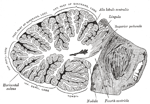
Vertical cross-section of the human cerebellum, showing folding pattern of the cortex, and interior structures
The unusual surface appearance of the cerebellum conceals the fact that most of its volume is made up of a very tightly folded layer of gray matter, the cerebellar cortex. It has been estimated that, if the human cerebellar cortex were completely unfolded, it would give rise to a layer of neural tissue about 1 meter long and averaging 5 centimeters wide — a total surface area of about 500 square cm, packed within a volume of dimensions 6 cm × 5 cm × 10 cm.[5] Underneath the gray matter of the cortex lies white matter, made up largely of myelinated nerve fibers running to and from the cortex. Embedded within the white matter — which is sometimes called the arbor vitae (Tree of Life) because of its branched, tree-like appearance in cross-section — are four deep cerebellar nuclei, composed of gray matter.[4]
Subdivisions
Based on surface appearance, three lobes can be distinguished in the cerebellum, called the flocculonodular lobe, anterior lobe (above the primary fissure), and posterior lobe (below the primary fissure). These lobes divide the cerebellum from rostral to caudal (in humans, top to bottom). In terms of function, however, there is a more important distinction along the medial-to-lateral dimension. Leaving out the flocculonodular part, which has distinct connections and functions, the cerebellum can be parsed functionally into a medial sector called the spinocerebellum and a larger lateral sector called the cerebrocerebellum.[4] A narrow strip of protruding tissue along the midline is called the vermis (Latin for "worm").[4]
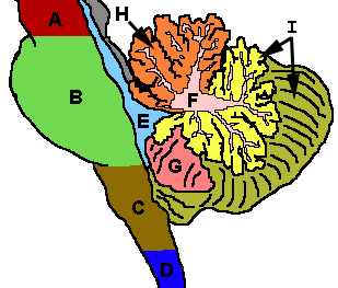 Cerebellum and surrounding regions; sagittal view of one hemisphere. A: Midbrain. B: Pons. C: Medulla. D: Spinal cord. E: Fourth ventricle. F: Arbor vitae. G: Tonsil. H: Anterior lobe. I: Posterior lobe. |
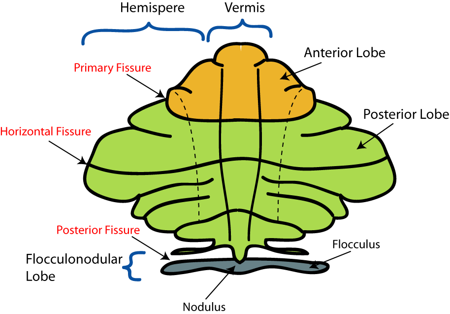
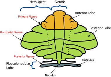
The smallest region, the flocculonodular lobe, is often called the vestibulocerebellum. It is the oldest part in evolutionary terms (archicerebellum) and participates mainly in balance and spatial orientation; its primary connections are with the vestibular nuclei, although it also receives visual and other sensory input. Damage to it causes disturbances of balance and gait.[4]
The medial zone of the anterior and posterior lobes constitutes the spinocerebellum, also known as paleocerebellum. This sector of the cerebellum functions mainly to fine-tune body and limb movements. It receives proprioception input from the dorsal columns of the spinal cord (including the spinocerebellar tract) and from the trigeminal nerve, as well as from visual and auditory systems. It sends fibres to deep cerebellar nuclei that, in turn, project to both the cerebral cortex and the brain stem, thus providing modulation of descending motor systems.[4]
The lateral zone, which in humans is by far the largest part, constitutes the cerebrocerebellum, also known as neocerebellum. It receives input exclusively from the cerebral cortex (especially the parietal lobe) via the pontine nuclei (forming cortico-ponto-cerebellar pathways), and sends output mainly to the ventrolateral thalamus (in turn connected to motor areas of the premotor cortex and primary motor area of the cerebral cortex) and to the red nucleus.[4] There is disagreement about the best way to describe the functions of the lateral cerebellum: It is thought to be involved in planning movement that is about to occur,[7] in evaluating sensory information for action,[4] and in a number of purely cognitive functions as well, such as determining the verb which best fits with a certain noun (as in 'sit' for 'chair').[8][9]
Cellular components

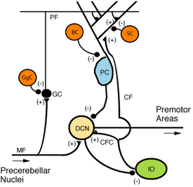
Microcircuitry of the cerebellum. (+): excitatory; (-): inhibitory; MF: Mossy fiber; DCN: Deep cerebellar nuclei; IO: Inferior olive; CF: Climbing fiber; GC: Granule cell; PF: Parallel fiber; PC: Purkinje cell; GgC: Golgi cell; SC: Stellate cell; BC: Basket cell
Transverse section of a cerebellar folium, showing principal cell types and connections.
Two types of neuron play dominant roles in the cerebellar circuit: Purkinje cells and granule cells. Three types of axons also play dominant roles: mossy fibers and climbing fibers (which enter the cerebellum from outside), and parallel fibers (which are the axons of granule cells). There are two main pathways through the cerebellar circuit, originating from mossy fibers and climbing fibers, both eventually terminating in the deep cerebellar nuclei.
Mossy fibers project directly to the deep nuclei, but also give rise to the pathway: mossy fiber → granule cells → parallel fibers → Purkinje cells → deep nuclei. Climbing fibers project to Purkinje cells and also send collaterals directly to the deep nuclei.[5] The mossy fiber and climbing fiber inputs each carry fiber-specific information; the cerebellum also receives dopaminergic, serotonergic, noradrenergic, and cholinergic inputs that presumably perform global modulation.[10]
The cerebellar cortex is divided into three layers. At the bottom lies the thick granular layer, densely packed with granule cells, along with interneurons, mainly Golgi cells but also including Lugaro cells and unipolar brush cells. In the middle lies the Purkinje layer, a narrow zone that contains only the cell bodies of Purkinje cells. At the top lies the molecular layer, which contains the flattened dendritic trees of Purkinje cells, along with the huge array of parallel fibers penetrating the Purkinje cell dendritic trees at right angles. This outermost layer of the cerebellar cortex also contains two types of inhibitory interneurons, stellate cells, and basket cells. Both stellate and basket cells form GABAergic synapses onto Purkinje cell dendrites.[5]
Purkinje cells
Drawing of a Purkinje cell from cat cerebellum
Purkinje cells are among the most distinctive neurons in the brain, and also among the earliest types to be recognized — they were first described by the Czech anatomist Jan Evangelista Purkyně in 1837. They are distinguished by the shape of the dendritic tree: The dendrites branch very profusely, but are severely flattened in a plane perpendicular to the cerebellar folds. Thus, the dendrites of a Purkinje cell form a dense planar net, through which parallel fibers pass at right angles.[5] The dendrites are covered with dendritic spines, each of which receives synaptic input from a parallel fiber. Purkinje cells receive more synaptic inputs than any other type of cell in the brain — estimates of the number of spines on a single human Purkinje cell run as high as 200,000.[5] The large, spherical cell bodies of Purkinje cells are packed into a narrow layer (one cell thick) of the cerebellar cortex, called the Purkinje layer. After emitting collaterals that innervate nearby parts of the cortex, their axons travel into the deep cerebellar nuclei, where they make on the order of 1,000 contacts each with several types of nuclear cells, all within a small domain. Purkinje cells use GABA as their neurotransmitter, and therefore exert inhibitory effects on their targets.[5]
Purkinje cells form the heart of the cerebellar circuit, and their large size and distinctive activity patterns have made it relatively easy to study their response patterns in behaving animals using extracellular recording techniques. Purkinje cells normally emit action potentials at a high rate even in the absence of synaptic input. In awake, behaving animals, mean rates averaging around 40 Hz are typical. The spike trains show a mixture of what are called simple and complex spikes. A simple spike is a single action potential followed by a refractory period of about 10 ms; a complex spike is a stereotyped sequence of action potentials with very short inter-spike intervals and declining amplitudes.[citation needed] Physiological studies have shown that complex spikes (which occur at baseline rates around 1 Hz and never at rates much higher than 10 Hz) are reliably associated with climbing fiber activation, while simple spikes are produced by a combination of baseline activity and parallel fiber input. Complex spikes are often followed by a pause of several hundred milliseconds during which simple spike activity is suppressed.[11]
Granule cells
Cerebellar granule cells, in contrast to Purkinje cells, are among the smallest neurons in the brain. They are also easily the most numerous neurons in the brain: In humans, estimates of their total number average around 50 billion, which means that about 3/4 of the brain's neurons are cerebellar granule cells.[5] Their cell bodies are packed into a thick layer at the bottom of the cerebellar cortex. A granule cell emits only four to five dendrites, each of which ends in an enlargement called a dendritic claw.[5] These enlargements are sites of excitatory input from mossy fibers and inhibitory input from Golgi cells.
Granule cells, parallel fibers, and Purkinje cells with flattened dendritic trees
The thin, unmyelinated axons of granule cells rise vertically to the upper (molecular) layer of the cortex, where they split in two, with each branch traveling horizontally to form a parallel fiber; the splitting of the vertical branch into two horizontal branches gives rise to a distinctive "T" shape. A parallel fiber runs for an average of 3 mm in each direction from the split, for a total length of about 6 mm (about 1/10 of the total width of the cortical layer).[5] As they run along, the parallel fibers pass through the dendritic trees of Purkinje cells, contacting one of every 3–5 that they pass, making a total of 80–100 synaptic connections with Purkinje cell dendritic spines.[5] Granule cells use glutamate as their neurotransmitter, and therefore exert excitatory effects on their targets.
Granule cells receive all of their input from mossy fibers, but outnumber them 200 to 1 (in humans). Thus, the information in the granule cell population activity state is the same as the information in the mossy fibers, but recoded in a much more expansive way. Because granule cells are so small and so densely packed, it has been very difficult to record their spike activity in behaving animals, so there is little data to use as a basis of theorizing. The most popular concept of their function was proposed by David Marr, who suggested that they could encode combinations of mossy fiber inputs. The idea is that with each granule cell receiving input from only 4–5 mossy fibers, a granule cell would not respond if only a single one of its inputs were active, but would respond if more than one were active. This combinatorial coding scheme would potentially allow the cerebellum to make much finer distinctions between input patterns than the mossy fibers alone would permit.[12]
Mossy fibers
Mossy fibers enter the granular layer from their points of origin, many arising from the pontine nuclei, others from the spinal cord, vestibular nuclei, etc. In the human cerebellum, the total number of mossy fibers has been estimated at about 200 million.[5] These fibers form excitatory synapses with the granule cells and the cells of the deep cerebellar nuclei. Within the granular layer, a mossy fiber generates a series of enlargements called rosettes. The contacts between mossy fibers and granule cell dendrites take place within structures called glomeruli. Each glomerulus has a mossy fiber rosette at its center, and up to 20 granule cell dendritic claws contacting it. Terminals from Golgi cells infiltrate the structure and make inhibitory synapses onto the granule cell dendrites. The entire assemblage is surrounded by a sheath of glial cells.[5] Each mossy fiber sends collateral branches to several cerebellar folia, generating a total of 20–30 rosettes; thus a single mossy fiber makes contact with an estimated 400–600 granule cells.[5]
Climbing fibers
Purkinje cells also receive input from the inferior olivary nucleus (IO) on the contralateral side of the brainstem, via climbing fibers. Although the IO lies in the medulla oblongata, and receives input from the spinal cord, brainstem, and cerebral cortex, its output goes entirely to the cerebellum. A climbing fiber gives off collaterals to the deep cerebellar nuclei before entering the cerebellar cortex, where it splits into about 10 terminal branches, each of which innervates a single Purkinje cell.[5] In striking contrast to the 100,000-plus inputs from parallel fibers, each Purkinje cell receives input from exactly one climbing fiber; but this single fiber "climbs" the dendrites of the Purkinje cell, winding around them and making a total of up to 300 synapses as it goes.[5] The net input is so strong that a single action potential from a climbing fiber is capable of producing an extended complex spike in the Purkinje cell: a burst of several spikes in a row, with diminishing amplitude, followed by a pause during which activity is suppressed. The climbing fiber synapses cover the cell body and proximal dendrites; this zone is devoid of parallel fiber inputs.[5]
Climbing fibers fire at low rates, but a single climbing fiber action potential induces a burst of several action potentials in a target Purkinje cell (a complex spike). The contrast between parallel fiber and climbing fiber inputs to Purkinje cells (over 100,000 of one type versus exactly one of the other type) is perhaps the most provocative feature of cerebellar anatomy, and has motivated much of the theorizing. In fact, the function of climbing fibers is the most controversial topic concerning the cerebellum. There are two schools of thought, one following Marr and Albus in holding that climbing fiber input serves primarily as a teaching signal, the other holding that its function is to shape cerebellar output directly. Both views have been defended in great length in numerous publications. In the words of one review, "In trying to synthesize the various hypotheses on the function of the climbing fibers, one has the sense of looking at a drawing by Escher. Each point of view seems to account for a certain collection of findings, but when one attempts to put the different views together, a coherent picture of what the climbing fibers are doing does not appear. For the majority of researchers, the climbing fibers signal errors in motor performance, either in the usual manner of discharge frequency modulation or as a single announcement of an 'unexpected event'. For other investigators, the message lies in the degree of ensemble synchrony and rhythmicity among a population of climbing fibers."[11]
Deep nuclei
- Main article: Deep cerebellar nuclei
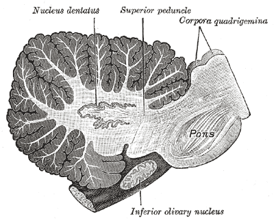
Cross-section of human cerebellum, showing the dentate nucleus, as well as the pons and inferior olivary nucleus
The deep nuclei of the cerebellum are clusters of gray matter lying within the white matter at the core of the cerebellum. They are, with the minor exception of the nearby vestibular nuclei, the sole sources of output from the cerebellum. These nuclei receive collateral projections from mossy fibers and climbing fibers, as well as inhibitory input from the Purkinje cells of the cerebellar cortex. The three nuclei (dentate, interpositus, and fastigial) each communicate with different parts of the brain and cerebellar cortex. The fastigial and interpositus nuclei belong to the spinocerebellum. The dentate nucleus, which in mammals is much larger than the others, is formed as a thin, convoluted layer of gray matter, and communicates exclusively with the lateral parts of the cerebellar cortex. The flocculonodular lobe is the only part of the cerebellar cortex that does not project to the deep nuclei — its output goes to the vestibular nuclei instead.[5]
The majority of neurons in the deep nuclei have large cell bodies and spherical dendritic trees with a radius of about 400 μm, and use glutamate as their neurotransmitter. These cells project to a variety of targets outside the cerebellum. Intermixed with them is a lesser number of small cells, which use GABA as neurotransmitter and project exclusively to the inferior olivary nucleus, the source of climbing fibers. Thus, the nucleo-olivary projection provides an inhibitory feedback to match the excitatory projection of climbing fibers to the nuclei. There is evidence that each small cluster of nuclear cells projects to the same cluster of olivary cells that send climbing fibers to it; there is strong and matching topography in both directions.[5]
When a Purkinje cell axon enters one of the deep nuclei, it branches to make contact with both large and small nuclear cells, but the total number of cells contacted is only about 35 (in cats). On the converse, a single deep nuclear cell receives input from approximately 860 Purkinje cells (again in cats).[5]
Compartmentalization
Schematic illustration of the structure of zones and microzones in the cerebellar cortex
From the viewpoint of gross anatomy, the cerebellar cortex appears to be a homogeneous sheet of tissue, and, from the viewpoint of microanatomy, all parts of this sheet appear to have the same internal structure. There are, however, a number of respects in which the structure of the cerebellum is compartmentalized. There are large compartments that are generally known as zones; these can be decomposed into smaller compartments known as microzones.[13]
The first indications of compartmental structure came from studies of the receptive fields of cells in various parts of the cerebellum cortex.[13] Each body part maps to specific points in the cerebellum, but there are numerous repetitions of the basic map, forming an arrangement that has been called "fractured somatotopy".[14] A clearer indication of compartmentalization is obtained by immunostaining the cerebellum for certain types of protein. The best-known of these markers are called "zebrins", because staining for them gives rise to a complex pattern reminiscent of the stripes on a zebra. The stripes generated by zebrins and other compartmentalization markers are oriented perpendicular to the cerebellar folds — that is, they are narrow in the mediolateral direction, but much more extended in the longitudinal direction. Different markers generate different sets of stripes, and the widths and lengths vary as a function of location, but they all have the same general shape.[13]
Oscarsson in the late 1970s proposed that these cortical zones can be partitioned into smaller units called microzones.[15] A microzone is defined as a group of Purkinje cells all having the same somatotopic receptive field. Microzones were found to contain on the order of 1000 Purkinje cells each, arranged in a long, narrow strip, oriented perpendicular to the cortical folds.[13] Thus, as the adjoining diagram illustrates, Purkinje cell dendrites are flattened in the same direction as the microzones extend, while parallel fibers cross them at right angles.
It is not only receptive fields that define the microzone structure: The climbing fiber input from the inferior olivary nucleus is equally important. The branches of a climbing fiber (usually numbering about 10) usually innervate Purkinje cells belonging to the same microzone. Moreover, olivary neurons that send climbing fibers to the same microzone tend to be coupled by gap junctions, which synchronize their activity, causing Purkinje cells within a microzone to show correlated complex spike activity on a millisecond time scale.[13] Also, the Purkinje cells belonging to a microzone all send their axons to the same small cluster of output cells within the deep cerebellar nuclei.[13] Finally, the axons of basket cells are much longer in the longitudinal direction than in the mediolateral direction, causing them to be confined largely to a single microzone.[13] The consequence of all this structure is that cellular interactions within a microzone are much stronger than interactions between different microzones.
In 2005, Richard Apps and Martin Garwicz summarized evidence that microzones themselves form part of a larger entity they call a multizonal microcomplex. Such a microcomplex includes several spatially separated cortical microzones, all of which project to the same group of deep cerebellar neurons, plus a group of coupled olivary neurons that project to all of the included microzones as well as to the deep nuclear area.[13]
Function
The strongest clues to the function of the cerebellum have come from examining the consequences of damage to it. Animals and humans with cerebellar dysfunction show, above all, problems with motor control, on the side of the body ipsilateral to the damaged cerebellum. They continue to be able to generate motor activity, but it loses precision, producing erratic, uncoordinated, or incorrectly timed movements. A standard test of cerebellar function is to reach with the tip of the finger for a target at arm's length: A healthy person will move the fingertip in a rapid straight trajectory, whereas a person with cerebellar damage will reach slowly and erratically, with many mid-course corrections. Deficits in non-motor functions are more difficult to detect. Thus, the general conclusion reached decades ago is that the basic function of the cerebellum is not to initiate movements, or to decide which movements to execute, but rather to calibrate the detailed form of a movement.[4]
Prior to the 1990s, the function of the cerebellum was almost universally believed to be purely motor-related, but newer findings have brought that view strongly into question. Functional imaging studies have shown cerebellar activation in relation to language, attention, and mental imagery; correlation studies have shown interactions between the cerebellum and non-motoric areas of the cerebral cortex; and a variety of non-motor symptoms have been recognized in people with damage that appears to be confined to the cerebellum.[16][17] In particular, the Cerebellar Cognitive Affective Syndrome has been described in adults [18] and children.[19]
Kenji Doya has argued that the function of the cerebellum is best understood not in terms of what behaviors it is involved in but rather in terms of what neural computations it performs; the cerebellum consists of a large number of more or less independent modules, all with the same geometrically regular internal structure, and therefore all, it is presumed, performing the same computation. If the input and output connections of a module are with motor areas (as many are), then the module will be involved in motor behavior; but, if the connections are with areas involved in non-motor cognition, the module will show other types of behavioral correlates. The cerebellum, Doya proposes, is best understood as a device for supervised learning, in contrast to the basal ganglia, which perform reinforcement learning, and the cerebral cortex, which performs unsupervised learning.[17]
Principles
The comparative simplicity and regularity of the cerebellar anatomy led to an early hope that it might imply a similar simplicity of computational function, as expressed in one of the first books on cerebellar electrophysiology, The Cerebellum as a Neuronal Machine by John C. Eccles, Masao Ito, and Janos Szentágothai.[20] Although a full understanding of cerebellar function has remained elusive, at least four principles have been identified as important: (1) feedforward processing, (2) divergence and convergence, (3) modularity, and (4) plasticity.
1. Feedforward processing: The cerebellum differs from most other parts of the brain (especially the cerebral cortex) in that the signal processing is almost entirely feedforward - that is, signals move unidirectionally through the system from input to output, with very little recurrent internal transmission. The small amount of recurrence that does exist consists of mutual inhibition; there are no mutually excitatory circuits. This feedforward mode of operation means that the cerebellum, in contrast to the cerebral cortex, cannot generate self-sustaining patterns of neural activity. Signals enter the circuit, are processed by each stage in sequential order, and then leave. As Eccles, Ito, and Szentágothai wrote, "This elimination in the design of all possibility of reverberatory chains of neuronal excitation is undoubtedly a great advantage in the performance of the cerebellum as a computer, because what the rest of the nervous system requires from the cerebellum is presumably not some output expressing the operation of complex reverberatory circuits in the cerebellum but rather a quick and clear response to the input of any particular set of information."[21]
2. Divergence and convergence: In the human cerebellum, information from 200 million mossy fiber inputs is expanded to 40 billion granule cells, whose parallel fiber outputs then converge onto 15 million Purkinje cells.[5] Because of the way that they are lined up longitudinally, the 1000 or so Purkinje cells belonging to a microzone may receive input from as many as 100 million parallel fibers, and focus their own output down to a group of less than 50 deep nuclear cells.[13] Thus, the cerebellar network receives a modest number of inputs, processes them very extensively through its rigorously structured internal network, and sends out the results via a very limited number of output cells.
3. Modularity: The cerebellar system is functionally divided into more or less independent modules, which probably number in the hundreds to thousands. All modules have a similar internal structure, but different inputs and outputs. A module (a multizonal microcompartment in the terminology of Apps and Garwicz) consists of a small cluster of neurons in the inferior olivary nucleus, a set of long narrow strips of Purkinje cells in the cerebellar cortex (microzones), and a small cluster of neurons in one of the deep cerebellar nuclei. Different modules share input from mossy fibers and parallel fibers, but in other respects they appear to function independently — the output of one module does not appear to significantly influence the activity of other modules.[13]
4. Plasticity: The synapses between parallel fibers and Purkinje cells, and the synapses between mossy fibers and deep nuclear cells, are both susceptible to modification of their strength. In a single cerebellar module, input from as many as a billion parallel fibers converges onto a group of less than 50 deep nuclear cells, and the influence of each parallel fiber on those nuclear cells is adjustable. This arrangement gives tremendous flexibility for fine-tuning the relationship between cerebellar inputs and outputs.[22]
Learning
There is considerable evidence that the cerebellum plays an essential role in some types of motor learning. The tasks where the cerebellum most clearly comes into play are those in which it is necessary to make fine adjustments to the way an action is performed. There has, however, been much dispute about whether learning takes place within the cerebellum itself, or whether it merely serves to provide signals that promote learning in other brain structures.[22] Most theories that assign learning to the circuitry of the cerebellum are derived from early ideas of David Marr and James Albus, who postulated that climbing fibers provide a teaching signal that induces synaptic modification in parallel fiber—Purkinje cell synapses.[23] Marr assumed that climbing fiber input would cause synchronously activated parallel fiber inputs to be strengthened. Most later cerebellar-learning models, however, have followed Albus in assuming that climbing fiber activity would be an error signal, and would cause synchronously activated parallel fiber inputs to be weakened. Some of these later models, such as the Adaptive Filter model of Fujita[24] made attempts to understand cerebellar function in terms of optimal control theory.
The idea that climbing fiber activity functions as an error signal has been examined in many experimental studies, with some supporting it but others casting doubt.[11] In a pioneering study by Gilbert and Thach from 1977, Purkinje cells from monkeys learning a reaching task showed increased complex spike activity — which is known to reliably indicate activity of the cell's climbing fiber input — during periods when performance was poor.[25] Several studies of motor learning in cats observed complex spike activity when there was a mismatch between an intended movement and the movement that was actually executed. Studies of the vestibulo-ocular reflex (which stabilizes the visual image on the retina when the head turns) found that climbing fiber activity indicated "retinal slip", although not in a very straightforward way.[11]
One of the most extensively studied cerebellar learning tasks is the eyeblink conditioning paradigm, in which a neutral conditioned stimulus such as a tone or a light is repeatedly paired with an unconditioned stimulus, such as an air puff, that elicits a blink response. After such repeated presentations of the CS and US, the CS will eventually elicit a blink before the US, a conditioned response or CR. Experiments showed that lesions localized either to a specific part of the interpositus nucleus (one of the deep cerebellar nuclei) or to a few specific points in the cerebellar cortex would abolish learning of a correctly timed blink response. If cerebellar outputs are pharmacologically inactivated while leaving the inputs and intracellular circuits intact, learning takes place even while the animal fails to show any response, whereas, if intracerebellar circuits are disrupted, no learning takes place — these facts taken together make a strong case that the learning, indeed, occurs inside the cerebellum.[26]
Theories and computational models
Model of a cerebellar perceptron, as formulated by James Albus
The large base of knowledge about the anatomical structure and behavioral functions of the cerebellum have made it a fertile ground for theorizing — there are perhaps more theories of the function of the cerebellum than of any other part of the brain. The most basic distinction among them is between "learning theories" and "performance theories" — that is, theories that make use of synaptic plasticity within the cerebellum to account for its role in learning, versus theories that account for aspects of ongoing behavior on the basis of cerebellar signal processing. Several theories of both types have been formulated as mathematical models and simulated using computers.[23]
Perhaps the earliest "performance" theory was the "delay line" hypothesis of Valentino Braitenberg. The original theory put forth by Braitenberg and Atwood in 1958 proposed that slow propagation of signals along parallel fibers imposes predictable delays that allow the cerebellum to detect time relationships within a certain window.[27] Experimental data did not support the original form of the theory, but Braitenberg continued to argue for modified versions.[28] The hypothesis that the cerebellum functions essentially as a timing system has also been advocated by Richard Ivry.[29] Another influential "performance" theory is the Tensor Network Theory of Pellionisz and Llinás, which provided an advanced mathematical formulation of the idea that the fundamental computation performed by the cerebellum is to transform sensory into motor coordinates.[30]
Theories in the "learning" category almost all derive from early publications by David Marr[12] and James Albus.[31] Marr's 1969 paper proposed that the cerebellum is a device for learning to associate elemental movements encoded by climbing fibers with mossy fiber inputs that encode the sensory context. Albus proposed that a cerebellar Purkinje cell functions as a perceptron, a neurally inspired abstract learning device. The most basic difference between the Marr and Albus theories is that Marr assumed that climbing fiber activity would cause parallel fiber synapses to be strengthened, whereas Albus proposed that they would be weakened. Albus also formulated his version as a software algorithm he called a CMAC (Cerebellar Model Articulation Controller), which has been tested in a number of applications.[32]
Pathology
- Main article: Ataxia
Altered walking gait of a woman with cerebellar disease
The most salient symptoms of cerebellar dysfunction are motor-related — the specific symptoms depend on which part of the cerebellum is involved and how it is disrupted. Damage to the flocculonodular lobe (the vestibular part) may show up as a loss of equilibrium and, in particular, an altered walking gait, with a wide stance that indicates difficulty in balancing.[4] Damage to the lateral zone, or the cerebrocerebellum, results in problems with skilled voluntary and planned movements. This can cause errors in the force, direction, speed and amplitude of movements. Some manifestations include hypotonia (decreased muscle tone), dysarthria (problems with speech articulation), dysmetria (problems judging distances or ranges of movement), dysdiadochokinesia (inability to perform rapid alternating movements), impaired check reflex or rebound phenomenon, and tremors (involuntary movement caused by alternating contractions of opposing muscle groups).[33] Damage to the midline portion may disrupt whole-body movements, whereas damage localized more laterally is more likely to disrupt fine movements of the hands or limbs. Damage to the upper part of the cerebellum tends to cause gait impairments and other problems with leg coordination; damage to the lower part is more likely to cause uncoordinated or poorly aimed movements of the arms and hands, as well as difficulties in speed.[4] This complex of motor symptoms is called "ataxia". To identify cerebellar problems, the neurological examination includes assessment of gait (a broad-based gait being indicative of ataxia), finger-pointing tests and assessment of posture.[2] If cerebellar dysfunction is indicated, a magnetic resonance imaging scan can be used to obtain a detailed picture of any structural alterations that may exist.[34]
The lower trace shows an attempt by a patient with cerebellar disease to reproduce the upper trace.
The list of medical problems that can produce cerebellar damage is long: it includes stroke; hemorrhage; tumors; alcoholism; physical trauma such as gunshot wounds; and chronic degenerative conditions such as olivopontocerebellar atrophy.[4] Some forms of migraine headache may also produce temporary dysfunction of the cerebellum, of variable severity.[35]
Aging
The human cerebellum changes with age. These changes may differ from those of other parts of the brain, for example the gene expression pattern in the human cerebellum shows less age-related alteration than in the cerebral cortex.[36] Some studies have reported reductions in numbers of cells or volume of tissue, but the amount of data relating to this question is not very large.[37][38]
Hemorrhage
Cerebellar hemorrhage is differentiated from other intracranial bleeding in that there is no hemiparesis. Symptoms can include occipitial headaches, ataxia, nystagmus, gaze palsy, facial weakness and vomiting. The most important risk factor is hypertension.[39] Early diagnosis is important since emergent decompression may be life-saving.[40]
Tumors
- Main article: Brain tumors
The cerebellum is occasionally afflicted by tumors. In adults, metastatic tumors are very common.
Tumors that commonly arise in the cerebellum include pilocytic astrocytomas, medulloblastomas (especially in children), ependymomas, and hemangioblastomas (often in the context of von Hippel-Lindau syndrome).
Comparative anatomy and evolution
Cross-section of the brain of a porbeagle shark, with the cerebellum highlighted
The circuits in the cerebellum are similar across all classes of vertebrates, including fish, reptiles, birds, and mammals.[41] There is also an analogous brain structure in cephalopods with well-developed brains, such as octopuses.[42] This has been taken as evidence that the cerebellum performs functions important to all animal species with a brain.
There is considerable variation in the size and shape of the cerebellum in different vertebrate species. In amphibians, lampreys, and hagfish, the cerebellum is little developed; in the latter two groups, it is barely distinguishable from the brain-stem. Although the spinocerebellum is present in these groups, the primary structures are small paired nuclei corresponding to the vestibulocerebellum.[43] The cerebellum is a bit larger in reptiles, considerably larger in birds, and larger yet in mammals. The large paired and convoluted lobes found in humans are typical of mammals, but the cerebellum is, in general, a single median lobe in other groups, and is either smooth or only slightly grooved. In mammals, the neocerebellum is the major part of the cerebellum by mass, but, in other vertebrates, it is typically the spinocerebellum.[43]
The cerebellum of cartilaginous and bony fishes is extraordinarily large and complex. In at least one important respect, it differs in internal structure from the mammalian cerebellum: The fish cerebellum does not contain discrete deep cerebellar nuclei. Instead, the primary targets of Purkinje cells are a distinct type of cell distributed across the cerebellar cortex, a type not seen in mammals. In mormyrid fish (a family of weakly electrosensitive freshwater fish), the cerebellum is considerably larger than the rest of the brain put together. The largest part of it is a special structure called the valvula, which has an unusually regular architecture and receives much of its input from the electrosensory system.[44]
The hallmark of the mammalian cerebellum is an expansion of the lateral lobes, whose main interactions are with the neocortex. As monkeys evolved into great apes, the expansion of the lateral lobes continued, in tandem with the expansion of the frontal lobes of the neocortex. In ancestral hominids, and in homo sapiens until the middle Pleistocene period, the cerebellum continued to expand, but the frontal lobes expanded more rapidly. The most recent period of human evolution, however, may actually have been associated with an increase in the relative size of the cerebellum, as the neocortex reduced its size somewhat while the cerebellum expanded.[45]
Cerebellum-like structures
Many vertebrate species have brain areas that resemble the cerebellum in terms of cellular architecture and neurochemistry.[41] The only one found in mammals is the dorsal cochlear nucleus (DCN), one of the two primary sensory nuclei that receive input directly from the auditory nerve. The DCN is a layered structure, with the bottom layer containing granule cells similar to those of the cerebellum, giving rise to parallel fibers that rise to the superficial layer and travel across it horizontally. The superficial layer contains a set of GABAergic neurons called cartwheel cells that resemble Purkinje cells anatomically and chemically — they receive parallel fiber input, but do not have any inputs that resemble climbing fibers. The output neurons of the DCN are called fusiform cells. They are glutamatergic, but also resemble Purkinje cells in some respects — they have spiny, flattened superficial dendritic trees that receive parallel fiber input, but they also have basal dendrites that receive input from auditory nerve fibers, which travel across the DCN in a direction at right angles to the parallel fibers. The DCN is most highly developed in rodents and other small animals, and is considerably reduced in primates. Its function is not well understood; the most popular speculations relate it to spatial hearing in one way or another.[46]
Most species of fish and amphibians possess a lateral line system that senses pressure waves in water. One of the brain areas that receives primary input from the lateral line organ, the medial octavolateral nucleus, has a cerebellum-like structure, with granule cells and parallel fibers. In electrosensitive fish, the input from the electrosensory system goes to the dorsal octavolateral nucleus, which also has a cerebellum-like structure. In ray-finned fishes (by far the largest group), the optic tectum has a layer — the marginal layer — that is cerebellum-like.[41]
All of these cerebellum-like structures appear to be primarily sensory-related rather than motor-related. All of them have granule cells that give rise to parallel fibers that connect to Purkinje-like neurons with modifiable synapses, but none have climbing fibers comparable to those of the cerebellum — instead they receive direct input from peripheral sensory organs. None has a demonstrated function, but the most influential speculation is that they serve to transform sensory inputs in some sophisticated way, perhaps to compensate for changes in body posture.[41] In fact, James Bower and others have argued, partly on the basis of these structures and partly on the basis of cerebellar studies, that the cerebellum itself is fundamentally a sensory structure, and that it contributes to motor control by moving the body in a way that controls the resulting sensory signals.[47]
History

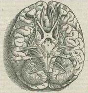
Base of the human brain, as drawn by Andreas Vesalius in 1543
The distinctive appearance of the cerebellum caused even the earliest anatomists to recognize it. Aristotle and Galen, however, did not consider it truly part of the brain: They called it the parencephalon ("same-as-brain"), as opposed to the encephalon or brain proper. Galen was the first to give an extensive description, noting that the cerebellar tissue seemed more solid than the rest of the brain, he speculated that its function is to strengthen the motor nerves.[48]
Further significant developments did not come until the Renaissance. Vesalius discussed the cerebellum briefly, and the anatomy was described more thoroughly by Thomas Willis in 1664. More anatomical work was done during the 18th century, but it was not until early in the 19th century that the first insights into the function of the cerebellum were obtained. Luigi Rolando in 1809 established the key insight that damage to the cerebellum results in motor disturbances. Jean Pierre Flourens in the first half of the 19th century carried out detailed experimental work, which revealed that animals with cerebellar damage can still move, but with a loss of coordination (strange movements, awkward gait, and muscular weakness), and that recovery after the lesion can be nearly complete unless the lesion is very extensive.[49] By the dawn of the 20th century, it was widely accepted that the primary function of the cerebellum relates to motor control; the first half of the 20th century produced several detailed descriptions of the clinical symptoms associated with cerebellar disease in humans.[2]
Additional images
")
")
")
")
")
")
")
See also
- Brain
- Central nervous system
- Cerebellar Model Articulation Controller
- Cerebellar peduncles
- Cerebellar theory of dyslexia
- Cerebellar stroke syndrome
- Regions in the human brain
- Paraneoplastic cerebellar degeneration
References
- ↑ Wolf U, Rapoport MJ, Schweizer TA (2009). Evaluating the affective component of the cerebellar cognitive affective syndrome. J. Neuropsychiatry Clin. Neurosci. 21 (3): 245–53.
- ↑ 2.0 2.1 2.2 2.3 2.4 Fine EJ, Ionita CC, Lohr L (2002). The history of the development of the cerebellar examination. Semin Neurol 22 (4): 375–84.
- ↑ Rapp, Brenda (2001). The Handbook of Cognitive Neuropsychology: What Deficits Reveal about the Human Mind, 481, Psychology Press.
- ↑ 4.00 4.01 4.02 4.03 4.04 4.05 4.06 4.07 4.08 4.09 4.10 4.11 Ghez C, Fahn S (1985). "The cerebellum" Principles of Neural Science, 2nd edition, 502–522, New York: Elsevier.
- ↑ 5.00 5.01 5.02 5.03 5.04 5.05 5.06 5.07 5.08 5.09 5.10 5.11 5.12 5.13 5.14 5.15 5.16 5.17 5.18 5.19 5.20 Llinas RR, Walton KD, Lang EJ (2004). "Ch. 7 Cerebellum" Shepherd GM The Synaptic Organization of the Brain, New York: Oxford University Press.
- ↑ Herculano-Houzel S (2010). Coordinated scaling of cortical and cerebellar numbers of neurons. Front. Neuroanat. 4:12.
- ↑ Kingsley RE (2000). Concise Text of Neuroscience, 2nd, Lippincott Williams and Wilkins.
- ↑ Timmann D, Daum I (2007). Cerebellar contributions to cognitive functions: a progress report after two decades of research. Cerebellum 6 (3): 159–62.
- ↑ Lenhoff, Howard, Paul Wang (December 1997). Williams Syndrome and the Brain. Scientific American 277 (6).
- ↑ Schweighofer N, Doya K, Kuroda S (2004). Cerebellar aminergic neuromodulation: towards a functional understanding. Brain Res. Brain Res. Rev. 44 (2–3): 103–116.
- ↑ 11.0 11.1 11.2 11.3 Simpson JI, Wylie DR, De Zeeuw CI (1996). On climbing fiber signals and their consequence(s). Behav. Brain Sci. 19 (3): 384–398.
- ↑ 12.0 12.1 Marr D (1969). A theory of cerebellar cortex. J. Physiol. 202 (2): 437–70.
- ↑ 13.0 13.1 13.2 13.3 13.4 13.5 13.6 13.7 13.8 13.9 Apps R, Garwicz M (2005). Anatomical and physiological foundations of cerebellar information processing. Nat. Rev. Neurosci. 6 (4): 297–311.
- ↑ Manni E, Petrosini L (2004). A century of cerebellar somatotopy: a debated representation. Nat. Rev. Neurosci. 5 (3): 241–9.
- ↑ Oscarsson O (1979). Functional units of the cerebellum-sagittal zones and microzones. Trends Neurosci. 2: 143–145.
- ↑ Rapp, Brenda (2001). The Handbook of Cognitive Neuropsychology: What Deficits Reveal about the Human Mind, Psychology Press.
- ↑ 17.0 17.1 Doya K (2000). Complementary roles of basal ganglia and cerebellum in learning and motor control. Curr. Op. Neurobiology 10 (6): 732–739.
- ↑ Schmahmann, J. D., & Sherman, J. C. "The cerebellar cognitive affective syndrome", Brain, 121(Pt 4), 561–579, 1998.
- ↑ Levisohn, L., Cronin-Golomb, A., & Schmahmann, J. D. "Neuropsychological consequences of cerebellar tumour resection in children: cerebellar cognitive affective syndrome in a paediatric population", Brain, 123(Pt 5), 1041–1050, 2000.
- ↑ Eccles JC, Ito M, Szentágothai J (1967). The Cerebellum as a Neuronal Machine, Springer-Verlag.
- ↑ The Cerebellum as a Neuronal Machine, p. 311
- ↑ 22.0 22.1 Boyden ES, Katoh A, Raymond JL (2004). Cerebellum-dependent learning: the role of multiple plasticity mechanisms. Annu. Rev. Neurosci. 27: 581–609. Cite error: Invalid
<ref>tag; name "Boyden" defined multiple times with different content - ↑ 23.0 23.1 Houk JC, Buckingham JT, Barto AG (1996). Models of the cerebellum and motor learning. Behav. Brain Sci. 19 (3): 368–383.
- ↑ Fujita M (1982). Adaptive filter model of the cerebellum. Biol Cybern 45 (3): 195–206.
- ↑ Gilbert PF, Thach WT (1977). Purkinje cell activity during motor learning. Brain Res. 128 (2): 309–28.
- ↑ Christian KM, Thompson RF (2003). Neural substrates of eyeblink conditioning: acquisition and retention. Learn. Mem. 10 (6): 427–55.
- ↑ Braitenberg V, Atwood RP (1958). Morphological observations on the cerebellar cortex. J. Comp. Neurol. 109 (1): 1–33.
- ↑ Braitenberg V, Heck D, Sultan F (1997). The detection and generation of sequences as a key to cerebellar function: Experiments and theory. Behav. Brain Sciences 20 (2): 229–277.
- ↑ Ivry RB, Spencer RM, Zelaznik HN, Diedrichsen J (2002). The cerebellum and event timing. Ann. N. Y. Acad. Sci. 978: 302–307.
- ↑ Pellionisz A, Llinas R (1982). Space-time representation in the brain. The cerebellum as a predictive space-time metric tensor. Neuroscience 7 (12): 2949–70.
- ↑ Albus JS (1971). A theory of cerebellar function. Math. Biosciences 10 (1–2): 25–61.
- ↑ Horváth G (2003). CMAC: Reconsidering an old neural network. Intelligent Control Systems and Signal Processing.
- ↑ Schmitz TJ (2007). "Examination of Coordination" Physical rehabilitation, 193–225, Philadelphia: F.A. Davis.
- ↑ Gilman S (1998). Imaging the brain. Second of two parts. N. Engl. J. Med. 338 (13): 889–96.
- ↑ Vincent M, Hadjikhani N (2007). The cerebellum and migraine. Headache 47 (6): 820–33.
- ↑ Fraser HB, Khaitovich P, Plotkin JB, Pääbo S, Eisen MB (2005). Aging and Gene Expression in the Primate Brain. PLoS Biology 3 (9).
- ↑ Andersen BB, Gunderson HJG, Pakkenberg B (2003). Aging of the human cerebellum: A stereological study. J. Comp. Neurol. 466 (3): 356–365.
- ↑ Raz N, Gunning-Dixon F, Head D, Williamson A, Acker JD (2001). Age and sex differences in the cerebellum and the ventral pons: A prospective MR study of healthy adults. Am. J. Neuroradiol. 22 (6): 1161–1167.
- ↑ Huff, MD, J Stephen Cerebellar Hemorrhage. Medscape. URL accessed on 9 September 2012.
- ↑ Towfighi, MD, Amytis, Jonathan Rosand, MD, MSc (Mar-Apr 2005). Stroke Clinical Updates. National Stroke Association XV (2): 1–5.
- ↑ 41.0 41.1 41.2 41.3 Bell CC, Han V, Sawtell NB (2008). Cerebellum-like structures and their implications for cerebellar function. Annu. Rev. Neurosci. 31: 1–24.
- ↑ Woodhams PL (1977). The ultrastructure of a cerebellar analogue in octopus. J Comp Neurol 174 (2): 329–45.
- ↑ 43.0 43.1 Romer AS, Parsons TS (1977). The Vertebrate Body, Philadelphia: Holt-Saunders International.
- ↑ Shi Z, Zhang Y, Meek J, Qiao J, Han VZ (2008). The neuronal organization of a unique cerebellar specialization: the valvula cerebelli of a mormyrid fish. J. Comp. Neurol. 509 (5): 449–73.
- ↑ Weaver AH (2005). Reciprocal evolution of the cerebellum and neocortex in fossil humans. Proc. Natl. Acad. Sci. USA. 102 (10): 3576–3580.
- ↑ Roberts PD, Portfors CV (2008). Design principles of sensory processing in cerebellum-like structures. Early stage processing of electrosensory and auditory objects. Biol Cybern 98 (6): 491–507.
- ↑ Bower JM (1997). Is the cerebellum sensory for motor's sake, or motor for sensory's sake: the view from the whiskers of a rat?. Prog Brain Res. 114: 463–96.
- ↑ Clarke E, O'Malley CD (1996). "Ch. 11: Cerebellum" The Human Brain and Spinal Cord, 2nd, Norman Publishing.
- ↑ Ito M (2002). Historical review of the significance of the cerebellum and the role of Purkinje cells in motor learning. Ann. N. Y. Acad. Sci. 978: 273–288.
Further reading
Books
- Ito M. Cerebellum and Neural Control. New York: Raven Press; 1984. ISBN 0890041067
- Kandel ER, Schwartz JH, Jessell TM. Principles of Neural Science, 4th ed. McGraw-Hill, New York (2000). ISBN 0838577016
- Llinás, R, Sotelo C. The Cerebellum Revisited. New York: Springer; 1992. ISBN 0387976930
- Parent A, Carpenter MB. Carpenter's Human Neuroanatomy. 9th ed. Philadelphia: Williams and Wilkins; 1995. ISBN 0683067524
Book chapters
- Miall R.C. (2001) Cerebellum. in Encyclopaedia of Neurological Science, Academic Press: in press.Full Text
- Miall R.C. & Wolpert D.M. (1995) The cerebellum as a predictive model of the motor system: A Smith Predictor hypothesis. in Neural Control of Movement. W.R. Ferrell & U. Proske (eds.), Plenum Press, New York, pp. 215-223. Full text
- Manto M-U, Pandolfo M (2002). The cerebellum and its disorders, Cambridge University Press.
- The Cerebellum (journal).
Papers
External links
- A worldwide list of laboratories that do research on the cerebellum
- Cerebellum and timing
- Cerebellum and multiple sclerosis
- Basal ganglia and cerebellum
- "The Treasure at the Bottom of the Brain”
- Cerebellum images
- Images of histological sections of the macaque monkey cerebellum
- Cerebellum - Cell Centered Database
- BrainMaps at UCDavis cerebellum
| This page uses Creative Commons Licensed content from Wikipedia (view authors). |