(New page: {{bioPsy}} thumb|300px|Blood flows from digestive system heart to [[artery|arteries, which narrow into arterioles, and then narrow further still into capil...) |
No edit summary |
||
| (One intermediate revision by the same user not shown) | |||
| Line 1: | Line 1: | ||
{{bioPsy}} |
{{bioPsy}} |
||
| + | {{Psyperspective}} |
||
| ⚫ | |||
| + | {{Infobox Anatomy | |
||
| ⚫ | '''Capillaries''' are the smallest of a body's [[blood vessel]]s, measuring 5-10 [[micrometre|μm]] in diameter, |
||
| + | Name = Capillary | |
||
| + | Latin = | |
||
| + | GraySubject = | |
||
| + | GrayPage = | |
||
| + | Image = A red blood cell in a capillary, pancreatic tissue - TEM.jpg | |
||
| + | Caption = Transmission electron microscope image of a capillary with a red blood cell within the pancreas. The capillary lining consists of long, thin endothelial cells, connected by tight junctions.| |
||
| + | Image2 = Illu capillary.jpg | |
||
| ⚫ | Caption2 = Blood flows away from the heart to [[artery|arteries]], which follow into [[arteriole]]s, and then narrow further into capillaries. After the tissue has been [[perfusion|perfused]], capillaries branch and widen to become venules and then widen more and connect to become veins, which return blood to the heart. | |
||
| + | Precursor = | |
||
| + | System = | |
||
| + | Artery = | |
||
| + | Vein = | |
||
| + | Nerve = | |
||
| + | Lymph = | |
||
| + | MeshName = | |
||
| + | MeshNumber = | |
||
| + | Code = {{TerminologiaHistologica|3|09|02.0.02001}} | |
||
| + | }} |
||
| ⚫ | '''Capillaries''' {{IPAc-en|ˈ|k|æ|p|ɨ|l|ɛr|i|z}} are part of the [[cardiovascular system]] and are the smallest of a body's [[blood vessel]]s and are parts of its [[microcirculation]]. Their endothelial linings are only one cell layer thick. These microvessels, measuring 5-10 [[micrometre|μm]] in diameter, connect [[arteriole]]s and [[venule]]s, and they help to enable the exchange of [[water]], [[oxygen]], [[carbon dioxide]], and many other [[nutrients]] and [[waste]] [[chemical]] substances between [[blood]] and the [[tissue (biology)|tissue]]s<ref>{{cite book |
||
| + | | last = Maton |
||
| + | | first = Anthea |
||
| + | | authorlink = |
||
| + | | coauthors = Jean Hopkins, Charles William McLaughlin, Susan Johnson, Maryanna Quon Warner, David LaHart, Jill D. Wright |
||
| + | | title = Human Biology and Health |
||
| + | | publisher = Prentice Hall |
||
| + | | year = 1993 |
||
| + | | location = Englewood Cliffs, New Jersey |
||
| + | | pages = |
||
| + | | url = |
||
| + | | doi = |
||
| + | | id = |
||
| + | | isbn = 0-13-981176-1}}{{page needed|date=November 2011}}</ref> surrounding them. During embryological development, new capillaries are formed through [[vasculogenesis]], the process of [[blood vessel]] formation that occurs by a ''[[wiktionary:de novo|de novo]]'' production of [[endothelial cell]]s and then their forming into vascular tubes.<ref name="Penn2008">{{cite book|author=John S. Penn|title=Retinal and Choroidal Angiogenesis|url=http://books.google.com/books?id=Y-26TIIROYwC&pg=PA119|accessdate=26 June 2010|date=11 March 2008|publisher=Springer|isbn=978-1-4020-6779-2|pages=119–}}</ref> The term [[angiogenesis]] denotes the formation of new capillaries from pre-existing blood vessels.<ref name="urlEndoderm -- Developmental Biology -- NCBI Bookshelf">{{cite web |url=http://www.ncbi.nlm.nih.gov/bookshelf/br.fcgi?book=dbio&part=A3745 |title=Endoderm -- Developmental Biology -- NCBI Bookshelf |format= |work= |accessdate=2010-04-07}}</ref> |
||
| − | == |
+ | ==Anatomy== |
| + | {{Unreferenced section|date=November 2011}} |
||
| − | The walls of capillaries are composed of only a single layer of [[cell (biology)|cells]], the [[endothelium]]. This layer is so thin that [[molecules]] such as [[oxygen]], [[water]] and [[lipids]] can pass through them by [[diffusion]] and enter the tissues. Waste products such as [[carbon dioxide]] and [[urea]] can diffuse back into the [[blood]] to be carried away for removal from the body. Capillaries are so small the red [[blood cell]]s need to partially fold into bullet-like shapes in order to pass through them in single file. |
||
| + | Blood flows away from a body's heart via [[arteries]], which branch and narrow into [[arterioles]], and then branch further still into capillaries. After their tissues have been perfused, capillaries then join and widen to become venules and then widen more and become [[veins]], which then return blood back to the body's heart. |
||
| ⚫ | |||
| ⚫ | Capillaries do not function on their own. The '''capillary bed''' is an interweaving network of capillaries that supply an [[organ (biology)|organ]]. The more [[Metabolism|metabolically]] active a cell is, the more capillaries they will require to supply nutrients to; and, to be able to carry away the waste products from an organ. |
||
| − | ==Immune response== |
||
| − | In an [[immune system|immune]] response, the [[endothelium|endothelial cells]] of the capillary will upregulate [[cell receptor|receptor]] molecules, thus it signals the need for an immune response by the site of [[infection]] and aids [[extravasion]] of these cells into the tissue. |
||
| + | A capillary bed can consist of two types of vessels: true capillaries which branch mainly from metarterioles and provide exchange between cells and a blood's circulation. Secondly, capillary beds also consist of a vascular shunt which is a short vessel that directly connects the arteriole and venule at opposite ends of the bed. |
||
| − | ==Capillary bed== |
||
| ⚫ | |||
| + | [[Metarteriole]]s provide direct communication between arterioles and venules and are important in bypassing the bloodflow through the capillaries. The internal diameter of 8 μm forces the red [[blood cell]]s to [[Precapillary sphincter]]s — rings of [[smooth muscle]]s at the origin of true capillaries that regulate blood flow into true capillaries — which thus control blood flow through a [[Tissue (biology)|tissue]]. |
||
| − | The capillary bed usually carries no more than 25% of the amount of blood it could contain, although this amount can be increased through [[autoregulation]] by inducing relaxation of [[smooth muscle]]. |
||
| + | {{plain image with caption|Capillary.svg|Cross section of a capillary.|300px|right}} |
||
| − | The capillaries do not possess this smooth muscle in their own walls, and so any change in their [[diameter]] is passive. Any signalling molecules they release (such as [[endothelin]] for constriction and [[nitric oxide]] for dilation) act on the smooth muscle cells in the walls of nearby, larger vessels, e.g. [[arterioles]]. |
||
| − | ==Types== |
+ | ===Types=== |
| − | + | There are three main types of capillaries: |
|
| − | *Continuous - |
+ | *''Continuous'' - They are continuous in the sense that the endothelial cells provide an uninterrupted lining, and they only allow small [[molecules]], like water and [[ions]] to diffuse through tight junctions which leave gaps of unjoined membrane which are called intercellular clefts. Tight junctions can be further divided into two subtypes: |
| + | :# Those with numerous transport vesicles that are primarily found in [[skeletal muscles]], finger, [[gonads]], and skin. |
||
| − | *Fenestrated - Fenestrated capillaries (derived from "fenestra," the [[Latin]] word for "window") have openings that allow small molecules to diffuse.<ref>{{BUHistology|22401lba}}</ref> |
||
| + | :# Those with few vesicles that are primarily found in the [[central nervous system]]. These capillaries are a constituent of the blood-brain-barrier. |
||
| − | *[[Sinusoid (blood vessel)|Sinusoidal]] - Sinusoidal (aka discontinuous)capillaries are special forms of fenestrated capillaries that have larger openings in the epithelium allowing [[red blood cell]]s and serum proteins to enter. |
||
| + | *''Fenestrated'' - Fenestrated capillaries (derived from "[[fenestra (disambiguation)|fenestra]]," [[Latin]] for "window") have pores in the endothelial cells (60-80 nm in diameter) that are spanned by a diaphragm of radially oriented [[fibrils]] and allow small molecules and limited amounts of protein to diffuse.<ref>{{BUHistology|22401lba}}</ref><ref>{{cite book | title = Functional Ultrastructure: An Atlas of Tissue Biology and Pathology| author = Pavelka, Margit; Jürgen Roth| publisher = Springer | year = 2005 | page = 232}}</ref> In the [[renal glomerulus]] there are cells with no diaphragms called podocyte foot processes or "pedicels," which have slit pores with an analogous function to the diaphragm of the capillaries. Both of these types of blood vessels have continuous basal lamina and are primarily located in the [[endocrine glands]], [[intestines]], [[pancreas]], and [[renal glomerulus|glomeruli]] of [[kidney]]. |
||
| + | *''[[Sinusoid (blood vessel)|Sinusoidal]]'' - Sinusoidal capillaries are a special type of fenestrated capillaries that have larger openings (30-40 μm in diameter) in the endothelium. These types of blood vessels allow red and white blood cells (7.5μm - 25μm diameter) and various [[Blood plasma|serum]] proteins to pass using a process that is aided by a discontinuous basal lamina. These capillaries lack [[pinocytosis|pinocytotic vesicles]], and therefore utilize gaps present in cell junctions to permit transfer between endothelial cells, and hence across the membrane. Sinusoid blood vessels are primarily located in the bone marrow, lymph nodes, and adrenal gland. Some sinusoids are special, in that they do not have the tight junctions between cells. They are called discontinuous sinusoidal capillaries, and are present in the liver and spleen where greater movement of cells and materials is necessary.{{citation needed|date=November 2011}} |
||
| + | The wall in the capillary is only 1 cell thick and is simple squamous epithelium.{{citation needed|date=November 2011}} |
||
| + | |||
| + | ==Physiology== |
||
| + | {{Unreferenced section|date=November 2011}} |
||
| + | The capillary wall is a one-layer endothelium that allows gas and lipophilic molecules to pass through without the need for special transport mechanisms. This transport mechanism allows bidirectional diffusion depending on osmotic gradients and is further explained by the [[Starling equation]]. |
||
| + | |||
| + | Capillary beds may control their blood flow via [[autoregulation]]. This allows an organ to maintain constant flow despite a change in central blood pressure. This is achieved by myogenic response, and in the kidney by tubuloglomerular feedback. When blood pressure increases, arterioles are stretched and subsequently constrict (a phenomenon known as the [[Bayliss effect]]) to counteract the increased tendency for high pressure to increase blood flow. |
||
| + | |||
| + | In the lungs special mechanisms have been adapted to meet the needs of increased necessity of blood flow during exercise. When the heart rate increases and more blood must flow through the lungs, capillaries are recruited and are also distended to make room for increased blood flow. This allows blood flow to increase while resistance decreases. |
||
| + | |||
| ⚫ | |||
| + | |||
| + | The Starling equation defines the forces across a semipermeable membrane and allows calculation of the net flux: |
||
| + | |||
| + | :<math>\ J_v = K_f ( [P_c - P_i] - \sigma[\pi_c - \pi_i] )</math> |
||
| + | |||
| + | where: |
||
| + | |||
| + | *<math> ( [P_c - P_i] - \sigma[\pi_c - \pi_i] )</math> is the net driving force, |
||
| + | *<math> K_f </math> is the proportionality constant, and |
||
| + | *<math> J_v </math> is the net fluid movement between compartments. |
||
| + | |||
| + | By convention, outward force is defined as positive, and inward force is defined as negative. The solution to the equation is known as the net filtration or net fluid movement (''J''<sub>''v''</sub>). If positive, fluid will tend to ''leave'' the capillary (filtration). If negative, fluid will tend to ''enter'' the capillary (absorption). This equation has a number of important physiologic implications, especially when pathologic processes grossly alter one or more of the variables. |
||
| + | |||
| + | ==The variables== |
||
| + | According to Starling's equation, the movement of fluid depends on six variables: |
||
| + | |||
| + | # Capillary [[hydrostatic pressure]] ( ''P''<sub>''c''</sub> ) |
||
| + | # Interstitial hydrostatic pressure ( ''P''<sub>''i''</sub> ) |
||
| + | # Capillary [[oncotic pressure]] ( π<sub>''z''</sub> ) |
||
| + | # Interstitial oncotic pressure ( π<sub>''i''</sub> ) |
||
| + | # Filtration coefficient ( ''K''<sub>''f''</sub> ) |
||
| + | # Reflection coefficient ( σ ) |
||
| + | |||
| + | [[Image:Illu capillary microcirculation.jpg|center]] |
||
| + | *Note that oncotic pressure is not illustrated in the image. |
||
| + | |||
| + | ==Pathophysiology== |
||
| + | Disorders of capillary formation as a developmental problem or acquired disorder are a feature in many common and serious disorders. Within a wide range of cellular factors and cytokines, problems with normal genetic expression and bioactivity of the vascular growth and permeability factor [[vascular endothelial growth factor]] (VEGF) appear to play a major role in many of these disorders. Cellular factors include reduced numbers and function of bone-marrow derived [[endothelial progenitor cell]]s.<ref>{{cite journal |pages=1056–60 |doi=10.1111/j.1582-4934.2010.01077.x |title=Epicardium derived cells (EPDCs) in development, cardiac disease and repair of ischemia |year=2010 |last1=Gittenberger-De Groot |first1=Adriana C. |last2=Winter |first2=Elizabeth M. |last3=Poelmann |first3=Robert E |journal=Journal of Cellular and Molecular Medicine |pmid=20646126 |volume=14 |issue=5}}</ref> and reduced ability of those cells to form blood vessels.<ref name="circ.ahajournals.org">{{cite journal |pages=2986–92 |doi=10.1161/01.CIR.0000130639.97284.EC |title=Circulating Humoral Factors and Endothelial Progenitor Cells in Patients with Differing Coronary Collateral Support |year=2004 |last1=Lambiase |first1=P. D. |journal=Circulation |volume=109 |issue=24 |pmid=15184289 |last2=Edwards |first2=RJ |last3=Anthopoulos |first3=P |last4=Rahman |first4=S |last5=Meng |first5=YG |last6=Bucknall |first6=CA |last7=Redwood |first7=SR |last8=Pearson |first8=JD |last9=Marber |first9=MS}}</ref> |
||
| + | * Formation of additional capillaries and larger blood vessels ([[angiogenesis]]) is a major mechanism by which a cancer may help to enhance its own growth. Disorders of retinal capillaries contribute to the pathogenesis of age-related [[macular degeneration]]. |
||
| + | * Reduced capillary density (capillary rarefaction) occurs in association with cardiovascular [[risk factors]]<ref>{{cite journal |pages=1873–9 |doi=10.1172/JCI119354 |pmc=508011 |title=Impaired microvascular dilatation and capillary rarefaction in young adults with a predisposition to high blood pressure |year=1997 |last1=Noon |first1=J P |last2=Walker |first2=B R |last3=Webb |first3=D J |last4=Shore |first4=A C |last5=Holton |first5=D W |last6=Edwards |first6=H V |last7=Watt |first7=G C |journal=Journal of Clinical Investigation |volume=99 |issue=8 |pmid=9109431}}</ref> and in patients with coronary heart disease<ref name="circ.ahajournals.org"/> |
||
| + | |||
| + | ==Therapeutics== |
||
| + | Major diseases where altering capillary formation could be helpful include conditions where there is excessive or abnormal capillary formation such as cancer and disorders harming eyesight; and medical conditions in which there is reduced capillary formation either for familial or genetic reasons, or as an acquired problem. |
||
| + | * In patients with the retinal disorder, neovascular age-related [[macular degeneration]], local anti-VEGF treatment to limit the bio-activity of [[vascular endothelial growth factor]] has been shown to protect vision by limiting progression.<ref>{{cite journal |pages=3033–41 |doi=10.1172/JCI42437 |title=Therapeutic targets in age-related macular disease |year=2010 |last1=Bird |first1=Alan C. |journal=Journal of Clinical Investigation |volume=120 |issue=9 |pmid=20811159 |pmc=2929720}}</ref> In a wide range of cancers, treatment approaches have been studied, or are in development, aimed at decreasing tumour growth by reducing [[angiogenesis]].<ref>{{cite journal |pages=3962–73 |doi=10.2741/3504 |title=Tumor angiogenesis and molecular targets for therapy |year=2009 |last1=Cao |first1=Yihai |journal=Frontiers in Bioscience |issue=14 |pmid=19273326 |volume=14}}</ref> |
||
| + | |||
| + | ==Blood sampling== |
||
| + | Capillary blood sampling can be used to test for, for example, [[Blood sugar|blood glucose]] (such as in [[blood glucose monitoring]]), [[hemoglobin]], [[pH]] and [[lactic acid|lactate]] (the two latter can be quantified in [[fetal scalp blood testing]] to check the [[acid base status]] of a fetus during [[childbirth]]). |
||
| + | |||
| + | Capillary blood sampling is generally performed by creating a small cut by a [[blood lancet]], followed by [[sampling (medicine)|sampling]] by [[capillary action]] on the cut with a [[Glucose meter|test strip]] or small pipe. |
||
==History== |
==History== |
||
| − | [[Ibn al-Nafis]] theorized a "premonition of the capillary circulation in his assertion that the [[pulmonary vein]] receives what comes out of the [[pulmonary artery]], this being the reason for the existence of perceptible passages between the two."<ref name=Paul>Dr. Paul Ghalioungui (1982), "The West denies Ibn Al Nafis's contribution to the discovery of the circulation", ''Symposium on Ibn al-Nafis'', Second International Conference on Islamic Medicine: Islamic Medical Organization, Kuwait ([[cf.]] [http://www.islamset.com/isc/nafis/drpaul.html The West denies Ibn Al Nafis's contribution to the discovery of the circulation], ''Encyclopedia of Islamic World'')</ref> |
+ | [[Ibn al-Nafis]] theorized a "premonition of the capillary circulation in his assertion that the [[pulmonary vein]] receives what comes out of the [[pulmonary artery]], this being the reason for the existence of perceptible passages between the two."<ref name=Paul>Dr. Paul Ghalioungui (1982), "The West denies Ibn Al Nafis's contribution to the discovery of the circulation", ''Symposium on Ibn al-Nafis'', Second International Conference on Islamic Medicine: Islamic Medical Organization, Kuwait ([[cf.]] [http://www.islamset.com/isc/nafis/drpaul.html The West denies Ibn Al Nafis's contribution to the discovery of the circulation], ''Encyclopedia of Islamic World'')</ref>{{Verify source|date=June 2010}} |
| − | [[Marcello Malpighi]] was the first to |
+ | [[Marcello Malpighi]] was the first to observe and correctly describe capillaries when he discovered them in a frog's lung in 1661.<ref>{{cite book | title = Blood Vessels | author = John Cliff, Walter | year = 1976 | publisher = CUP Archives | page = 14}}</ref> |
==See also== |
==See also== |
||
| Line 38: | Line 120: | ||
==External links== |
==External links== |
||
| − | |||
* {{BUHistology|00903loa}} |
* {{BUHistology|00903loa}} |
||
| + | *{http://microcirc.org Microcirculatory Society, Inc} |
||
| − | |||
| + | *{http://www.bishoujyunkan.co.jp/bisyoujyunkan1.htm Microcirculation Research Institute Ltd.(Japan)} |
||
| + | *{http://histology.leeds.ac.uk/circulatory/capillaries.php} |
||
{{cardiovascular_system}} |
{{cardiovascular_system}} |
||
[[Category:Cardiovascular system]] |
[[Category:Cardiovascular system]] |
||
| + | [[Category:Capillaries]] |
||
| − | |||
<!-- |
<!-- |
||
[[bs:Kapilari]] |
[[bs:Kapilari]] |
||
Latest revision as of 19:36, 15 August 2013
Assessment |
Biopsychology |
Comparative |
Cognitive |
Developmental |
Language |
Individual differences |
Personality |
Philosophy |
Social |
Methods |
Statistics |
Clinical |
Educational |
Industrial |
Professional items |
World psychology |
Biological: Behavioural genetics · Evolutionary psychology · Neuroanatomy · Neurochemistry · Neuroendocrinology · Neuroscience · Psychoneuroimmunology · Physiological Psychology · Psychopharmacology (Index, Outline)
| Capillary | ||
|---|---|---|
| Transmission electron microscope image of a capillary with a red blood cell within the pancreas. The capillary lining consists of long, thin endothelial cells, connected by tight junctions. | ||
| Latin | ' | |
| Gray's | subject # | |
| System | ||
| MeSH | [1] | |
| Blood flows away from the heart to arteries, which follow into arterioles, and then narrow further into capillaries. After the tissue has been perfused, capillaries branch and widen to become venules and then widen more and connect to become veins, which return blood to the heart. | ||
Capillaries /ˈkæpɨlɛriz/ are part of the cardiovascular system and are the smallest of a body's blood vessels and are parts of its microcirculation. Their endothelial linings are only one cell layer thick. These microvessels, measuring 5-10 μm in diameter, connect arterioles and venules, and they help to enable the exchange of water, oxygen, carbon dioxide, and many other nutrients and waste chemical substances between blood and the tissues[1] surrounding them. During embryological development, new capillaries are formed through vasculogenesis, the process of blood vessel formation that occurs by a de novo production of endothelial cells and then their forming into vascular tubes.[2] The term angiogenesis denotes the formation of new capillaries from pre-existing blood vessels.[3]
Anatomy
Blood flows away from a body's heart via arteries, which branch and narrow into arterioles, and then branch further still into capillaries. After their tissues have been perfused, capillaries then join and widen to become venules and then widen more and become veins, which then return blood back to the body's heart.
Capillaries do not function on their own. The capillary bed is an interweaving network of capillaries that supply an organ. The more metabolically active a cell is, the more capillaries they will require to supply nutrients to; and, to be able to carry away the waste products from an organ.
A capillary bed can consist of two types of vessels: true capillaries which branch mainly from metarterioles and provide exchange between cells and a blood's circulation. Secondly, capillary beds also consist of a vascular shunt which is a short vessel that directly connects the arteriole and venule at opposite ends of the bed.
Metarterioles provide direct communication between arterioles and venules and are important in bypassing the bloodflow through the capillaries. The internal diameter of 8 μm forces the red blood cells to Precapillary sphincters — rings of smooth muscles at the origin of true capillaries that regulate blood flow into true capillaries — which thus control blood flow through a tissue.
Template:Plain image with caption
Types
There are three main types of capillaries:
- Continuous - They are continuous in the sense that the endothelial cells provide an uninterrupted lining, and they only allow small molecules, like water and ions to diffuse through tight junctions which leave gaps of unjoined membrane which are called intercellular clefts. Tight junctions can be further divided into two subtypes:
- Those with numerous transport vesicles that are primarily found in skeletal muscles, finger, gonads, and skin.
- Those with few vesicles that are primarily found in the central nervous system. These capillaries are a constituent of the blood-brain-barrier.
- Fenestrated - Fenestrated capillaries (derived from "fenestra," Latin for "window") have pores in the endothelial cells (60-80 nm in diameter) that are spanned by a diaphragm of radially oriented fibrils and allow small molecules and limited amounts of protein to diffuse.[4][5] In the renal glomerulus there are cells with no diaphragms called podocyte foot processes or "pedicels," which have slit pores with an analogous function to the diaphragm of the capillaries. Both of these types of blood vessels have continuous basal lamina and are primarily located in the endocrine glands, intestines, pancreas, and glomeruli of kidney.
- Sinusoidal - Sinusoidal capillaries are a special type of fenestrated capillaries that have larger openings (30-40 μm in diameter) in the endothelium. These types of blood vessels allow red and white blood cells (7.5μm - 25μm diameter) and various serum proteins to pass using a process that is aided by a discontinuous basal lamina. These capillaries lack pinocytotic vesicles, and therefore utilize gaps present in cell junctions to permit transfer between endothelial cells, and hence across the membrane. Sinusoid blood vessels are primarily located in the bone marrow, lymph nodes, and adrenal gland. Some sinusoids are special, in that they do not have the tight junctions between cells. They are called discontinuous sinusoidal capillaries, and are present in the liver and spleen where greater movement of cells and materials is necessary.[citation needed]
The wall in the capillary is only 1 cell thick and is simple squamous epithelium.[citation needed]
Physiology
The capillary wall is a one-layer endothelium that allows gas and lipophilic molecules to pass through without the need for special transport mechanisms. This transport mechanism allows bidirectional diffusion depending on osmotic gradients and is further explained by the Starling equation.
Capillary beds may control their blood flow via autoregulation. This allows an organ to maintain constant flow despite a change in central blood pressure. This is achieved by myogenic response, and in the kidney by tubuloglomerular feedback. When blood pressure increases, arterioles are stretched and subsequently constrict (a phenomenon known as the Bayliss effect) to counteract the increased tendency for high pressure to increase blood flow.
In the lungs special mechanisms have been adapted to meet the needs of increased necessity of blood flow during exercise. When the heart rate increases and more blood must flow through the lungs, capillaries are recruited and are also distended to make room for increased blood flow. This allows blood flow to increase while resistance decreases.
Capillary permeability can be increased by the release of certain cytokines, anaphylatoxins, or other mediators (such as leukotrienes, prostaglandins, histamine, bradykinin, etc.) highly influenced by the immune system.
The Starling equation defines the forces across a semipermeable membrane and allows calculation of the net flux:
![{\displaystyle \ J_{v}=K_{f}([P_{c}-P_{i}]-\sigma [\pi _{c}-\pi _{i}])}](https://services.fandom.com/mathoid-facade/v1/media/math/render/svg/0c649678f8997bbd27c434df913a4f692f612bae)
where:
- is the net driving force,
- is the proportionality constant, and
- is the net fluid movement between compartments.
![{\displaystyle ([P_{c}-P_{i}]-\sigma [\pi _{c}-\pi _{i}])}](https://services.fandom.com/mathoid-facade/v1/media/math/render/svg/95e94cffffd5ee0b84b0a467692a0d8e27dd275c)


By convention, outward force is defined as positive, and inward force is defined as negative. The solution to the equation is known as the net filtration or net fluid movement (Jv). If positive, fluid will tend to leave the capillary (filtration). If negative, fluid will tend to enter the capillary (absorption). This equation has a number of important physiologic implications, especially when pathologic processes grossly alter one or more of the variables.
The variables
According to Starling's equation, the movement of fluid depends on six variables:
- Capillary hydrostatic pressure ( Pc )
- Interstitial hydrostatic pressure ( Pi )
- Capillary oncotic pressure ( πz )
- Interstitial oncotic pressure ( πi )
- Filtration coefficient ( Kf )
- Reflection coefficient ( σ )
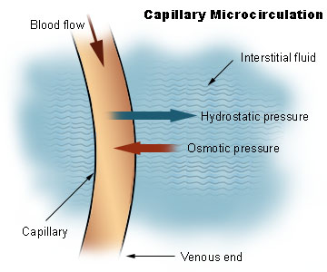
- Note that oncotic pressure is not illustrated in the image.
Pathophysiology
Disorders of capillary formation as a developmental problem or acquired disorder are a feature in many common and serious disorders. Within a wide range of cellular factors and cytokines, problems with normal genetic expression and bioactivity of the vascular growth and permeability factor vascular endothelial growth factor (VEGF) appear to play a major role in many of these disorders. Cellular factors include reduced numbers and function of bone-marrow derived endothelial progenitor cells.[6] and reduced ability of those cells to form blood vessels.[7]
- Formation of additional capillaries and larger blood vessels (angiogenesis) is a major mechanism by which a cancer may help to enhance its own growth. Disorders of retinal capillaries contribute to the pathogenesis of age-related macular degeneration.
- Reduced capillary density (capillary rarefaction) occurs in association with cardiovascular risk factors[8] and in patients with coronary heart disease[7]
Therapeutics
Major diseases where altering capillary formation could be helpful include conditions where there is excessive or abnormal capillary formation such as cancer and disorders harming eyesight; and medical conditions in which there is reduced capillary formation either for familial or genetic reasons, or as an acquired problem.
- In patients with the retinal disorder, neovascular age-related macular degeneration, local anti-VEGF treatment to limit the bio-activity of vascular endothelial growth factor has been shown to protect vision by limiting progression.[9] In a wide range of cancers, treatment approaches have been studied, or are in development, aimed at decreasing tumour growth by reducing angiogenesis.[10]
Blood sampling
Capillary blood sampling can be used to test for, for example, blood glucose (such as in blood glucose monitoring), hemoglobin, pH and lactate (the two latter can be quantified in fetal scalp blood testing to check the acid base status of a fetus during childbirth).
Capillary blood sampling is generally performed by creating a small cut by a blood lancet, followed by sampling by capillary action on the cut with a test strip or small pipe.
History
Ibn al-Nafis theorized a "premonition of the capillary circulation in his assertion that the pulmonary vein receives what comes out of the pulmonary artery, this being the reason for the existence of perceptible passages between the two."[11][verification needed]
Marcello Malpighi was the first to observe and correctly describe capillaries when he discovered them in a frog's lung in 1661.[12]
See also
- Alveolar-capillary barrier
- Blood brain barrier
References
- ↑ Maton, Anthea; Jean Hopkins, Charles William McLaughlin, Susan Johnson, Maryanna Quon Warner, David LaHart, Jill D. Wright (1993). Human Biology and Health, Englewood Cliffs, New Jersey: Prentice Hall.Template:Page needed
- ↑ John S. Penn (11 March 2008). Retinal and Choroidal Angiogenesis, 119–, Springer. URL accessed 26 June 2010.
- ↑ Endoderm -- Developmental Biology -- NCBI Bookshelf. URL accessed on 2010-04-07.
- ↑ Histology at Boston University 22401lba
- ↑ Pavelka, Margit; Jürgen Roth (2005). Functional Ultrastructure: An Atlas of Tissue Biology and Pathology, Springer.
- ↑ (2010). Epicardium derived cells (EPDCs) in development, cardiac disease and repair of ischemia. Journal of Cellular and Molecular Medicine 14 (5): 1056–60.
- ↑ 7.0 7.1 (2004). Circulating Humoral Factors and Endothelial Progenitor Cells in Patients with Differing Coronary Collateral Support. Circulation 109 (24): 2986–92.
- ↑ (1997). Impaired microvascular dilatation and capillary rarefaction in young adults with a predisposition to high blood pressure. Journal of Clinical Investigation 99 (8): 1873–9.
- ↑ (2010). Therapeutic targets in age-related macular disease. Journal of Clinical Investigation 120 (9): 3033–41.
- ↑ (2009). Tumor angiogenesis and molecular targets for therapy. Frontiers in Bioscience 14 (14): 3962–73.
- ↑ Dr. Paul Ghalioungui (1982), "The West denies Ibn Al Nafis's contribution to the discovery of the circulation", Symposium on Ibn al-Nafis, Second International Conference on Islamic Medicine: Islamic Medical Organization, Kuwait (cf. The West denies Ibn Al Nafis's contribution to the discovery of the circulation, Encyclopedia of Islamic World)
- ↑ John Cliff, Walter (1976). Blood Vessels, CUP Archives.
External links
- Histology at Boston University 00903loa
- {http://microcirc.org Microcirculatory Society, Inc}
- {http://www.bishoujyunkan.co.jp/bisyoujyunkan1.htm Microcirculation Research Institute Ltd.(Japan)}
- {http://histology.leeds.ac.uk/circulatory/capillaries.php}
Cardiovascular system |
|---|
|
Blood | Heart → Aorta → Arteries → Arterioles → Capillaries → Venules → Veins → Vena cava → Heart → Pulmonary arteries → Lungs → Pulmonary vein |
| This page uses Creative Commons Licensed content from Wikipedia (view authors). |