No edit summary |
|||
| Line 6: | Line 6: | ||
GraySubject = | |
GraySubject = | |
||
GrayPage = | |
GrayPage = | |
||
| − | Image = |
+ | Image = Basal Ganglia and Related Structures.svg | |
Caption = Basal ganglia labeled at top right. | |
Caption = Basal ganglia labeled at top right. | |
||
| − | Image2 = |
+ | Image2 = Basal ganglia 1.jpg | |
| − | Caption2 = |
+ | Caption2 = Basal ganglia on frontal section of brain | |
| − | + | IsPartOf = [[Cerebrum]] | |
|
| − | IsPartOf = | |
||
Components = | |
Components = | |
||
Artery = | |
Artery = | |
||
| Line 19: | Line 18: | ||
MeshName = Basal+Ganglia | |
MeshName = Basal+Ganglia | |
||
MeshNumber = A08.186.211.730.885.105 | |
MeshNumber = A08.186.211.730.885.105 | |
||
| ⚫ | |||
| + | NeuroLexID = birnlex_826 | |
||
DorlandsPre = n_11 | |
DorlandsPre = n_11 | |
||
DorlandsSuf = 12580456 | |
DorlandsSuf = 12580456 | |
||
}} |
}} |
||
| − | The '''basal ganglia''' are a group of [[Nucleus (neuroanatomy)|nuclei]] in the [[brain]] interconnected with the [[cerebral cortex]], [[thalamus]] and [[brainstem]]. Mammalian basal ganglia are associated with a variety of functions: [[motor control]], [[cognition]], [[emotions]] and [[learning]]. |
||
| + | The '''basal ganglia''' (or '''basal nuclei''') are a group of [[Nucleus (neuroanatomy)|nuclei]] of varied origin in the [[brain]]s of vertebrates that act as a cohesive functional unit. They are situated at the base of the [[telencephalon|forebrain]] and are strongly connected with the [[cerebral cortex]], [[thalamus]], and other brain areas. The basal ganglia are associated with a variety of functions, including voluntary motor control, procedural [[learning]] relating to routine behaviors or "habits" such as [[bruxism]], eye movements, cognitive,<ref name=a1>{{cite journal |last1=Stocco |first1=Andrea |last2=Lebiere |first2=Christian |last3=Anderson |first3=John R. |title=Conditional Routing of Information to the Cortex: A Model of the Basal Ganglia's Role in Cognitive Coordination |journal=Psychological Review |volume=117 |issue=2 |pages=541–74 |year=2010 |pmid=20438237 |doi=10.1037/a0019077 |pmc=3064519}}</ref> and emotional functions.<ref name=rapid>{{cite book |first1=James A. |last1=Weyhenmeyer |first2=Eve. A. |last2=Gallman |title=Rapid Review of Neuroscience |publisher=Mosby Elsevier |year=2007 |page=102 |isbn=0-323-02261-8}}</ref> Currently popular theories implicate the basal ganglia primarily in [[action selection]], that is, the decision of which of several possible behaviors to execute at a given time.<ref name=a1 /><ref>{{cite journal |last1=Chakravarthy |first1=V. S. |last2=Joseph |first2=Denny |last3=Bapi |first3=Raju S. |title=What do the basal ganglia do? A modeling perspective |journal=Biological Cybernetics |volume=103 |issue=3 |pages=237–53 |year=2010 |pmid=20644953 |doi=10.1007/s00422-010-0401-y}}</ref> Experimental studies show that the basal ganglia exert an inhibitory influence on a number of [[motor control|motor systems]], and that a release of this inhibition permits a motor system to become active. The "behavior switching" that takes place within the basal ganglia is influenced by signals from many parts of the brain, including the [[prefrontal cortex]], which plays a key role in [[executive functions]].<ref name=rapid/><ref>{{cite journal|title=Executive impairment in Parkinson's disease: response automaticity and task switching|author=Cameron IG, Watanabe M, Pari G, Munoz DP.|publisher=Neuropsychologia|date=2010 June|issue=7|volume=48|pmid=20303998|doi=10.1016/j.neuropsychologia.2010.03.015|pages=1948–57|journal=Neuropsychologia}}</ref> |
||
| ⚫ | |||
| − | The acceptance that the basal ganglia system constitutes one major cerebral system has been slow to appear. The first [[anatomy|anatomical]] identification of distinct subcortical structures was published by Thomas Willis in 1664. For many years, the term [[striatum|corpus striatum]] was used to describe a large group of subcortical elements, some of which were later discovered to be functionally unrelated. Additionally, the [[putamen]] and the [[caudate nucleus]] were not linked together. The putamen was thought to be associated to the [[pallidum]] in what used to be called the "nucleus lenticularis" (see [[lentiform nucleus]] on the fig.). Pioneering work by Cécile and Oskar Vogt (1941) greatly simplified the description of the basal ganglia by proposing the term [[striatum]] to describe the group of structures consisting of the caudate nucleus, the putamen and the mass linking them [[anatomical terms of location|ventrally]], the [[fundus]]. The striatum gets its name from the striated appearance created by radiating dense bundles of striato-pallido-nigral [[axons]], described by anatomist Kinnear Wilson as "pencil-like". The anatomical link of the striatum with its primary targets, the [[pallidum]] and the [[substantia nigra]] was later discovered. Together, these structures constitute, the striato-pallido-nigral bundle, which is the core of the basal ganglia. This nerve bundle forms the so-called "comb bundle of Edinger" when it crosses the [[internal capsule]]. Additional structures that later became associated with the basal ganglia are the "body of Luys" (1865) (nucleus of Luys on the figure) or [[subthalamic nucleus]], whose lesion was known to produce movement disorders. More recently, other areas such as the central complex (centre médian-parafascicular) and the pedunculopontine complex have been thought to be regulators of the basal ganglia. At the beginning of the 20th century, the basal ganglia system was associated with motor functions, as lesions of these areas would often result in disordered movement in humans ([[chorea]], athetosis, [[Parkinson's disease]]). |
||
| + | The main components of the basal ganglia are the [[striatum]] ([[caudate nucleus]] and [[putamen]]), the [[globus pallidus]], the [[substantia nigra]], the [[nucleus accumbens]], and the [[subthalamic nucleus]].<ref name=brs>{{cite book |first1=James D. |last1=Fix |title=Neuroanatomy (Board Review Series) |edition=4th |location=Baltimore |publisher=Wulters Kluwer & Lippincott Wiliams & Wilkins |chapter=Basal Ganglia and the Striatal Motor System |year=2008 |pages=274–281 |isbn=0-7817-7245-1}}</ref> The largest component, the striatum, receives input from many brain areas but sends output only to other components of the basal ganglia. The pallidum receives input from the striatum, and sends inhibitory output to a number of motor-related areas. The substantia nigra is the source of the striatal input of the [[neurotransmitter]] [[dopamine]], which plays an important role in basal ganglia function. The subthalamic nucleus receives input mainly from the striatum and cerebral cortex, and projects to the globus pallidus. Each of these areas has a complex internal anatomical and neurochemical organization. |
||
| − | ==Anatomical subdivisions== |
||
| ⚫ | |||
| ⚫ | |||
| + | The basal ganglia play a central role in a number of neurological conditions, including several [[movement disorder]]s. The most notable are [[Parkinson's disease]], which involves degeneration of the dopamine-producing cells in the substantia nigra pars compacta, and [[Huntington's disease]], which primarily involves damage to the striatum.<ref name=a1 /><ref name=brs /> Basal ganglia dysfunction is also implicated in some other disorders of behavior control such as [[Tourette syndrome]], [[hemiballismus]], and [[obsessive–compulsive disorder]]. |
||
| − | The five individual nuclei that make up the [[primate]] basal ganglia, along with their major subdivisions, are: |
||
| − | * ''[[rostral]]'' |
||
| − | ** the [[striatum]] |
||
| − | *** [[putamen]] |
||
| − | *** [[caudate nucleus]] |
||
| − | *** [[nucleus accumbens]] |
||
| − | ** external segment of the [[globus pallidus]] (GPe) |
||
| − | ** internal segment of the globus pallidus (GPi) |
||
| − | * ''[[caudal]]'' |
||
| − | ** [[subthalamic nucleus]] (STN) |
||
| − | ** [[substantia nigra]] (SN) |
||
| − | *** pars compacta (SNc) |
||
| − | *** pars reticulata (SNr) |
||
| − | *** pars lateralis (SNl) |
||
| + | The basal ganglia have a [[limbic system|limbic]] sector whose components are assigned distinct names: the [[nucleus accumbens]], [[ventral pallidum]], and [[ventral tegmental area]] (VTA). There is considerable evidence that this limbic part plays a central role in [[reinforcement|reward learning]], particularly a pathway from the VTA to the nucleus accumbens that uses the neurotransmitter dopamine. A number of highly addictive drugs, including [[cocaine]], [[amphetamine]], and [[nicotine]], are thought to work by increasing the efficacy of this dopamine signal. There is also evidence implicating overactivity of the VTA dopaminergic projection in [[schizophrenia]].<ref>{{cite journal |last1=Inta |first1=D. |last2=Meyer-Lindenberg |first2=A. |last3=Gass |first3=P. |title=Alterations in Postnatal Neurogenesis and Dopamine Dysregulation in Schizophrenia: A Hypothesis |journal=Schizophrenia Bulletin |year=2010 |doi=10.1093/schbul/sbq134 |volume=37 |issue=4 |pages=674–80 |pmid=21097511 |pmc=3122276}}</ref> |
||
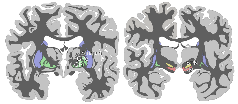
| − | There are 2 sets of basal ganglia in the mammalian brain, mirrored in the left and right hemispheres. Two coronal sections are used to show the basal ganglia; the STN and substantia nigra lie deeper back in the brain (more [[Anatomical terms of location|caudal]]). Images show two schematic [[Anatomical terms of location|coronal]] cross-sections of the [[human]] brain with nuclei of the basal ganglia labeled on the right side. |
||
| + | ==Anatomy== |
||
| − | As it refers to a group of nuclei, the term "basal ganglia" is plural (the singular of ganglia is ''[[ganglion]]''). However this is a [[misnomer]], as "ganglion" refers to a [[soma (biology)|somatic]] cluster within the [[peripheral nervous system]], whereas the basal ganglia are within the [[central nervous system]] (CNS). A somatic cluster within the CNS is referred to as a nucleus, so some [[neuroanatomy|neuroanatomists]] refer to the basal ganglia as the "basal nuclei". |
||
| ⚫ | |||
| + | In terms of development, the human nervous system is often classified based on the original 3 primitive vesicles from which it develops: These primary vesicles form in the normal development of the [[neural tube]] of the human fetus and initially include [[prosencephalon]], [[mesencephalon]], and [[rhombencephalon]], in rostral to caudal (from head to toe) orientation. Later in development of the nervous system each section itself turns into smaller components. During development, the cells that migrate tangentially to form the basal ganglia are directed by the lateral and medial [[ganglionic eminence]]s.<ref>Marín & Rubenstein. (2001). A Long, Remarkable Journey: Tangential Migration in the Telencephalon. ''Nature Reviews Neuroscience, 2.''</ref> The following table demonstrates this developmental classification and traces it to the anatomic structures found in the basal ganglia.<ref name=a1 /><ref name=brs /><ref>{{cite web|url=http://biology.about.com/library/organs/brain/blprosenceph.htm|title=Divisions of the Brain|author=Regina Bailey |publisher=about.com|accessdate=2010-11-30| archiveurl= http://web.archive.org/web/20101202223928/http://biology.about.com/library/organs/brain/blprosenceph.htm| archivedate= 2 December 2010 <!--DASHBot-->| deadurl= no}}</ref> The structures relevant to the basal ganglia are shown in '''bold'''. |
||
| ⚫ | |||
| − | "Basal ganglia"-like areas are found in the central nervous systems of many species. The striatal and pallidal components can be clearly identified in all [[amniote]]s (mammals, birds, and reptiles) and [[amphibian]]s. The anatomical connections of these nuclei and their [[pharmacology]] also appear relatively conserved. Non-[[tetrapod]] vertebrates such as fish also display basal ganglia-like structures, although the data are less clear in this case. |
||
| + | {| class="wikitable" |
||
| ⚫ | |||
| + | ! scope="col" | '''Primary division of the [[neural tube]]''' |
||
| + | ! scope="col" | '''Secondary subdivision ''' |
||
| + | ! scope="col" | '''Final segments in a human adult''' |
||
| + | |- |
||
| + | ! scope="row" | [[Prosencephalon]] |
||
| + | | <ol><li>[[Telencephalon]]<li>[[Diencephalon]]</ol> |
||
| + | | <ol><li>On each side of the brain: the cerebral cortices, '''caudate''', '''putamen''', '''globus pallidus (pallidum)'''<li>Thalamus, hypothalamus, subthalamus, epithalamus, '''subthalamic nuclei'''</ol> |
||
| + | |- |
||
| + | ! scope="row" | [[Mesencephalon]] |
||
| + | | <ol><li>Mesencephalon</ol> |
||
| + | | <ol><li>Mesencephalon (midbrain): '''substantia nigra pars compacta (SNc)''', '''substantia nigra pars reticulata (SNr)'''</ol> |
||
| + | |- |
||
| + | ! scope="row" | [[Rhombencephalon]] |
||
| + | | <ol><li>[[Metencephalon]]<li>[[Myelencephalon]]</ol> |
||
| + | | <ol><li>Pons and cerebellum<li>Medulla</ol> |
||
| + | |- |
||
| + | |} |
||
| ⚫ | [[Image:Basal-ganglia-coronal-sections-large.png|thumb|400px|[[Anatomical terms of location|Coronal]] slices of human brain showing the basal ganglia. White matter is shown in dark gray, gray matter is show in light gray.<BR>Anterior: [[striatum]], [[globus pallidus]] (GPe and GPi)<BR>Posterior: [[subthalamic nucleus]] (STN), [[substantia nigra]] (SN)]] |
||
| − | ==Neuronal Classes in the Striatum== |
||
| − | The '''striatum''' is a large nucleus located deep in the [[telencephalon]] in proximity to the lateral [[brain ventricle]]s. In primates, it is composed of 4 neuronal classes: spiny (96%), leptodendritic (2%), spidery (1%) neurons and microneurons(1%) (Yelnik et al. 1991). In ''spiny neurons'' [[dendrite|dendritic]] arborisations are spherical. Most of the [[dendritic spine]]s in this cell type receive [[synapse|synaptic]] input from cortical [[afferent]]s. Their axons have abundant collateral projections participating in local circuitry. The neurons are [[GABA]]ergic. The ''leptodendritic neurons'' (or Deiter's neurons) have all the morphological properties of the pallidal neurons. The ''spidery neurons'' are specific to primates. They have a big [[soma (biology)|soma]] and short dendritic and axonal branches. They are the [[acetylcholine|cholinergic]] [[interneuron]]s and are characterized by the fact that they are tonically active. The ''microneurons'' are local circuit neurons similar to those found in the [[thalamus]] and are [[GABAergic]]. Some may be also be [[dopaminergic]] (Rochette et al.). |
||
| + | The basal ganglia form a fundamental component of the [[cerebrum]]. In contrast to the [[cerebral cortex|cortical]] layer that lines the surface of the forebrain, the basal ganglia are a collection of distinct masses of gray matter lying deep in the brain not far from the junction of the [[thalamus]]. Like most parts of the brain, the basal ganglia consist of left and right sides that are virtual mirror images of each other. |
||
| ⚫ | |||
| + | In terms of anatomy, the basal ganglia are divided by anatomists into four distinct structures, depending on how [[Anatomical terms of location#Superior and inferior|superior]] or [[Anatomical terms of location#Directional terms|rostral]] they are (in other words depending on how close to the top of the head they are): Two of them, the [[striatum]] and the [[pallidum]], are relatively large; the other two, the [[substantia nigra]] and the [[subthalamic nucleus]], are smaller. In the illustration to the right, two [[Anatomical terms of location|coronal]] sections of the human brain show the location of the basal ganglia components. Of note, and not seen in this section, the subthalamic nucleus and substantia nigra lie farther back ([[Posterior (anatomy)|posteriorly]]) in the brain than the striatum and pallidum. |
||
| − | The different types of neuron of the basal ganglia biosynthesize different [[neurotransmitter]]s. |
||
| − | === |
+ | ===Striatum=== |
| + | {{Main|Striatum}} |
||
| − | Medium neurons, the principal cells of the neostriatum, are inhibitory; they produce transmitter [[GABA]] (connections using GABA are shown in blue in the diagram below). |
||
| + | [[File:Basal ganglia 2.jpg|thumb|right|300px|Basal ganglia]] |
||
| + | The striatum is the largest component of the basal ganglia. The term "striatum" comes from the observation that this structure has a striped appearance when sliced in certain directions, arising from numerous large and small bundles of nerve fibers (white matter) that traverse it. Early anatomists, examining the human brain, perceived the striatum as two distinct masses of gray matter separated by a large tract of white matter called the [[internal capsule]]. They named these two masses the "caudate nucleus" and "putamen". More recent anatomists have concluded, on the basis of microscopic and neurochemical studies, that it is more appropriate to consider these masses as two separated parts of a single entity, the "striatum", in the same way that a city may be separated into two parts by a river. Numerous functional differences between the caudate and putamen have been identified, but these are taken to be consequences of the fact that each sector of the striatum is preferentially connected to specific parts of the cerebral cortex. |
||
| + | The internal organization of the striatum is extraordinarily complex. The great majority of neurons (about 96%) are of a type called "medium spiny neurons".<ref name=a1 /> These are GABAergic cells (meaning that they inhibit their targets) with small cell bodies and dendrites densely covered with [[dendritic spine]]s, which receive synaptic input primarily from the cortex and thalamus. Medium spiny neurons can be divided into subtypes in a number of ways, on the basis of neurochemistry and connectivity. The next most numerous type (around 2%) are a class of large [[acetylcholine|cholinergic]] interneurons with smooth dendrites. There are also several other types of interneurons making up smaller fractions of the neural population. |
||
| ⚫ | |||
| − | [[Dopamine]] is biosynthesized in the dopaminergic neurons, primarily in the [[substantia nigra]] pars compacta. Disruption in the biosynthesis or transmission of [[dopamine]] can lead to serious motor and cognitive deficits, such as occurs in [[Parkinson's disease]]. The substantia nigra pars compacta (SNc) primarily targets the striatum with this neurotransmitter (shown as the magenta connection in the classic connectivity diagram below). |
||
| + | Numerous studies have shown that the connections between cortex and striatum are, in general, topographic; that is, each part of the cortex sends stronger input to some parts of the striatum than to others. The nature of the topography has been difficult to understand, however—perhaps in part because the striatum is organized in three dimensions, whereas the cortex, as a layered structure, is organized in two. This dimensional discrepancy entails a great deal of distortion and discontinuity in mapping one structure to the other. It is interesting to note that the same topography applies to the striatal connections to the thalamus.<ref>{{cite journal |last1=Kamishina |first1=H |last2=Yurcisin |first2=G |last3=Corwin |first3=J |last4=Reep |first4=R |title=Striatal projections from the rat lateral posterior thalamic nucleus |journal=Brain Research |volume=1204 |pages=24–39 |year=2008 |pmid=18342841 |doi=10.1016/j.brainres.2008.01.094}}</ref> |
||
| − | === Globus pallidus === |
||
| − | === |
+ | ===Pallidum=== |
| + | {{Main|Pallidum}} |
||
| + | The ''pallidum'' consists of a large structure called the [[globus pallidus]] ("pale globe") together with a smaller ventral extension called the [[ventral pallidum]]. The globus pallidus appears as a single neural mass, but can be divided into two functionally distinct parts, called the internal (or medial) and external (lateral) segments, abbreviated GPi and GPe.<ref name=a1 /> Both segments contain primarily GABAergic neurons, which therefore have inhibitory effects on their targets. The two segments participate in distinct neural circuits. The external segment, or GPe, receives input mainly from the striatum, and projects to the subthalamic nucleus. The internal segment, or GPi, receives signals from the striatum via two pathways, called "direct" and "indirect". Pallidal neurons operate using a disinhibition principle. These neurons fire at steady high rates in the absence of input, and signals from the striatum cause them to pause or reduce their rate of firing. Because pallidal neurons themselves have inhibitory effects on their targets, the net effect of striatal input to the pallidum is a reduction of the tonic inhibition exerted by pallidal cells on their targets (disinhibition) with an increased rate of firing in the targets. |
||
| − | ==Connections== |
||
| − | Basal ganglia connectivity is illustrated in the figure. |
||
| ⚫ | |||
| − | The striatum is the main (but not the only) input zone for other brain areas to connect to the basal ganglia. Via the striatum, the basal ganglia receives input from the [[Cerebral cortex|cortex]], mainly from the [[motor cortex|motor]] and [[prefrontal cortex|prefrontal]] cortices. |
||
| + | {{Main|Substantia nigra}} |
||
| + | The substantia nigra is a mesencephalic gray matter portion of the basal ganglia that is divided into SNr (reticulata) and SNc (compacta). SNr often works in unison with GPi, and the SNr-GPi complex inhibits the thalamus. Substantia nigra pars compacta (SNc) however, produces the neurotransmitter [[dopamine]], which is very significant in maintaining balance in the striatal pathway. The circuit portion below explains the role and circuit connections of each of the components of the basal ganglia. |
||
| + | ===Subthalamic nucleus=== |
||
| − | The [[neural network|circuitry]] of the basal ganglia is often divided into two major pathways, the '''direct pathway''' and the '''indirect pathway''': |
||
| + | {{Main|Subthalamic nucleus}} |
||
| + | The subthalamic nucleus (STN) is a diencephalic gray matter portion of the basal ganglia, and the only portion of the ganglia that actually produces an excitatory neurotransmitter, [[glutamate]]. The role of the subthalamic nucleus is to stimulate the SNr-GPi complex and it is part of the [[Indirect pathway of movement|indirect pathway]]. |
||
| + | ===Circuit connections=== |
||
| − | :* '''Direct pathway''': striatum <sup>-</sup>→ GPi/SNr <sup>-</sup>→ thalamus <sup>+</sup>→ cortex |
||
| + | [[Image:basal-ganglia-classic.png|thumb|200px|right|Connectivity diagram showing excitatory [[glutamatergic]] pathways as <span style="color:red;">red</span>, inhibitory [[GABA]]ergic pathways as <span style="color:blue;">blue</span>, and modulatory [[dopaminergic]] pathways as <span style="color:magenta;">magenta</span>. (Abbreviations: GPe: globus pallidus external; GPi: globus pallidus internal; STN: subthalamic nucleus; SNc: substantia nigra compacta; SNr: substantia nigra reticulata)]] |
||
| − | :* '''Indirect pathway''': striatum <sup>-</sup>→ GPe <sup>-</sup>→ STN <sup>+</sup>→ GPi/SNr <sup>-</sup>→ thalamus <sup>+</sup>→ cortex |
||
| + | In order to understand the complex circuitry of the basal ganglia, one has to first understand the important participants in this circuit. Parts of the basal ganglia are in direct communication with the thalamus and the cortex. The [[Cerebral cortex|cortex]], [[thalamus]], and the basal ganglia are, therefore, the three main participants in the circuit created by the basal ganglia. |
||
| + | At the top of the hierarchy lies the cerebral cortex. The cortex has many different areas with different functions. One such cortical area is called the [[primary motor cortex]] (along the [[pre-central gyrus]]). Specialized neurons from the primary motor cortex extend their [[axons]] all the way to the striatum portion of the basal ganglia. These cortical neurons release the neurotransmitter [[glutamate]], which is excitatory in nature. Once excited by glutamate, the cells in the striatum project in two different directions giving rise to two major pathways: the "''direct''" and the "''indirect''" pathways: |
||
| − | Cortical activity that excites cells in the striatum that participate in the direct pathway leads to inhibition of areas of the GPi and SNr, which in turn removes their tonic inhibition from the thalamus. This removal of inhibition by inhibition is called '''disinhibition'''. In contrast, cortical activity that excites the striatal cells in the indirect pathway is thought to inhibit the thalamus. This is due to an odd number of the pathways in the '''indirect pathway''' being inhibition pathways (blue arrows), as opposed to an even number of inhibition pathways in the '''direct pathway'''. |
||
| + | In the [[direct pathway]], cortical cells project excitatory inputs to the striatum, which in turn projects inhibitory neurons onto the cells of the SNr-GPi complex. The SNr-GPi complex projects directly onto the thalamus through the inhibitory [[ansa lenticularis]] pathway. The striatal inhibition of the SNr-GPi complex coupled with SNr-GPi inhibition of the thalamus therefore results in a net '''reduction''' of inhibition of the thalamus via the striatum. The thalamus projects excitatory glutamatergic neurons to the cortex itself. The direct pathway, therefore, results in the excitation of the motor cortex by the thalamus. Once stimulated, the cortex projects its own excitatory outputs to the brain stem and ultimately muscle fibers via the [[lateral corticospinal tract]]. The following diagram depicts the direct pathway: |
||
| − | Dopamine from the [[substantia nigra]] pars compacta stimulates all of the dopamine receptors : D1, D2, D3, D4, and D5. However, via the G proteins (Gs, Gi, Go) the dopamine receptors D2, D3 and D4 are inhibitory, and the dopamine receptors D1 and D5 are stimulatory. Striatal cells of the direct pathway have D1 receptors, while those of the indirect pathway have D2 receptors. Through this mechanism, the presence of dopamine selects for the direct pathway. And the absence of dopamine selects for the indirect pathway. |
||
| + | :'''Cortex''' (stimulates) → '''Striatum''' (inhibits) → '''"SNr-GPi" complex''' (less inhibition of thalamus) → '''Thalamus''' (stimulates) → '''Cortex''' (stimulates) → '''Muscles, etc.''' → (<u>hyperkinetic state</u>) |
||
| ⚫ | |||
| + | |||
| ⚫ | |||
| + | The [[indirect pathway]] also starts from neurons in the striatum. Once stimulated by the cortex, striatal neurons in the indirect pathway project inhibitory axons onto the cells of the globus pallidus externa (GPe), which tonically inhibits the subthalamic nucleus (STN). This inhibition (by the striatum) of the inhibitory projections of the GPe, results in the net reduction of inhibition of the STN. The STN, in turn, projects excitatory inputs to the SNr-GPi complex (which inhibits the thalamus). The end-result is inhibition of the thalamus and, therefore, decreased stimulation of the motor cortex by the thalamus and reduced muscle activity. The direct and indirect pathways are therefore antagonist in their functions. Following is a diagram of the indirect pathway: |
||
| + | |||
| + | :'''Cortex''' (stimulates) → '''Striatum''' (inhibits) → '''GPe''' (less inhibition of STN) → '''STN''' (stimulates) → '''"SNr-GPi" complex''' (inhibits) → '''Thalamus''' (is stimulating less) → '''Cortex''' (is stimulating less) → '''Muscles, etc.''' → (<u>hypokinetic state</u>) |
||
| + | |||
| + | [[File:Basal ganglia circuits.svg|thumb|right|Main circuits of the basal ganglia. This diagram shows 2 coronal slices that have been superimposed to include the involved basal ganglia structures. The + and - signs at the point of the arrows indicate whether the pathway is excitatory or inhibitory, respectively, in effect. <span style="color:green;">Green arrows</span> refer to excitatory [[Glutamic acid|glutamatergic]] pathways, <span style="color:red;">red arrows</span> refer to inhibitory [[gamma-Aminobutyric acid|GABAergic]] pathways and <span style="color:turquoise;">turquoise arrows</span> refer to [[dopamine]]rgic pathways that are excitatory on the direct pathway and inhibitory on the indirect pathway.]] |
||
| + | |||
| + | The antagonistic functions of the direct and indirect pathways are modulated by the substantia nigra pars compacta (SNc), which produces dopamine. In the presence of dopamine, D1-receptors in the basal ganglia stimulate the GABAergic neurons, favoring the direct pathway, and thus increasing movement. The GABAergic neurons of the indirect pathway are stimulated by excitatory neurotransmitters acetylcholine and glutamate. This sets off the indirect pathway that ultimately results in inhibition of upper motor neurons, and less movement. In the presence of dopamine, D2-receptors in the basal ganglia inhibit these GABAergic neurons, which reduces the indirect pathways inhibitory effect. Dopamine therefore increases the excitatory effect of the direct pathway (causing movement) and reduces the inhibitory effect of the indirect pathway (preventing full inhibition of movement). Through these mechanisms the body is able to maintain balance between excitation and inhibition of motion. Lack of balance in this delicate system leads to pathologies such as Parkinson's disease. Parkinson's disease involves the loss of dopamine which means the direct pathway is less able to function (so no movement is initiated) and the indirect pathway is in overdrive (causing too much inhibition of movement). |
||
| + | |||
| + | ===A note concerning terminology=== |
||
| + | The nomenclature of the basal ganglia system and its components has always been problematic. Early anatomists, seeing the macroscopic anatomical structure but knowing nothing of the cellular architecture or neurochemistry, grouped together components that are now believed to have distinct functions (such as the internal and external segments of the globus pallidus), and gave distinct names to components that are now thought to be functionally parts of a single structure (such as the caudate nucleus and putamen). |
||
| + | |||
| + | The term "basal" comes from the fact that most of its elements are located in the basal part of the forebrain. The term [[ganglion|ganglia]] is a misnomer: In modern usage, neural clusters are called "ganglia" only in the [[peripheral nervous system]]; in the [[central nervous system]] they are called "nuclei". For this reason, the basal ganglia are also occasionally known as the "basal nuclei".<ref>{{cite book|last=Soltanzadeh|first=Akbar|title= Neurologic Disorders|isbn=964-6088-03-1|publisher=Jafari|year=2004|location=Tehran}}{{Page needed|date=December 2010}}</ref> [[Terminologia anatomica]] (1998), the international authority for anatomical naming, retained "nuclei basales", but this is not commonly used. |
||
| + | |||
| + | The International Basal Ganglia Society (IBAGS) informally considers the basal ganglia to be made up of the [[striatum]], the pallidum (with two nuclei), the [[substantia nigra]] (with its two distinct parts), and the [[subthalamic nucleus]]. Percheron ''et al.'' in 1991 and Parent and Parent in 2005 included the [[Primate basal ganglia system#Central region of the thalamus (C)|central region]] (centre median-parafascicular) of the thalamus as part of the basal ganglia,<ref name="Percheron1991">{{cite journal |last1=Percheron |first1=G |last2=Filion |first2=M |title=Parallel processing in the basal ganglia: up to a point |journal=Trends in Neurosciences |volume=14 |issue=2 |pages=55–9 |year=1991 |pmid=1708537 |doi=10.1016/0166-2236(91)90020-U}}</ref><ref>{{cite journal |last1=Parent |first1=Martin |last2=Parent |first2=Andre |title=Single-axon tracing and three-dimensional reconstruction of centre median-parafascicular thalamic neurons in primates |journal=The Journal of Comparative Neurology |volume=481 |issue=1 |pages=127–44 |year=2005 |pmid=15558721 |doi=10.1002/cne.20348}}</ref> while Mena-Segovia ''et al.'' in 2004 included the [[pedunculopontine complex]] as well.<ref>{{cite journal |last1=Menasegovia |first1=J |last2=Bolam |first2=J |last3=Magill |first3=P |title=Pedunculopontine nucleus and basal ganglia: distant relatives or part of the same family? |journal=Trends in Neurosciences |volume=27 |issue=10 |pages=585–8 |year=2004 |pmid=15374668 |doi=10.1016/j.tins.2004.07.009}}</ref> |
||
| + | |||
| + | Also, the names given to the various nuclei of the basal ganglia are different in different species. In particular, the internal segment of the globus pallidus in primates is called the entopeduncular nucleus in [[rodent]]s. The "striatum" and "external segment of the globus pallidus" in primates are called the "paleostriatum augmentatum" and "paleostriatum primitivum," respectively, in birds. |
||
| + | |||
| + | ==Function== |
||
| + | Information about the functions of the basal ganglia comes from anatomical studies, from physiological studies carried out mainly in rats and monkeys, and from the study of diseases that damage them. |
||
| + | |||
| + | The greatest source of insight into the functions of the basal ganglia has come from the study of two neurological disorders, [[Parkinson's disease]] and [[Huntington's disease]]. For both of these disorders, the nature of the neural damage is well understood and can be correlated with the resulting symptoms. Parkinson's disease involves major loss of dopaminergic cells in the substantia nigra; Huntington's disease involves massive loss of medium spiny neurons in the striatum. The symptoms of the two diseases are virtually opposite: Parkinson's disease is characterized by gradual loss of the ability to initiate movement, whereas Huntington's disease is characterized by an inability to prevent parts of the body from moving unintentionally. It is noteworthy that, although both diseases have cognitive symptoms, especially in their advanced stages, the most salient symptoms relate to the ability to initiate and control movement. Thus, both are classified primarily as [[movement disorder]]s. A different movement disorder, called [[hemiballismus]], may result from damage restricted to the subthalamic nucleus. Hemiballismus is characterized by violent and uncontrollable flinging movements of the arms and legs. |
||
| + | |||
| + | ===Eye movements=== <!-- Please preserve this link. It is used elsewhere --> |
||
| + | One of the most intensively studied functions of the basal ganglia (BG) is their role in controlling eye movements.<ref name=Hikosaka2000>{{cite journal |last1=Hikosaka |first1=O |last2=Takikawa |first2=Y |last3=Kawagoe |first3=R |title=Role of the basal ganglia in the control of purposive saccadic eye movements |journal=Physiological reviews |volume=80 |issue=3 |pages=953–78 |year=2000 |pmid=10893428}}</ref> Eye movement is influenced by an extensive network of brain regions that converge on a [[diencephalon|midbrain]] area called the [[superior colliculus]] (SC). The SC is a layered structure whose layers form two-dimensional [[retinotopic]] maps of visual space. A "bump" of neural activity in the deep layers of the SC drives an eye movement directed toward the corresponding point in space. |
||
| + | |||
| + | The SC receives a strong inhibitory projection from the BG, originating in the [[substantia nigra]] ''pars reticulata'' (SNr).<ref name=Hikosaka2000 /> Neurons in the SNr usually fire continuously at high rates, but at the onset of an eye movement they "pause", thereby releasing the SC from inhibition. Eye movements of all types are associated with "pausing" in the SNr; however, individual SNr neurons may be more strongly associated with some types of movements than others. Neurons in some parts of the caudate nucleus also show activity related to eye movements. Since the great majority of caudate cells fire at very low rates, this activity almost always shows up as an increase in firing rate. Thus, eye movements begin with activation in the caudate nucleus, which inhibits the SNr via the direct GABAergic projections, which in turn disinhibits the SC. |
||
| + | |||
| + | ===Role in motivation=== |
||
| + | Although the role of the basal ganglia in motor control is clear, there are also many indications that it is involved in the control of behavior in a more fundamental way, at the level of [[motivation]]. In [[Parkinson's disease]], the ability to execute the components of movement is not greatly affected, but motivational factors such as hunger fail to cause movements to be initiated or switched at the proper times. The immobility of Parkinsonian patients has sometimes been described as a "paralysis of the will".<ref name=Niv2007>{{cite journal |last1=Niv |first1=Y. |last2=Rivlin-Etzion |first2=M. |title=Parkinson's Disease: Fighting the Will? |journal=Journal of Neuroscience |volume=27 |issue=44 |pages=11777–9 |year=2007 |pmid=17978012 |doi=10.1523/JNEUROSCI.4010-07.2007}}</ref> These patients have occasionally been observed to show a phenomenon called ''kinesia paradoxica'', in which a person who is otherwise immobile responds to an emergency in a coordinated and energetic way, then lapses back into immobility once the emergency has passed. |
||
| + | |||
| + | The role in motivation of the "limbic" part of the basal ganglia—the [[nucleus accumbens]] (NA), [[ventral pallidum]], and [[ventral tegmental area]] (VTA)—is particularly well established. Thousands of experimental studies combine to demonstrate that the dopaminergic projection from the VTA to the NA plays a central role in the brain's reward system. Animals with stimulating electrodes implanted along this pathway will bar-press very energetically if each press is followed by a brief pulse of electrical current. Numerous things that people find rewarding, including addictive drugs, good-tasting food, and sex, have been shown to elicit activation of the VTA dopamine system. Damage to the NA or VTA can produce a state of profound torpor. |
||
| + | |||
| + | Although it is not universally accepted, some theorists have proposed a distinction between "appetitive" behaviors, which are initiated by the basal ganglia, and "consummatory" behaviors, which are not. For example, an animal with severe basal ganglia damage will not move toward food even if it is placed a few inches away, but, if the food is placed directly in the mouth, the animal will chew it and swallow it. |
||
| + | |||
| ⚫ | |||
| + | The basal ganglia form one of the basic components of the [[telencephalon|forebrain]], and can be recognized in all species of vertebrates.<ref name="Parent">{{cite book|title=Comparative Neurobiology of the Basal Ganglia |author=Parent A |publisher=Wiley |year=1986 |isbn=978-0-471-80348-5}}{{Page needed|date=December 2010}}</ref> Even in the lamprey (generally considered one of the most primitive of vertebrates), striatal, pallidal, and nigral elements can be identified on the basis of anatomy and histochemistry.<ref name=Grillner1998>{{cite journal |last1=Grillner |first1=S |last2=Ekeberg |first2=O |last3=Elmanira |first3=A |last4=Lansner |first4=A |last5=Parker |first5=D |last6=Tegner |first6=J |last7=Wallen |first7=P |title=Intrinsic function of a neuronal network — a vertebrate central pattern generator1 |journal=Brain Research Reviews |volume=26 |issue=2–3 |pages=184–97 |year=1998 |pmid=9651523 |doi=10.1016/S0165-0173(98)00002-2}}</ref> |
||
| + | |||
| ⚫ | A clear emergent issue in comparative anatomy of the basal ganglia is the development of this system through phylogeny as a convergent cortically re-entrant loop in conjunction with the development and expansion of the cortical mantle. There is controversy, however, regarding the extent to which convergent selective processing occurs versus segregated parallel processing within re-entrant closed loops of the basal ganglia. Regardless, the transformation of the basal ganglia into a cortically re-entrant system in mammalian evolution occurs through a re-direction of pallidal (or "paleostriatum primitivum") output from midbrain targets such as the superior colliculus, as occurs in [[sauropsid]] brain, to specific regions of the ventral thalamus and from there back to specified regions of the cerebral cortex that form a subset of those cortical regions projecting into the striatum. The abrupt rostral re-direction of the pathway from the internal segment of the globus pallidus into the ventral thalamus—via the path of the [[ansa lenticularis]]—could be viewed as a footprint of this evolutionary transformation of basal ganglia outflow and targeted influence. |
||
| + | |||
| ⚫ | |||
| + | In most regions of the brain, the predominant classes of neurons use [[glutamate]] as neurotransmitter and have excitatory effects on their targets. In the basal ganglia, however, the great majority of neurons use [[GABA]] as neurotransmitter and have inhibitory effects on their targets. The inputs from the cortex and thalamus to the striatum and STN are glutamatergic, but the outputs from the striatum, pallidum, and substantia nigra ''pars reticulata'' all use GABA. Thus, following the initial excitation of the striatum, the internal dynamics of the basal ganglia are dominated by inhibition and disinhibition. |
||
| + | |||
| + | Other neurotransmitters have important [[neuromodulator|modulatory]] effects. The most intensively studied is [[dopamine]], which is used by the projection from the substantia nigra ''pars compacta'' to the striatum, and also in the analogous projection from the ventral tegmental area to the nucleus accumbens. [[Acetylcholine]] also plays an important role, being used both by several external inputs to the striatum, and by a group of striatal interneurons. Although cholinergic cells make up only a small fraction of the total population, the striatum has one of the highest acetylcholine concentrations of any brain structure. |
||
| + | |||
| ⚫ | |||
| + | {{Main|Basal ganglia disease}} |
||
| + | {{columns-list|2| |
||
| + | * [[Athetosis]] |
||
* [[Athymhormic syndrome]] ([[PAP syndrome]]) |
* [[Athymhormic syndrome]] ([[PAP syndrome]]) |
||
| ⚫ | |||
| ⚫ | |||
| + | * [[Blepharospasm]] |
||
| + | * [[Bruxism]] |
||
* [[Cerebral palsy]]: basal ganglia damage during second and third trimester of pregnancy |
* [[Cerebral palsy]]: basal ganglia damage during second and third trimester of pregnancy |
||
| + | * [[Chorea]] |
||
| + | * [[Dystonia]] |
||
* [[Fahr's disease]] |
* [[Fahr's disease]] |
||
| − | * [[Foreign accent syndrome (FAS) |
+ | * [[Foreign accent syndrome]] (FAS) |
* [[Huntington's disease]] |
* [[Huntington's disease]] |
||
| + | * [[Kernicterus]] |
||
| − | * [[Obsessive-compulsive disorder]] |
||
| + | * [[Lesch-Nyhan syndrome]] |
||
| + | * [[Major Depressive Disorder]] <ref>{{cite journal |author=Kempton MJ, Salvador Z, Munafò MR, Geddes JR, Simmons A, Frangou S, Williams SC. |title=Structural Neuroimaging Studies in Major Depressive Disorder: Meta-analysis and Comparison With Bipolar Disorder |journal=Arch Gen Psychiatry |volume=68 |issue=7 |pages=675–90 | year=2011 |url=http://archpsyc.ama-assn.org/cgi/content/full/68/7/675 | doi=10.1001/archgenpsychiatry.2011.60 |pmid=21727252}} see also MRI database at [http://sites.google.com/site/depressiondatabase/ www.depressiondatabase.org]</ref> |
||
| + | * [[Obsessive-compulsive disorder]]<ref name="Radua and Mataix-Cols, 2009">{{cite journal|last1=Radua|first1=Joaquim|last2=Mataix-Cols|first2=David|year=2009|month=November|title=Voxel-wise meta-analysis of grey matter changes in obsessive–compulsive disorder|journal=British Journal of Psychiatry|volume=195|pages=393–402|doi=10.1192/bjp.bp.108.055046|pmid=19880927|issue=5}}</ref><ref name=radua2010>{{Cite journal |
||
| + | | last1 = Radua | first1 = Joaquim |
||
| + | | last2 = van den Heuvel | first2 = Odile A. |
||
| + | | last3 = Surguladze | first3 = Simon |
||
| + | | last4 = Mataix-Cols | first4 = David |
||
| + | | title = Meta-analytical comparison of voxel-based morphometry studies in obsessive-compulsive disorder vs other anxiety disorders |
||
| + | | journal = Archives of General Psychiatry |
||
| + | | volume = 67 |
||
| + | | issue = 7 |
||
| + | | pages = 701–711 |
||
| + | | date = 5 July 2010 |
||
| + | | doi = 10.1001/archgenpsychiatry.2010.70 |
||
| + | | pmid = 20603451 |
||
| + | }} |
||
| + | </ref> |
||
| + | * Other [[anxiety disorder]]s <ref name=radua2010 /> |
||
| + | * [[PANDAS]] |
||
* [[Parkinson's disease]] |
* [[Parkinson's disease]] |
||
| ⚫ | |||
| − | * [[Progressive supranuclear palsy]] |
||
| + | * [[Stuttering]]<ref>{{cite journal |first1=Per A. |last1=Alm |title=Stuttering and the basal ganglia circuits: a critical review of possible relations |journal=Journal of communication disorders |volume=37 |issue=4 |pages=325–69 |year=2004 |pmid=15159193 |doi=10.1016/j.jcomdis.2004.03.001}}</ref> |
||
| ⚫ | |||
| + | * [[Sydenham's chorea]] |
||
* [[Tardive dyskinesia]], caused by chronic [[antipsychotic]] treatment |
* [[Tardive dyskinesia]], caused by chronic [[antipsychotic]] treatment |
||
| ⚫ | |||
| − | * [[Stuttering]] |
||
| ⚫ | |||
* [[Wilson's disease]] |
* [[Wilson's disease]] |
||
| + | }} |
||
| + | |||
| ⚫ | |||
| + | The acceptance that the basal ganglia system constitutes one major cerebral system took long to arise. The first [[anatomy|anatomical]] identification of distinct subcortical structures was published by [[Thomas Willis]] in 1664.<ref>Andrew Gilies, ''[http://www.anc.ed.ac.uk/~anaru/research/history/ A brief history of the basal ganglia]'', retrieved on 27 June 2005</ref> For many years, the term [[striatum|corpus striatum]]<ref>Vieussens (1685){{Verify source|needs more bibliographic info|date=December 2010}}</ref> was used to describe a large group of subcortical elements, some of which were later discovered to be functionally unrelated.<ref>{{cite journal |last1=Percheron |first1=G |last2=Fénelon |first2=G |last3=Leroux-Hugon |first3=V |last4=Fève |first4=A |title=History of the basal ganglia system. Slow development of a major cerebral system |journal=Revue neurologique |volume=150 |issue=8–9 |pages=543–54 |year=1994 |pmid=7754290}}</ref> For many years, the [[putamen]] and the [[caudate nucleus]] were not associated with each other. Instead, the putamen was associated with the [[pallidum]] in what was called the [[Lentiform nucleus|nucleus lenticularis]] or [[Lentiform nucleus|nucleus lentiformis]]. |
||
| + | |||
| + | A thorough reconsideration by [[Cecile Vogt-Mugnier|Cécile]] and [[Oskar Vogt]] (1941) simplified the description of the basal ganglia by proposing the term [[striatum]] to describe the group of structures consisting of the caudate nucleus, the putamen, and the mass linking them [[anatomical terms of location|ventrally]], the [[nucleus accumbens]]. The striatum was named on the basis of the striated (striped) appearance created by radiating dense bundles of striato-pallido-nigral [[axons]], described by anatomist [[Samuel Alexander Kinnier Wilson]] (1912) as "pencil-like". |
||
| + | |||
| + | The anatomical link of the striatum with its primary targets, the [[pallidum]] and the [[substantia nigra]], was discovered later. The name ''[[globus pallidus]]'' was attributed by Déjerine to [[Karl Friedrich Burdach|Burdach]] (1822). For this, the Vogts proposed the simpler "[[pallidum]]". The term "locus niger" was introduced by [[Félix Vicq-d'Azyr]] as ''tache noire'' in (1786), though that structure has since become known as the substantia nigra, due to contributions by [[Samuel Thomas von Sömmering|Von Sömmering]] in 1788. The structural similarity between the [[substantia nigra]] and [[globus pallidus]] was noted by Mirto in 1896. Together, the two are known as the pallidonigral ensemble, which represents the core of the basal ganglia. Altogether, the main structures of the basal ganglia are linked to each other by the striato-pallido-nigral bundle, which passes through the [[pallidum]], crosses the [[internal capsule]] as the "comb bundle of Edinger", then finally reaches the [[substantia nigra]]. |
||
| + | |||
| + | Additional structures that later became associated with the basal ganglia are the "body of Luys" (1865) (nucleus of Luys on the figure) or [[subthalamic nucleus]], whose lesion was known to produce movement disorders. More recently, other areas such as the [[central complex]] (centre médian-parafascicular) and the [[pedunculopontine complex]] have been thought to be regulators of the basal ganglia. |
||
| + | |||
| + | Near the beginning of the 20th century, the basal ganglia system was first associated with motor functions, as lesions of these areas would often result in disordered movement in humans ([[chorea]], [[athetosis]], [[Parkinson's disease]]). |
||
==See also== |
==See also== |
||
* [[Amygdaloid body]] |
* [[Amygdaloid body]] |
||
| + | * [[Anatomical subdivisions and connections of the basal ganglia]] |
||
* [[Caudate nucleus]] |
* [[Caudate nucleus]] |
||
* [[Extrapyramidal symptoms]] |
* [[Extrapyramidal symptoms]] |
||
Latest revision as of 11:32, 29 October 2013
Assessment |
Biopsychology |
Comparative |
Cognitive |
Developmental |
Language |
Individual differences |
Personality |
Philosophy |
Social |
Methods |
Statistics |
Clinical |
Educational |
Industrial |
Professional items |
World psychology |
Biological: Behavioural genetics · Evolutionary psychology · Neuroanatomy · Neurochemistry · Neuroendocrinology · Neuroscience · Psychoneuroimmunology · Physiological Psychology · Psychopharmacology (Index, Outline)
| Brain: Basal ganglia | ||
|---|---|---|
| Basal ganglia labeled at top right. | ||
| Basal ganglia on frontal section of brain | ||
| Latin | nuclei basales | |
| Gray's | subject # | |
| Part of | Cerebrum | |
| Components | ||
| Artery | ||
| Vein | ||
| BrainInfo/UW | hier-206 | |
| MeSH | A08.186.211.730.885.105 | |
The basal ganglia (or basal nuclei) are a group of nuclei of varied origin in the brains of vertebrates that act as a cohesive functional unit. They are situated at the base of the forebrain and are strongly connected with the cerebral cortex, thalamus, and other brain areas. The basal ganglia are associated with a variety of functions, including voluntary motor control, procedural learning relating to routine behaviors or "habits" such as bruxism, eye movements, cognitive,[1] and emotional functions.[2] Currently popular theories implicate the basal ganglia primarily in action selection, that is, the decision of which of several possible behaviors to execute at a given time.[1][3] Experimental studies show that the basal ganglia exert an inhibitory influence on a number of motor systems, and that a release of this inhibition permits a motor system to become active. The "behavior switching" that takes place within the basal ganglia is influenced by signals from many parts of the brain, including the prefrontal cortex, which plays a key role in executive functions.[2][4]
The main components of the basal ganglia are the striatum (caudate nucleus and putamen), the globus pallidus, the substantia nigra, the nucleus accumbens, and the subthalamic nucleus.[5] The largest component, the striatum, receives input from many brain areas but sends output only to other components of the basal ganglia. The pallidum receives input from the striatum, and sends inhibitory output to a number of motor-related areas. The substantia nigra is the source of the striatal input of the neurotransmitter dopamine, which plays an important role in basal ganglia function. The subthalamic nucleus receives input mainly from the striatum and cerebral cortex, and projects to the globus pallidus. Each of these areas has a complex internal anatomical and neurochemical organization.
The basal ganglia play a central role in a number of neurological conditions, including several movement disorders. The most notable are Parkinson's disease, which involves degeneration of the dopamine-producing cells in the substantia nigra pars compacta, and Huntington's disease, which primarily involves damage to the striatum.[1][5] Basal ganglia dysfunction is also implicated in some other disorders of behavior control such as Tourette syndrome, hemiballismus, and obsessive–compulsive disorder.
The basal ganglia have a limbic sector whose components are assigned distinct names: the nucleus accumbens, ventral pallidum, and ventral tegmental area (VTA). There is considerable evidence that this limbic part plays a central role in reward learning, particularly a pathway from the VTA to the nucleus accumbens that uses the neurotransmitter dopamine. A number of highly addictive drugs, including cocaine, amphetamine, and nicotine, are thought to work by increasing the efficacy of this dopamine signal. There is also evidence implicating overactivity of the VTA dopaminergic projection in schizophrenia.[6]
Anatomy
In terms of development, the human nervous system is often classified based on the original 3 primitive vesicles from which it develops: These primary vesicles form in the normal development of the neural tube of the human fetus and initially include prosencephalon, mesencephalon, and rhombencephalon, in rostral to caudal (from head to toe) orientation. Later in development of the nervous system each section itself turns into smaller components. During development, the cells that migrate tangentially to form the basal ganglia are directed by the lateral and medial ganglionic eminences.[7] The following table demonstrates this developmental classification and traces it to the anatomic structures found in the basal ganglia.[1][5][8] The structures relevant to the basal ganglia are shown in bold.
| Primary division of the neural tube | Secondary subdivision | Final segments in a human adult |
|---|---|---|
| Prosencephalon |
| |
| Mesencephalon |
|
|
| Rhombencephalon |
|
Coronal slices of human brain showing the basal ganglia. White matter is shown in dark gray, gray matter is show in light gray.
Anterior: striatum, globus pallidus (GPe and GPi)
Posterior: subthalamic nucleus (STN), substantia nigra (SN)
The basal ganglia form a fundamental component of the cerebrum. In contrast to the cortical layer that lines the surface of the forebrain, the basal ganglia are a collection of distinct masses of gray matter lying deep in the brain not far from the junction of the thalamus. Like most parts of the brain, the basal ganglia consist of left and right sides that are virtual mirror images of each other.
In terms of anatomy, the basal ganglia are divided by anatomists into four distinct structures, depending on how superior or rostral they are (in other words depending on how close to the top of the head they are): Two of them, the striatum and the pallidum, are relatively large; the other two, the substantia nigra and the subthalamic nucleus, are smaller. In the illustration to the right, two coronal sections of the human brain show the location of the basal ganglia components. Of note, and not seen in this section, the subthalamic nucleus and substantia nigra lie farther back (posteriorly) in the brain than the striatum and pallidum.
Striatum
- Main article: Striatum
Basal ganglia
The striatum is the largest component of the basal ganglia. The term "striatum" comes from the observation that this structure has a striped appearance when sliced in certain directions, arising from numerous large and small bundles of nerve fibers (white matter) that traverse it. Early anatomists, examining the human brain, perceived the striatum as two distinct masses of gray matter separated by a large tract of white matter called the internal capsule. They named these two masses the "caudate nucleus" and "putamen". More recent anatomists have concluded, on the basis of microscopic and neurochemical studies, that it is more appropriate to consider these masses as two separated parts of a single entity, the "striatum", in the same way that a city may be separated into two parts by a river. Numerous functional differences between the caudate and putamen have been identified, but these are taken to be consequences of the fact that each sector of the striatum is preferentially connected to specific parts of the cerebral cortex.
The internal organization of the striatum is extraordinarily complex. The great majority of neurons (about 96%) are of a type called "medium spiny neurons".[1] These are GABAergic cells (meaning that they inhibit their targets) with small cell bodies and dendrites densely covered with dendritic spines, which receive synaptic input primarily from the cortex and thalamus. Medium spiny neurons can be divided into subtypes in a number of ways, on the basis of neurochemistry and connectivity. The next most numerous type (around 2%) are a class of large cholinergic interneurons with smooth dendrites. There are also several other types of interneurons making up smaller fractions of the neural population.
Numerous studies have shown that the connections between cortex and striatum are, in general, topographic; that is, each part of the cortex sends stronger input to some parts of the striatum than to others. The nature of the topography has been difficult to understand, however—perhaps in part because the striatum is organized in three dimensions, whereas the cortex, as a layered structure, is organized in two. This dimensional discrepancy entails a great deal of distortion and discontinuity in mapping one structure to the other. It is interesting to note that the same topography applies to the striatal connections to the thalamus.[9]
Pallidum
- Main article: Pallidum
The pallidum consists of a large structure called the globus pallidus ("pale globe") together with a smaller ventral extension called the ventral pallidum. The globus pallidus appears as a single neural mass, but can be divided into two functionally distinct parts, called the internal (or medial) and external (lateral) segments, abbreviated GPi and GPe.[1] Both segments contain primarily GABAergic neurons, which therefore have inhibitory effects on their targets. The two segments participate in distinct neural circuits. The external segment, or GPe, receives input mainly from the striatum, and projects to the subthalamic nucleus. The internal segment, or GPi, receives signals from the striatum via two pathways, called "direct" and "indirect". Pallidal neurons operate using a disinhibition principle. These neurons fire at steady high rates in the absence of input, and signals from the striatum cause them to pause or reduce their rate of firing. Because pallidal neurons themselves have inhibitory effects on their targets, the net effect of striatal input to the pallidum is a reduction of the tonic inhibition exerted by pallidal cells on their targets (disinhibition) with an increased rate of firing in the targets.
Substantia nigra
- Main article: Substantia nigra
The substantia nigra is a mesencephalic gray matter portion of the basal ganglia that is divided into SNr (reticulata) and SNc (compacta). SNr often works in unison with GPi, and the SNr-GPi complex inhibits the thalamus. Substantia nigra pars compacta (SNc) however, produces the neurotransmitter dopamine, which is very significant in maintaining balance in the striatal pathway. The circuit portion below explains the role and circuit connections of each of the components of the basal ganglia.
Subthalamic nucleus
- Main article: Subthalamic nucleus
The subthalamic nucleus (STN) is a diencephalic gray matter portion of the basal ganglia, and the only portion of the ganglia that actually produces an excitatory neurotransmitter, glutamate. The role of the subthalamic nucleus is to stimulate the SNr-GPi complex and it is part of the indirect pathway.
Circuit connections
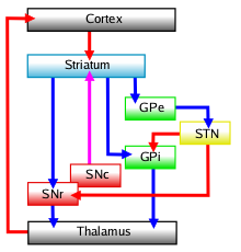
Connectivity diagram showing excitatory glutamatergic pathways as red, inhibitory GABAergic pathways as blue, and modulatory dopaminergic pathways as magenta. (Abbreviations: GPe: globus pallidus external; GPi: globus pallidus internal; STN: subthalamic nucleus; SNc: substantia nigra compacta; SNr: substantia nigra reticulata)
In order to understand the complex circuitry of the basal ganglia, one has to first understand the important participants in this circuit. Parts of the basal ganglia are in direct communication with the thalamus and the cortex. The cortex, thalamus, and the basal ganglia are, therefore, the three main participants in the circuit created by the basal ganglia.
At the top of the hierarchy lies the cerebral cortex. The cortex has many different areas with different functions. One such cortical area is called the primary motor cortex (along the pre-central gyrus). Specialized neurons from the primary motor cortex extend their axons all the way to the striatum portion of the basal ganglia. These cortical neurons release the neurotransmitter glutamate, which is excitatory in nature. Once excited by glutamate, the cells in the striatum project in two different directions giving rise to two major pathways: the "direct" and the "indirect" pathways:
In the direct pathway, cortical cells project excitatory inputs to the striatum, which in turn projects inhibitory neurons onto the cells of the SNr-GPi complex. The SNr-GPi complex projects directly onto the thalamus through the inhibitory ansa lenticularis pathway. The striatal inhibition of the SNr-GPi complex coupled with SNr-GPi inhibition of the thalamus therefore results in a net reduction of inhibition of the thalamus via the striatum. The thalamus projects excitatory glutamatergic neurons to the cortex itself. The direct pathway, therefore, results in the excitation of the motor cortex by the thalamus. Once stimulated, the cortex projects its own excitatory outputs to the brain stem and ultimately muscle fibers via the lateral corticospinal tract. The following diagram depicts the direct pathway:
- Cortex (stimulates) → Striatum (inhibits) → "SNr-GPi" complex (less inhibition of thalamus) → Thalamus (stimulates) → Cortex (stimulates) → Muscles, etc. → (hyperkinetic state)
The indirect pathway also starts from neurons in the striatum. Once stimulated by the cortex, striatal neurons in the indirect pathway project inhibitory axons onto the cells of the globus pallidus externa (GPe), which tonically inhibits the subthalamic nucleus (STN). This inhibition (by the striatum) of the inhibitory projections of the GPe, results in the net reduction of inhibition of the STN. The STN, in turn, projects excitatory inputs to the SNr-GPi complex (which inhibits the thalamus). The end-result is inhibition of the thalamus and, therefore, decreased stimulation of the motor cortex by the thalamus and reduced muscle activity. The direct and indirect pathways are therefore antagonist in their functions. Following is a diagram of the indirect pathway:
- Cortex (stimulates) → Striatum (inhibits) → GPe (less inhibition of STN) → STN (stimulates) → "SNr-GPi" complex (inhibits) → Thalamus (is stimulating less) → Cortex (is stimulating less) → Muscles, etc. → (hypokinetic state)
Main circuits of the basal ganglia. This diagram shows 2 coronal slices that have been superimposed to include the involved basal ganglia structures. The + and - signs at the point of the arrows indicate whether the pathway is excitatory or inhibitory, respectively, in effect. Green arrows refer to excitatory glutamatergic pathways, red arrows refer to inhibitory GABAergic pathways and turquoise arrows refer to dopaminergic pathways that are excitatory on the direct pathway and inhibitory on the indirect pathway.
The antagonistic functions of the direct and indirect pathways are modulated by the substantia nigra pars compacta (SNc), which produces dopamine. In the presence of dopamine, D1-receptors in the basal ganglia stimulate the GABAergic neurons, favoring the direct pathway, and thus increasing movement. The GABAergic neurons of the indirect pathway are stimulated by excitatory neurotransmitters acetylcholine and glutamate. This sets off the indirect pathway that ultimately results in inhibition of upper motor neurons, and less movement. In the presence of dopamine, D2-receptors in the basal ganglia inhibit these GABAergic neurons, which reduces the indirect pathways inhibitory effect. Dopamine therefore increases the excitatory effect of the direct pathway (causing movement) and reduces the inhibitory effect of the indirect pathway (preventing full inhibition of movement). Through these mechanisms the body is able to maintain balance between excitation and inhibition of motion. Lack of balance in this delicate system leads to pathologies such as Parkinson's disease. Parkinson's disease involves the loss of dopamine which means the direct pathway is less able to function (so no movement is initiated) and the indirect pathway is in overdrive (causing too much inhibition of movement).
A note concerning terminology
The nomenclature of the basal ganglia system and its components has always been problematic. Early anatomists, seeing the macroscopic anatomical structure but knowing nothing of the cellular architecture or neurochemistry, grouped together components that are now believed to have distinct functions (such as the internal and external segments of the globus pallidus), and gave distinct names to components that are now thought to be functionally parts of a single structure (such as the caudate nucleus and putamen).
The term "basal" comes from the fact that most of its elements are located in the basal part of the forebrain. The term ganglia is a misnomer: In modern usage, neural clusters are called "ganglia" only in the peripheral nervous system; in the central nervous system they are called "nuclei". For this reason, the basal ganglia are also occasionally known as the "basal nuclei".[10] Terminologia anatomica (1998), the international authority for anatomical naming, retained "nuclei basales", but this is not commonly used.
The International Basal Ganglia Society (IBAGS) informally considers the basal ganglia to be made up of the striatum, the pallidum (with two nuclei), the substantia nigra (with its two distinct parts), and the subthalamic nucleus. Percheron et al. in 1991 and Parent and Parent in 2005 included the central region (centre median-parafascicular) of the thalamus as part of the basal ganglia,[11][12] while Mena-Segovia et al. in 2004 included the pedunculopontine complex as well.[13]
Also, the names given to the various nuclei of the basal ganglia are different in different species. In particular, the internal segment of the globus pallidus in primates is called the entopeduncular nucleus in rodents. The "striatum" and "external segment of the globus pallidus" in primates are called the "paleostriatum augmentatum" and "paleostriatum primitivum," respectively, in birds.
Function
Information about the functions of the basal ganglia comes from anatomical studies, from physiological studies carried out mainly in rats and monkeys, and from the study of diseases that damage them.
The greatest source of insight into the functions of the basal ganglia has come from the study of two neurological disorders, Parkinson's disease and Huntington's disease. For both of these disorders, the nature of the neural damage is well understood and can be correlated with the resulting symptoms. Parkinson's disease involves major loss of dopaminergic cells in the substantia nigra; Huntington's disease involves massive loss of medium spiny neurons in the striatum. The symptoms of the two diseases are virtually opposite: Parkinson's disease is characterized by gradual loss of the ability to initiate movement, whereas Huntington's disease is characterized by an inability to prevent parts of the body from moving unintentionally. It is noteworthy that, although both diseases have cognitive symptoms, especially in their advanced stages, the most salient symptoms relate to the ability to initiate and control movement. Thus, both are classified primarily as movement disorders. A different movement disorder, called hemiballismus, may result from damage restricted to the subthalamic nucleus. Hemiballismus is characterized by violent and uncontrollable flinging movements of the arms and legs.
Eye movements
One of the most intensively studied functions of the basal ganglia (BG) is their role in controlling eye movements.[14] Eye movement is influenced by an extensive network of brain regions that converge on a midbrain area called the superior colliculus (SC). The SC is a layered structure whose layers form two-dimensional retinotopic maps of visual space. A "bump" of neural activity in the deep layers of the SC drives an eye movement directed toward the corresponding point in space.
The SC receives a strong inhibitory projection from the BG, originating in the substantia nigra pars reticulata (SNr).[14] Neurons in the SNr usually fire continuously at high rates, but at the onset of an eye movement they "pause", thereby releasing the SC from inhibition. Eye movements of all types are associated with "pausing" in the SNr; however, individual SNr neurons may be more strongly associated with some types of movements than others. Neurons in some parts of the caudate nucleus also show activity related to eye movements. Since the great majority of caudate cells fire at very low rates, this activity almost always shows up as an increase in firing rate. Thus, eye movements begin with activation in the caudate nucleus, which inhibits the SNr via the direct GABAergic projections, which in turn disinhibits the SC.
Role in motivation
Although the role of the basal ganglia in motor control is clear, there are also many indications that it is involved in the control of behavior in a more fundamental way, at the level of motivation. In Parkinson's disease, the ability to execute the components of movement is not greatly affected, but motivational factors such as hunger fail to cause movements to be initiated or switched at the proper times. The immobility of Parkinsonian patients has sometimes been described as a "paralysis of the will".[15] These patients have occasionally been observed to show a phenomenon called kinesia paradoxica, in which a person who is otherwise immobile responds to an emergency in a coordinated and energetic way, then lapses back into immobility once the emergency has passed.
The role in motivation of the "limbic" part of the basal ganglia—the nucleus accumbens (NA), ventral pallidum, and ventral tegmental area (VTA)—is particularly well established. Thousands of experimental studies combine to demonstrate that the dopaminergic projection from the VTA to the NA plays a central role in the brain's reward system. Animals with stimulating electrodes implanted along this pathway will bar-press very energetically if each press is followed by a brief pulse of electrical current. Numerous things that people find rewarding, including addictive drugs, good-tasting food, and sex, have been shown to elicit activation of the VTA dopamine system. Damage to the NA or VTA can produce a state of profound torpor.
Although it is not universally accepted, some theorists have proposed a distinction between "appetitive" behaviors, which are initiated by the basal ganglia, and "consummatory" behaviors, which are not. For example, an animal with severe basal ganglia damage will not move toward food even if it is placed a few inches away, but, if the food is placed directly in the mouth, the animal will chew it and swallow it.
Comparative anatomy and naming
The basal ganglia form one of the basic components of the forebrain, and can be recognized in all species of vertebrates.[16] Even in the lamprey (generally considered one of the most primitive of vertebrates), striatal, pallidal, and nigral elements can be identified on the basis of anatomy and histochemistry.[17]
A clear emergent issue in comparative anatomy of the basal ganglia is the development of this system through phylogeny as a convergent cortically re-entrant loop in conjunction with the development and expansion of the cortical mantle. There is controversy, however, regarding the extent to which convergent selective processing occurs versus segregated parallel processing within re-entrant closed loops of the basal ganglia. Regardless, the transformation of the basal ganglia into a cortically re-entrant system in mammalian evolution occurs through a re-direction of pallidal (or "paleostriatum primitivum") output from midbrain targets such as the superior colliculus, as occurs in sauropsid brain, to specific regions of the ventral thalamus and from there back to specified regions of the cerebral cortex that form a subset of those cortical regions projecting into the striatum. The abrupt rostral re-direction of the pathway from the internal segment of the globus pallidus into the ventral thalamus—via the path of the ansa lenticularis—could be viewed as a footprint of this evolutionary transformation of basal ganglia outflow and targeted influence.
Neurotransmitters
In most regions of the brain, the predominant classes of neurons use glutamate as neurotransmitter and have excitatory effects on their targets. In the basal ganglia, however, the great majority of neurons use GABA as neurotransmitter and have inhibitory effects on their targets. The inputs from the cortex and thalamus to the striatum and STN are glutamatergic, but the outputs from the striatum, pallidum, and substantia nigra pars reticulata all use GABA. Thus, following the initial excitation of the striatum, the internal dynamics of the basal ganglia are dominated by inhibition and disinhibition.
Other neurotransmitters have important modulatory effects. The most intensively studied is dopamine, which is used by the projection from the substantia nigra pars compacta to the striatum, and also in the analogous projection from the ventral tegmental area to the nucleus accumbens. Acetylcholine also plays an important role, being used both by several external inputs to the striatum, and by a group of striatal interneurons. Although cholinergic cells make up only a small fraction of the total population, the striatum has one of the highest acetylcholine concentrations of any brain structure.
Disorders associated with the basal ganglia
- Main article: Basal ganglia disease
- Athetosis
- Athymhormic syndrome (PAP syndrome)
- Attention-deficit hyperactivity disorder (ADHD)
- Blepharospasm
- Bruxism
- Cerebral palsy: basal ganglia damage during second and third trimester of pregnancy
- Chorea
- Dystonia
- Fahr's disease
- Foreign accent syndrome (FAS)
- Huntington's disease
- Kernicterus
- Lesch-Nyhan syndrome
- Major Depressive Disorder [18]
- Obsessive-compulsive disorder[19][20]
- Other anxiety disorders [20]
- PANDAS
- Parkinson's disease
- Spasmodic dysphonia
- Stuttering[21]
- Sydenham's chorea
- Tardive dyskinesia, caused by chronic antipsychotic treatment
- Tourette's disorder
- Wilson's disease
History
The acceptance that the basal ganglia system constitutes one major cerebral system took long to arise. The first anatomical identification of distinct subcortical structures was published by Thomas Willis in 1664.[22] For many years, the term corpus striatum[23] was used to describe a large group of subcortical elements, some of which were later discovered to be functionally unrelated.[24] For many years, the putamen and the caudate nucleus were not associated with each other. Instead, the putamen was associated with the pallidum in what was called the nucleus lenticularis or nucleus lentiformis.
A thorough reconsideration by Cécile and Oskar Vogt (1941) simplified the description of the basal ganglia by proposing the term striatum to describe the group of structures consisting of the caudate nucleus, the putamen, and the mass linking them ventrally, the nucleus accumbens. The striatum was named on the basis of the striated (striped) appearance created by radiating dense bundles of striato-pallido-nigral axons, described by anatomist Samuel Alexander Kinnier Wilson (1912) as "pencil-like".
The anatomical link of the striatum with its primary targets, the pallidum and the substantia nigra, was discovered later. The name globus pallidus was attributed by Déjerine to Burdach (1822). For this, the Vogts proposed the simpler "pallidum". The term "locus niger" was introduced by Félix Vicq-d'Azyr as tache noire in (1786), though that structure has since become known as the substantia nigra, due to contributions by Von Sömmering in 1788. The structural similarity between the substantia nigra and globus pallidus was noted by Mirto in 1896. Together, the two are known as the pallidonigral ensemble, which represents the core of the basal ganglia. Altogether, the main structures of the basal ganglia are linked to each other by the striato-pallido-nigral bundle, which passes through the pallidum, crosses the internal capsule as the "comb bundle of Edinger", then finally reaches the substantia nigra.
Additional structures that later became associated with the basal ganglia are the "body of Luys" (1865) (nucleus of Luys on the figure) or subthalamic nucleus, whose lesion was known to produce movement disorders. More recently, other areas such as the central complex (centre médian-parafascicular) and the pedunculopontine complex have been thought to be regulators of the basal ganglia.
Near the beginning of the 20th century, the basal ganglia system was first associated with motor functions, as lesions of these areas would often result in disordered movement in humans (chorea, athetosis, Parkinson's disease).
See also
- Amygdaloid body
- Anatomical subdivisions and connections of the basal ganglia
- Caudate nucleus
- Extrapyramidal symptoms
- Globus palladius
- Nucleus basalis magnocellularis
- Primate basal ganglia system
References
- Nolte, John, The Human Brain: An Introduction to its Functional Anatomy (Fifth Edition). (St. Louis: Mosby, Inc., 2002), 464-484. ISBN 0-323-01320-1
- Parent, André, Comparative Neurobiology of the Basal Ganglia (Wiley, New York, 1986), ISBN 0-471-80348-0
- Reynolds, J. et, al., "A cellular mechanism of reward-related learning", Nature (2001) volume 413, pages 67-70.
- Andrew Gilies, A brief history of the basal ganglia, retrieved on 27 June 2005
- Alm, Per A., Stuttering and the basal ganglia circuits, Journal of Communication Disorders (2004) volume 37, pages 325-369.
Additional images
")
Human brain, cerebrum, Interior of the cerebral hemispheres—Rostral Basal ganglia and associated structures (TA A14.1.09.321–552, GA 9.832–837) | |||||||
|---|---|---|---|---|---|---|---|
| Basal ganglia |
| ||||||
| Rhinencephalon |
| ||||||
| Other basal forebrain |
| ||||||
| Archicortex: Hippocampal formation/ Hippocampus anatomy |
| ||||||
| {| class="navbox collapsible nowraplinks" style="margin:auto; " | |||||||
| |||||||
|}
External links
- Basal ganglia disease, adult-onset - Geneva Foundation for Medical Education and Research
- Dictionary at eMedicine Basal+ganglia
- Dictionary at eMedicine basal+nuclei
| Telencephalon (cerebrum, cerebral cortex, cerebral hemispheres) - edit |
|
primary sulci/fissures: medial longitudinal, lateral, central, parietoöccipital, calcarine, cingulate frontal lobe: precentral gyrus (primary motor cortex, 4), precentral sulcus, superior frontal gyrus (6, 8), middle frontal gyrus (46), inferior frontal gyrus (Broca's area, 44-pars opercularis, 45-pars triangularis), prefrontal cortex (orbitofrontal cortex, 9, 10, 11, 12, 47) parietal lobe: postcentral sulcus, postcentral gyrus (1, 2, 3, 43), superior parietal lobule (5), inferior parietal lobule (39-angular gyrus, 40), precuneus (7), intraparietal sulcus occipital lobe: primary visual cortex (17), cuneus, lingual gyrus, 18, 19 (18 and 19 span whole lobe) temporal lobe: transverse temporal gyrus (41-42-primary auditory cortex), superior temporal gyrus (38, 22-Wernicke's area), middle temporal gyrus (21), inferior temporal gyrus (20), fusiform gyrus (36, 37) limbic lobe/fornicate gyrus: cingulate cortex/cingulate gyrus, anterior cingulate (24, 32, 33), posterior cingulate (23, 31), subcortical/insular cortex: rhinencephalon, olfactory bulb, corpus callosum, lateral ventricles, septum pellucidum, ependyma, internal capsule, corona radiata, external capsule hippocampal formation: dentate gyrus, hippocampus, subiculum basal ganglia: striatum (caudate nucleus, putamen), lentiform nucleus (putamen, globus pallidus), claustrum, extreme capsule, amygdala, nucleus accumbens Some categorizations are approximations, and some Brodmann areas span gyri. |
| This page uses Creative Commons Licensed content from Wikipedia (view authors). |
- ↑ 1.0 1.1 1.2 1.3 1.4 1.5 (2010). Conditional Routing of Information to the Cortex: A Model of the Basal Ganglia's Role in Cognitive Coordination. Psychological Review 117 (2): 541–74.
- ↑ 2.0 2.1 (2007) Rapid Review of Neuroscience, Mosby Elsevier.
- ↑ (2010). What do the basal ganglia do? A modeling perspective. Biological Cybernetics 103 (3): 237–53.
- ↑ Cameron IG, Watanabe M, Pari G, Munoz DP. (2010 June). Executive impairment in Parkinson's disease: response automaticity and task switching. Neuropsychologia 48 (7): 1948–57.
- ↑ 5.0 5.1 5.2 (2008) "Basal Ganglia and the Striatal Motor System" Neuroanatomy (Board Review Series), 4th, 274–281, Baltimore: Wulters Kluwer & Lippincott Wiliams & Wilkins.
- ↑ (2010). Alterations in Postnatal Neurogenesis and Dopamine Dysregulation in Schizophrenia: A Hypothesis. Schizophrenia Bulletin 37 (4): 674–80.
- ↑ Marín & Rubenstein. (2001). A Long, Remarkable Journey: Tangential Migration in the Telencephalon. Nature Reviews Neuroscience, 2.
- ↑ Regina Bailey. Divisions of the Brain. about.com. URL accessed on 2010-11-30.
- ↑ (2008). Striatal projections from the rat lateral posterior thalamic nucleus. Brain Research 1204: 24–39.
- ↑ Soltanzadeh, Akbar (2004). Neurologic Disorders, Tehran: Jafari.Template:Page needed
- ↑ (1991). Parallel processing in the basal ganglia: up to a point. Trends in Neurosciences 14 (2): 55–9.
- ↑ (2005). Single-axon tracing and three-dimensional reconstruction of centre median-parafascicular thalamic neurons in primates. The Journal of Comparative Neurology 481 (1): 127–44.
- ↑ (2004). Pedunculopontine nucleus and basal ganglia: distant relatives or part of the same family?. Trends in Neurosciences 27 (10): 585–8.
- ↑ 14.0 14.1 (2000). Role of the basal ganglia in the control of purposive saccadic eye movements. Physiological reviews 80 (3): 953–78.
- ↑ (2007). Parkinson's Disease: Fighting the Will?. Journal of Neuroscience 27 (44): 11777–9.
- ↑ Parent A (1986). Comparative Neurobiology of the Basal Ganglia, Wiley.Template:Page needed
- ↑ (1998). Intrinsic function of a neuronal network — a vertebrate central pattern generator1. Brain Research Reviews 26 (2–3): 184–97.
- ↑ Kempton MJ, Salvador Z, Munafò MR, Geddes JR, Simmons A, Frangou S, Williams SC. (2011). Structural Neuroimaging Studies in Major Depressive Disorder: Meta-analysis and Comparison With Bipolar Disorder. Arch Gen Psychiatry 68 (7): 675–90. see also MRI database at www.depressiondatabase.org
- ↑ (November 2009). Voxel-wise meta-analysis of grey matter changes in obsessive–compulsive disorder. British Journal of Psychiatry 195 (5): 393–402.
- ↑ 20.0 20.1 (5 July 2010) Meta-analytical comparison of voxel-based morphometry studies in obsessive-compulsive disorder vs other anxiety disorders. Archives of General Psychiatry 67 (7): 701–711.
- ↑ (2004). Stuttering and the basal ganglia circuits: a critical review of possible relations. Journal of communication disorders 37 (4): 325–69.
- ↑ Andrew Gilies, A brief history of the basal ganglia, retrieved on 27 June 2005
- ↑ Vieussens (1685)[verification needed]
- ↑ (1994). History of the basal ganglia system. Slow development of a major cerebral system. Revue neurologique 150 (8–9): 543–54.