m (Reverted edits by 109.156.211.68 (talk | block) to last version by Dr Joe Kiff) |
|||
| (3 intermediate revisions by 2 users not shown) | |||
| Line 76: | Line 76: | ||
==See also== |
==See also== |
||
| + | *[[Amygdalohippocampectomy]] |
||
*[[Medial forebrain bundle]] |
*[[Medial forebrain bundle]] |
||
==References== |
==References== |
||
{{Reflist|2}} |
{{Reflist|2}} |
||
| + | |||
===Books=== |
===Books=== |
||
*Aggleton, J. P.(2000)(ed.), The Amygdala: A Functional Analysis, Oxford University Press, London. |
*Aggleton, J. P.(2000)(ed.), The Amygdala: A Functional Analysis, Oxford University Press, London. |
||
Latest revision as of 17:09, 18 September 2016
Assessment |
Biopsychology |
Comparative |
Cognitive |
Developmental |
Language |
Individual differences |
Personality |
Philosophy |
Social |
Methods |
Statistics |
Clinical |
Educational |
Industrial |
Professional items |
World psychology |
Biological: Behavioural genetics · Evolutionary psychology · Neuroanatomy · Neurochemistry · Neuroendocrinology · Neuroscience · Psychoneuroimmunology · Physiological Psychology · Psychopharmacology (Index, Outline)
| Brain: Amygdala | ||
|---|---|---|
 | ||
| Location of the amygdala in the human brain | ||
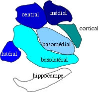 | ||
| Subdivision of the amygdala | ||
| Latin | corpus amygdaloideum | |
| Gray's | subject #189 835 | |
| Part of | ||
| Components | ||
| Artery | ||
| Vein | ||
| BrainInfo/UW | hier-219 | |
| MeSH | [1] | |
Located deep in the brain's medial temporal lobe, the almond-shaped amygdala (in Latin, corpus amygdaloideum) is believed to play a key role in the emotions. It forms part of the limbic system. In humans and other animals, this subcortical brain structure is linked to both fear responses and pleasure. Its size is positively correlated with aggressive behavior across species. In humans it is the most sexually dimorphic brain structure, and shrinks by more than 30% in males upon castration. Conditions such as anxiety, autism, depression, post-traumatic stress disorder, and phobias are suspected of being linked to abnormal functioning of the amygdala owing to damage, developmental problems, or neurotransmitter imbalance.
Anatomical subdivisions
The regions described as amygdalae encompass several nuclei with distinct functional traits. Among these nuclei are the:
- basolateral complex, which can be further subdivided[1][2][3] :
- Lateral basal nuclei
- Accessory basal nuclei
- centromedial nucleus
- cortical nucleus. The basolateral complex can be further subdivided into the lateral, the basal and the accessory basal nuclei.
Connections
The amygdalae send impulses to the hypothalamus for important activation of the sympathetic nervous system, to the reticular nucleus for increased reflexes, to the nuclei of the trigeminal nerve and facial nerve for facial expressions of fear, and to the ventral tegmental area, locus coeruleus, and laterodorsal tegmental nucleus for activation of dopamine, norepinephrine and epinephrine.[2]
The cortical nucleus is involved in the sense of smell and pheromone-processing. It receives input from the olfactory bulb and olfactory cortex. The lateral amygdalae, which send impulses to the rest of the basolateral complexes and to the centromedial nuclei, receive input from the sensory systems. The centromedial nuclei are the main outputs for the basolateral complexes, and are involved in emotional arousal in rats and cats.[2][4]
Memory modulation
The amygdala plays a key role in the modulation of memory consolidation. Following any learning event, the long-term memory for the event is not instantaneously formed. Rather, information regarding the event is slowly put into long-term storage over time, a process referred to as "memory consolidation", until it reaches a relatively permanent state.
- Main article: The role of the amygdala in memory
Drug addiction
Experiments with rats also suggest that the amygdala is involved in learning about various cues with the consumption of drugs of abuse. It is well known that one of the major problems in drug addiction is that drug-associated cues induce significant craving in individuals, even if the individuals have not taken the drugs in a long time. The amygdala appears to play a key role in the initial learning of the association between the cues and the drugs. In addition, inactivation of the amygdala prevents the ability of cues to induce reinstatement in rats in a drug self-administration paradigm.
Learnt fear
- Main article: role of the amygdala in emotion
- Main article: The role of the amygdala in learnt fear
Kluver-Bucy syndrome
- Removal of the amygdala[2] from monkeys results in Kluver-Bucy syndrome.
Neuropsychological correlates of amygdala activity
Early research on primates provided explanations as to the functions of the amygdala, as well as a basis for further research. As early as 1888, rhesus monkeys with a lesioned temporal cortex (including the amygdala) were observed to have significant social and emotional deficits.[5] Heinrich Klüver and Paul Bucy later expanded upon this same observation by showing that large lesions to the anterior temporal lobe produced noticeable changes, including overreaction to all objects, hypoemotionality, loss of fear, hypersexuality, and hyperorality, a condition in which inappropriate objects are placed in the mouth. Some monkeys also displayed an inability to recognize familiar objects and would approach animate and inanimate objects indiscriminately, exhibiting a loss of fear towards the experimenters. This behavioral disorder was later named Klüver-Bucy syndrome accordingly.[6] Later studies served to focus on the amygdala specifically, as the temporal cortex encompasses a broad set of brain structures, making it difficult to find which ones specifically may have correlated with certain symptoms. Monkey mothers who had amygdala damage showed a reduction in maternal behaviors towards their infants, often physically abusing or neglecting them.[7] In 1981, researchers found that selective radio frequency lesions of the whole amygdala caused Klüver-Bucy Syndrome.[8]
With advances in neuroimaging technology such as MRI, neuroscientists have made significant findings concerning the amygdala in the human brain. Consensus of data shows the amygdala has a substantial role in mental states, and is related to many psychological disorders. In a 2003 study, subjects with Borderline Personality Disorder showed significantly greater left amygdala activity than normal control subjects. Some borderline patients even had difficulties classifying neutral faces or saw them as threatening.[9] In 2006, researchers observed hyperactivity in the amygdala when patients were shown threatening faces or confronted with frightening situations. Patients with more severe social phobia showed a correlation with increased response in the amygdala.[10] Similarly, depressed patients showed exaggerated left amygdala activity when interpreting emotions for all faces, and especially for fearful faces. Interestingly, this hyperactivity was normalized when patients went on antidepressants.[11] By contrast, the amygdala has been observed to relate differently in people with Bipolar Disorder. A 2003 study found that adult and adolescent bipolar patients tended to have considerably smaller amygdala volumes and somewhat smaller hippocampal volumes.[12] Many studies have focused on the connections between the amygdala and autism.[13]
Recent research suggests that parasites, in particular toxoplasma, form cysts in the brain, often taking up residence in the amygdala. This may provide clues as to how specific parasites manipulate behavior and may contribute to the development of disorders, including paranoia.[14]
Serotonin and the amygdala
- Main article: Serotonin and the amygdala
See also
References
- ↑ Cite error: Invalid
<ref>tag; no text was provided for refs namedrd - ↑ 2.0 2.1 2.2 Ben Best (2004). The Amygdala and the Emotions. URL accessed on 2007-03-15.
- ↑ Solano-Castiella E, Anwander A, Lohmann G, Weiss M, Docherty C, Geyer S, Reimer E, Friederici AD, Turner R (2010). Diffusion tensor imaging segments the human amygdala in vivo. Neuroimage 49 (4): 2958–65.
- ↑ Michael McDannald, Erin Kerfoot, Michela Gallagher, and Peter C. Holland, John Hopkins University (2005). Amygdala central nucleus function is necessary for learning but not expression of conditioned visual orienting. URL accessed on 2007-03-15.
- ↑ Brown, S. & Shafer, E. (1888). An investigation into the functions of the occipital and temporal lobes of the monkey's brain.. Philosophical Transactions of the Royal Society of London: Biological Sciences 179: 303-327.
- ↑ Kluver, H. & Bucy, P. (1939). Preliminary analysis of function of the temporal lobe in monkeys.. Archives of Neurology 42: 979-1000.
- ↑ Bucher, K., Myersn, R., Southwick, C. (1970). Anterior temporal cortex and maternal behaviour in monkey.. Neurology 20: 415.
- ↑ Aggleton, JP. & Passingham, RE. (1981). Syndrome produced by lesions of the amygdala in monkeys (Macaca mulatta).. Journal of Comparative and Physiological Psychology 95: 961-977.
- ↑ Donegan et al. (2003). Amygdala hyperreactivity in borderline personality disorder: implications for emotional dysregulation.. Biological Psychiatry 54 (11): 1284-1293.
- ↑ Studying Brain Activity Could Aid Diagnosis Of Social Phobia. Monash University. January 19, 2006.
- ↑ Sheline et al. (2001). Increased amygdala response to masked emotional faces in depressed subjects resolves with antidepressant treatment: an fMRI study.. Biological Psychiatry 50 (9): 651-658.
- ↑ Blumberg et al. (2003). Amygdala and hippocampal volumes in adolescents and adults with bipolar disorder. Arch Gen Psychiatry 60 (12): 1201-8.
- ↑ Schultz RT (2005). Developmental deficits in social perception in autism: the role of the amygdala and fusiform face area. Int J Dev Neurosci 23 (2–3): 125–41.
- ↑ Vyas et al. (2007). Behavioral changes induced by Toxoplasma infection of rodents are highly specific to aversion of cat odors. Proc Natl Acad Sci U S A. 104 (15): 6442-7.
Books
- Aggleton, J. P.(2000)(ed.), The Amygdala: A Functional Analysis, Oxford University Press, London.
- Ben-Air, Y.(1981)(ed.). The Amygdaloid Complex, Amsterdam, Elsevier/N. Holland.
Papers
- Kandel ER, Schwartz JH, Jessell TM. Principles of Neural Science, 4th ed. McGraw-Hill, New York (2000). ISBN 0838577016
External links:
- BrainMaps at UCDavis amygdala
- Amygdala, Panic and Post Traumatic Stress
- BrainInfo at the University of Washington hier-219
| Human brain: Limbic system | |
| Amygdala - Cingulate gyrus - Fornicate gyrus - Hippocampus - Hypothalamus - Mammillary body - Nucleus accumbens - Orbitofrontal cortex - Parahippocampal gyrus |
| This page uses Creative Commons Licensed content from Wikipedia (view authors). |