No edit summary |
No edit summary |
||
| Line 1: | Line 1: | ||
{{BioPsy}} |
{{BioPsy}} |
||
| − | {{PsyPerspective} |
+ | {{PsyPerspective}} |
{{Infobox Anatomy | |
{{Infobox Anatomy | |
||
Name = {{PAGENAME}} | |
Name = {{PAGENAME}} | |
||
| Line 17: | Line 17: | ||
DorlandsSuf = | |
DorlandsSuf = | |
||
}} |
}} |
||
| − | The '''abdominal wall''' represents the boundaries of the [[abdominal cavity]]. The abdominal wall is split into the posterior (back), lateral (sides) and anterior (front) walls. |
+ | The '''abdominal wall''' is part of the [[abdomen ]] and represents the boundaries of the [[abdominal cavity]]. The abdominal wall is split into the posterior (back), lateral (sides) and anterior (front) walls. |
There is a common set of layers covering and forming all the walls: the deepest being the [[extraperitoneal fat]], the [[parietal peritoneum]], and a layer of [[fascia]] which has different names over where it covers (eg transversalis, psoas fascia). |
There is a common set of layers covering and forming all the walls: the deepest being the [[extraperitoneal fat]], the [[parietal peritoneum]], and a layer of [[fascia]] which has different names over where it covers (eg transversalis, psoas fascia). |
||
| Line 86: | Line 86: | ||
[[Category:Abdomen]] |
[[Category:Abdomen]] |
||
| + | [[Category:Muscles]] |
||
| + | |||
{{enWP|Abdominal wall}} |
{{enWP|Abdominal wall}} |
||
Revision as of 19:55, 18 December 2009
Assessment |
Biopsychology |
Comparative |
Cognitive |
Developmental |
Language |
Individual differences |
Personality |
Philosophy |
Social |
Methods |
Statistics |
Clinical |
Educational |
Industrial |
Professional items |
World psychology |
Biological: Behavioural genetics · Evolutionary psychology · Neuroanatomy · Neurochemistry · Neuroendocrinology · Neuroscience · Psychoneuroimmunology · Physiological Psychology · Psychopharmacology (Index, Outline)
| Abdominal wall | ||
|---|---|---|
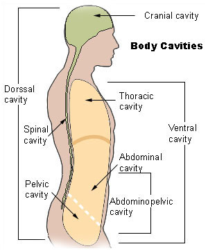 | ||
| Body cavities | ||
| Latin | ' | |
| Gray's | subject #118 408 | |
| System | ||
| MeSH | [1] | |
| Diagram of sheath of Rectus above the arcuate line. | ||
The abdominal wall is part of the abdomen and represents the boundaries of the abdominal cavity. The abdominal wall is split into the posterior (back), lateral (sides) and anterior (front) walls.
There is a common set of layers covering and forming all the walls: the deepest being the extraperitoneal fat, the parietal peritoneum, and a layer of fascia which has different names over where it covers (eg transversalis, psoas fascia).
Superficial to these, but not present in the posterior wall are the three layers of muscle, the transversus abdominis (tranvserse abdominal muscle), the internal (obliquus internus) and the external oblique (obliquus externus).
Muscles of the abdominal wall

Henry Gray (1825–1861). Anatomy of the Human Body.
| Muscle | Origin and insertion |
| The obliquus externus (external oblique) muscle is the outermost muscle covering the side of the abdomen. It is broad, flat, and irregularly quadrilateral. | It originates on the lower eight ribs, and then curves down and forward towards its insertion on the outer anterior crest of the ilium and (via the sheath of the rectus abdominis muscle) the midline linea alba. |
| The obliquus internus (internal oblique) muscle is triangularly shaped and is smaller and thinner than the external oblique muscle that overlies it. | It originates from Poupart's ligament/inguinal ligament and the inner anterior crest of the ilium. The lower two-thirds of it insert, in common with fibers of the external oblique and the underlying transversus abdominis, into the linea alba. The upper third inserts into the lower six ribs. |
| The transversus abdominis muscle is flat and triangular, with its fibers running horizontally. It lies between the internal oblique and the underlying transversalis fascia. | It originates from Poupart's ligament, the inner lip of the ilium, the lumbar fascia and the inner surface of the cartilages of the six lower ribs. It inserts into the linea alba behind the rectus abdominis. |
| The rectus abdominis muscles are long and flat. The muscle is crossed by three tendinous intersections called the linae transversae. The rectus abdominis is enclosed in a thick sheath formed, as described above, by fibers from each of the three muscles of the lateral abdominal wall. | They originate at the pubic bone, run up the abdomen on either side of the linea alba, and insert into the cartilages of the fifth, sixth, and seventh ribs. |
| The pyramidalis muscle is small and triangular. It is located in the lower abdomen in front of the rectus abdominis. | It originates at the pubic bone and is inserted into the linea alba half way up to the umbilicus (belly button). |
Layers of anterior abdominal wall
In human anatomy, the layers of the abdominal wall are (from superficial to deep):
- Skin
- Fascia
- Camper's fascia - fatty superficial layer
- Scarpa's fascia - deep fibrous layer
- Muscle
- External oblique muscle
- Internal oblique muscle
- Transverse abdominal muscle
- Deep fascia or subserous fascia
Inner surface
The surface contains several ligaments separated by fossae:
| Ligament/fold | Remnant of | Lateral fossa | Hernia |
| median umbilical ligament | urachus | supravesical fossa | - |
| medial umbilical ligament | umbilical artery | medial inguinal fossa | direct inguinal hernia |
| lateral umbilical fold | inferior epigastric vessels | lateral inguinal fossa | indirect inguinal hernia |
See also
References
External links
- Memorial University of Newfoundland - Anatomy at MUN digest/abwall
- Norman/Georgetown skel&wallsabd - "Skeleton of the Abdomen", Wesley Norman, PhD, DSc
- MeSH Abdominal+Wall
- Anterolateral Abdominal Wall - University of Edinburgh Faculty of Medicine
- Muscles of the Anterior Abdominal Wall - University of Arkansas
| This page uses Creative Commons Licensed content from Wikipedia (view authors). |