No edit summary Tag: Visual edit |
|||
| (2 intermediate revisions by 2 users not shown) | |||
| Line 8: | Line 8: | ||
The [[abdominal wall]] is split into the posterior (back), lateral (sides) and anterior (front) walls. |
The [[abdominal wall]] is split into the posterior (back), lateral (sides) and anterior (front) walls. |
||
| − | |||
| − | |||
==[[Muscles]] of the [[abdominal wall]]== |
==[[Muscles]] of the [[abdominal wall]]== |
||
[[Image:Grays_Anatomy_image392.png|thumb|RIGHT|200px|''Henry Gray (1825–1861). Anatomy of the Human Body.'']] |
[[Image:Grays_Anatomy_image392.png|thumb|RIGHT|200px|''Henry Gray (1825–1861). Anatomy of the Human Body.'']] |
||
| Line 18: | Line 16: | ||
| The obliquus externus ([[external oblique muscle|external oblique]]) [[muscle]] is the outermost muscle covering the side of the abdomen. It is broad, flat, and irregularly quadrilateral. || It originates on the lower eight [[rib]]s, and then curves down and forward towards its insertion on the outer anterior crest of the [[Ilium (bone)|ilium]] and (via the sheath of the [[rectus abdominis]] muscle) the midline [[linea alba]]. |
| The obliquus externus ([[external oblique muscle|external oblique]]) [[muscle]] is the outermost muscle covering the side of the abdomen. It is broad, flat, and irregularly quadrilateral. || It originates on the lower eight [[rib]]s, and then curves down and forward towards its insertion on the outer anterior crest of the [[Ilium (bone)|ilium]] and (via the sheath of the [[rectus abdominis]] muscle) the midline [[linea alba]]. |
||
|- |
|- |
||
| − | | The obliquus internus ([[internal oblique muscle|internal oblique]]) muscle is triangularly shaped and is smaller and thinner than the external oblique muscle that overlies it. || It originates from [[Poupart's ligament]]/[[inguinal ligament]] and the inner anterior crest of the ilium. The lower two-thirds of it insert, in common with fibers of the external oblique and the underlying transversus abdominis, into the [[linea alba]]. |
+ | | The obliquus internus ([[internal oblique muscle|internal oblique]]) muscle is triangularly shaped and is smaller and thinner than the external oblique muscle that overlies it. || It originates from [[Poupart's ligament]]/[[inguinal ligament]] and the inner anterior crest of the ilium. The lower two-thirds of it insert, in common with fibers of the external oblique and the underlying transversus abdominis, into the [[linea alba]]. The upper third inserts into the lower six ribs. |
|- |
|- |
||
| The [[transversus abdominis]] muscle is flat and triangular, with its fibers running horizontally. It lies between the internal oblique and the underlying transversalis fascia. || It originates from Poupart's ligament, the inner lip of the ilium, the lumbar fascia and the inner surface of the [[cartilage]]s of the six lower ribs. It inserts into the linea alba behind the [[rectus abdominis]]. |
| The [[transversus abdominis]] muscle is flat and triangular, with its fibers running horizontally. It lies between the internal oblique and the underlying transversalis fascia. || It originates from Poupart's ligament, the inner lip of the ilium, the lumbar fascia and the inner surface of the [[cartilage]]s of the six lower ribs. It inserts into the linea alba behind the [[rectus abdominis]]. |
||
|- |
|- |
||
| − | | The [[rectus abdominis muscle]]s are long and flat. The muscle is crossed by three [[tendon|tendinous]] intersections called the [[linae transversae]]. |
+ | | The [[rectus abdominis muscle]]s are long and flat. The muscle is crossed by three [[tendon|tendinous]] intersections called the [[linae transversae]]. The rectus abdominis is enclosed in a thick sheath formed, as described above, by fibers from each of the three muscles of the lateral abdominal wall. || They originate at the [[pubic bone]], run up the abdomen on either side of the linea alba, and insert into the cartilages of the fifth, sixth, and seventh ribs. |
|- |
|- |
||
| The [[pyramidalis muscle]] is small and triangular. It is located in the lower abdomen in front of the rectus abdominis. || It originates at the pubic bone and is inserted into the linea alba half way up to the [[umbilicus]] (belly button). |
| The [[pyramidalis muscle]] is small and triangular. It is located in the lower abdomen in front of the rectus abdominis. || It originates at the pubic bone and is inserted into the linea alba half way up to the [[umbilicus]] (belly button). |
||
| Line 30: | Line 28: | ||
== Abdominal organs == |
== Abdominal organs == |
||
[[Image:Gray1120.png|thumb|RIGHT|200px|The relations of the viscera and large vessels of the abdomen.]] |
[[Image:Gray1120.png|thumb|RIGHT|200px|The relations of the viscera and large vessels of the abdomen.]] |
||
| − | The abdomen contains most of the tubelike organs of the digestive tract, as well as several solid organs. Hollow abdominal organs include the [[stomach]], the [[small intestine]], and the [[colon (anatomy)|colon]] with its attached [[Vermiform appendix|appendix]]. Organs such as the [[liver]], its attached [[gallbladder]], and the [[pancreas]] function in close association with the digestive tract and communicate with it via ducts. The [[spleen]], [[kidney]]s, and [[adrenal gland]]s also lie within the abdomen, along with many blood vessels including the [[aorta]] and [[venae cavae|inferior vena cava]]. Anatomists may consider the [[urinary bladder]], [[uterus]], [[fallopian tube]]s, and [[ovary|ovaries]] as either abdominal organs or as pelvic organs. Finally, the abdomen contains an extensive membrane called the [[peritoneum]]. A fold of peritoneum may completely cover certain organs, whereas it may cover only one side of organs that usually lie closer to the abdominal wall. Anatomists call the latter type of |
+ | The abdomen contains most of the tubelike organs of the digestive tract, as well as several solid organs. Hollow abdominal organs include the [[stomach]], the [[small intestine]], and the [[colon (anatomy)|colon]] with its attached [[Vermiform appendix|appendix]]. Organs such as the [[liver]], its attached [[gallbladder]], and the [[pancreas]] function in close association with the digestive tract and communicate with it via ducts. The [[spleen]], [[kidney]]s, and [[adrenal gland]]s also lie within the abdomen, along with many blood vessels including the [[aorta]] and [[venae cavae|inferior vena cava]]. Anatomists may consider the [[urinary bladder]], [[uterus]], [[fallopian tube]]s, and [[ovary|ovaries]] as either abdominal organs or as pelvic organs. Finally, the abdomen contains an extensive membrane called the [[peritoneum]]. A fold of peritoneum may completely cover certain organs, whereas it may cover only one side of organs that usually lie closer to the abdominal wall. Anatomists call the latter type of organs ''retroperitoneal.'' |
== Surface landmarks of the abdomen == |
== Surface landmarks of the abdomen == |
||
| − | In the mid-line a slight furrow extends from the ensiform cartilage/[[xiphoid process]] above to the [[symphysis pubis]] below, representing the [[linea alba]] in the abdominal wall. At about its midpoint sits the umbilicus or navel. |
+ | In the mid-line a slight furrow extends from the ensiform cartilage/[[xiphoid process]] above to the [[symphysis pubis]] below, representing the [[linea alba]] in the abdominal wall. At about its midpoint sits the umbilicus or navel. On each side of it the broad recti muscles stand out in muscular people. The outline of these muscles is interrupted by three or more transverse depressions indicating the [[lineae transversae]]. There is usually one about the [[ensiform cartilage]], one at the [[umbilicus]], and one between. It is the combination of the linea alba and the linea transversae which form the abdominal "six-pack" sought after by many people. A body fat of around 10% or below with a bodyweight that is not underweight is required to see them. |
| ⚫ | The upper lateral limit of the abdomen is the subcostal margin formed by the cartilages of the false ribs (8, 9, 10) joining one another. The lower lateral limit is the anterior crest of the [[ilium]] and [[Poupart's ligament]], which runs from the anterior superior spine of the ilium to the spine of the [[pubis]]. These lower limits are marked by visible grooves. Just above the pubic spines on either side are the external abdominal rings, which are openings in the muscular wall of the abdomen through which the [[spermatic cord]] emerges in the male, and through which an [[inguinal hernia]] may rupture. |
||
| − | |||
| − | The upper lateral limit of the abdomen is the subcostal margin formed by the cartilages of the false ribs (8, 9, 10) joining one another. The lower lateral limit is the anterior |
||
| ⚫ | crest of the [[ilium]] and [[Poupart's ligament]], which runs from the anterior superior spine of the ilium to the spine of the [[pubis]]. |
||
One method by which the location of the abdominal contents can be appreciated is to draw three horizontal and two vertical lines. |
One method by which the location of the abdominal contents can be appreciated is to draw three horizontal and two vertical lines. |
||
| Line 43: | Line 39: | ||
===Horizontal lines=== |
===Horizontal lines=== |
||
[[Image:Gray1225.png|thumb|Front of abdomen, showing surface markings for duodenum, pancreas, and kidneys.]] |
[[Image:Gray1225.png|thumb|Front of abdomen, showing surface markings for duodenum, pancreas, and kidneys.]] |
||
| − | * The highest of the former is the [[transpyloric line]] of C. Addison, which is situated half-way between the [[suprasternal notch]] and the top of the symphysis pubis, and often cuts the pyloric opening of the stomach an inch to the right of the mid-line. The [[hilum]] of each [[kidney]] is a little below it, while its left end approximately touches the lower limit of the [[spleen]]. |
+ | * The highest of the former is the [[transpyloric line]] of C. Addison, which is situated half-way between the [[suprasternal notch]] and the top of the symphysis pubis, and often cuts the pyloric opening of the stomach an inch to the right of the mid-line. The [[hilum]] of each [[kidney]] is a little below it, while its left end approximately touches the lower limit of the [[spleen]]. It corresponds to the first lumbar vertebra behind. |
| − | * The second line is the ''subcostal'', drawn from the lowest point of the [[subcostal arch]] ([[tenth rib]]). |
+ | * The second line is the ''subcostal'', drawn from the lowest point of the [[subcostal arch]] ([[tenth rib]]). It corresponds to the upper part of the third lumbar vertebra, and it is an inch or so above the umbilicus. It indicates roughly the [[transverse colon]], the lower ends of the kidneys, and the upper limit of the transverse (3rd) part of the [[duodenum]]. |
| − | * The third line is called the ''intertubercular'', and runs across between the two rough [[Tubercle (anatomy)|tubercles]], which can be felt on the outer lip of the crest of the ilium about two and a half inches (60 mm) from the anterior superior spine. |
+ | * The third line is called the ''intertubercular'', and runs across between the two rough [[Tubercle (anatomy)|tubercles]], which can be felt on the outer lip of the crest of the ilium about two and a half inches (60 mm) from the anterior superior spine. This line corresponds to the body of the fifth lumbar vertebra, and passes through or just above the [[ileo-caecal valve]], where the [[small intestine]] joins the [[large intestine|large]]. |
===Vertical lines=== |
===Vertical lines=== |
||
The two vertical or mid-Poupart lines are drawn from the point midway between the anterior superior spine and the pubic symphysis on each side, vertically upward to the costal margin. |
The two vertical or mid-Poupart lines are drawn from the point midway between the anterior superior spine and the pubic symphysis on each side, vertically upward to the costal margin. |
||
| − | * The right one is the most valuable, as the [[ileo-caecal valve]] is situated where it cuts the intertubercular line. The orifice of the [[vermiform appendix]] lies an inch lower, at [[McBurney's point]]. |
+ | * The right one is the most valuable, as the [[ileo-caecal valve]] is situated where it cuts the intertubercular line. The orifice of the [[vermiform appendix]] lies an inch lower, at [[McBurney's point]]. In its upper part, the vertical line meets the transpyloric line at the lower margin of the ribs, usually the ninth, and here the [[gallbladder]] is situated. |
* The left mid-Poupart line corresponds in its upper three-quarters to the inner edge of the [[descending colon]]. |
* The left mid-Poupart line corresponds in its upper three-quarters to the inner edge of the [[descending colon]]. |
||
| Line 60: | Line 56: | ||
==Regions of the abdomen== |
==Regions of the abdomen== |
||
[[Image:Gray1220.png|thumb|Surface lines of the front of the thorax and abdomen.]] |
[[Image:Gray1220.png|thumb|Surface lines of the front of the thorax and abdomen.]] |
||
| − | These three horizontal and two vertical lines divide the abdomen into nine "regions." (Note that "hypo" means "below" and "epi" means "above", while "chond" means "cartilage" (in this case, the cartilage of the rib) and "gast" means stomach. The reversal of "left" and "right" is intentional, because the anatomical designations reflect the position on the patient. |
+ | These three horizontal and two vertical lines divide the abdomen into nine "regions." (Note that "hypo" means "below" and "epi" means "above", while "chond" means "cartilage" (in this case, the cartilage of the rib) and "gast" means stomach. The reversal of "left" and "right" is intentional, because the anatomical designations reflect the position on the patient.) |
{| class="wikitable" |
{| class="wikitable" |
||
| right hypochondriac/[[hypochondrium]] || epigastric/[[epigastrium]] || left hypochondriac/hypochondrium |
| right hypochondriac/[[hypochondrium]] || epigastric/[[epigastrium]] || left hypochondriac/hypochondrium |
||
| Line 77: | Line 73: | ||
==Animal abdomen== |
==Animal abdomen== |
||
| − | In [[vertebrate]]s such as [[mammal]]s the '''abdomen''' (belly) constitutes the part of the body between the [[thorax]] (chest) and [[pelvis]]. The region enclosed by the abdomen is termed the [[abdominal cavity]]. |
+ | In [[vertebrate]]s such as [[mammal]]s the '''abdomen''' (belly) constitutes the part of the body between the [[thorax]] (chest) and [[pelvis]]. The region enclosed by the abdomen is termed the [[abdominal cavity]]. In [[arthropod]]s it is the most distal section of the body which lies behind the thorax or [[cephalothorax]] <ref>Abdomen. (n.d.). Dictionary.com Unabridged (v 1.1). URL: http://dictionary.reference.com/browse/abdomen [Accessed: 22 Oct 2007]</ref><ref>Abdomen. Dictionary.com. The American Heritage Dictionary of the English Language, 4th Edition. URL: http://dictionary.reference.com/browse/abdomen [Accessed: 22 October 2007].</ref>. |
==Vertebrates== |
==Vertebrates== |
||
| − | In vertebrates, the abdomen is a large cavity enclosed by the abdominal muscles, [[ventral]]ly and [[Anatomical terms of location|laterally]], and by the [[vertebral column]] dorsally. Lower ribs can also enclose ventral and lateral walls. The abdominal cavity is continuous with the pelvic cavity. It is separated from the [[thoracic cavity]] by the [[diaphragm]]. Structures such as the [[aorta]], inferior [[vena cava]] and [[esophagus]] pass through the diaphragm. Both the abdominal and pelvic cavities are lined by a serous membrane known as the [[parietal peritoneum]]. This membrane is continuous with the [[visceral peritoneum]] lining the organs<ref>Peritoneum. The Veterinary Dictionary. Elsevier, 2007. http://www.answers.com/topic/peritoneum [Accessed: 22 |
+ | In vertebrates, the abdomen is a large cavity enclosed by the abdominal muscles, [[ventral]]ly and [[Anatomical terms of location|laterally]], and by the [[vertebral column]] dorsally. Lower ribs can also enclose ventral and lateral walls. The abdominal cavity is continuous with the pelvic cavity. It is separated from the [[thoracic cavity]] by the [[diaphragm]]. Structures such as the [[aorta]], inferior [[vena cava]] and [[esophagus]] pass through the diaphragm. Both the abdominal and pelvic cavities are lined by a serous membrane known as the [[parietal peritoneum]]. This membrane is continuous with the [[visceral peritoneum]] lining the organs<ref>Peritoneum. The Veterinary Dictionary. Elsevier, 2007. http://www.answers.com/topic/peritoneum [Accessed: 22 October 2007)</ref>. The abdomen in vertebrates contains a number of [[organ (anatomy)|organs]] belonging, for instance, to the [[digestive tract]] and [[urinary system]]. |
===Abdominal organs=== |
===Abdominal organs=== |
||
| Line 87: | Line 83: | ||
* '''Urinary system''': [[Kidney]]s and [[ureters]] |
* '''Urinary system''': [[Kidney]]s and [[ureters]] |
||
* '''Other organs''': [[Spleen]] |
* '''Other organs''': [[Spleen]] |
||
| − | Abdominal organs can be highly specialized in some animals. For example the stomach of [[ruminant]]s (a [[suborder]] of mammals) is divided into four chambers - [[rumen]], [[reticulum]], [[omasum]] and [[abomasum]].<ref>"Ruminant." The Veterinary Dictionary. Elsevier, 2007. http://www.answers.com/topic/ruminant [Accessed: 22 |
+ | Abdominal organs can be highly specialized in some animals. For example the stomach of [[ruminant]]s (a [[suborder]] of mammals) is divided into four chambers - [[rumen]], [[reticulum]], [[omasum]] and [[abomasum]].<ref>"Ruminant." The Veterinary Dictionary. Elsevier, 2007. http://www.answers.com/topic/ruminant [Accessed: 22 October 2007].</ref> |
==Invertebrates== |
==Invertebrates== |
||
| Line 96: | Line 92: | ||
The abdomen contains the insect's digestive tract and reproductive organs, it consists of eleven segments in most orders of insects though the eleventh segment is absent in the adult of most higher orders. The number of these segments does vary from species to species with the number of segments visible reduced to only seven in the common [[honeybee]]. In the [[Collembola]] (Springtails) the abdomen has only six segments. |
The abdomen contains the insect's digestive tract and reproductive organs, it consists of eleven segments in most orders of insects though the eleventh segment is absent in the adult of most higher orders. The number of these segments does vary from species to species with the number of segments visible reduced to only seven in the common [[honeybee]]. In the [[Collembola]] (Springtails) the abdomen has only six segments. |
||
| − | The abdomen is sometimes highly modified. |
+ | The abdomen is sometimes highly modified. In [[ant]]s, the first segment of the abdomen is fused to the [[thorax]] and called the [[propodeum]]. The second segment forms the narrow [[petiole (insect)|petiole]]. Some ants have an additional [[postpetiole]] segment, and the remaining segments form the bulbous [[gaster]].[https://web.archive.org/web/20041024084744/http://www.desertants.org/indexpages/terminology.html] The petiole and gaster (abdominal segments 2 and onward) are collectively called the [[metasoma]]. |
Unlike other Arthropods, insects possess no legs on the abdomen in adult form, though the [[Protura]] do have rudimentary leg-like appendages on the first three abdominal segments, and [[Archaeognatha]] possess small, articulated "styli" which are sometimes considered to be rudimentary appendages. Many larval insects including the [[Lepidoptera]] and the [[Symphyta]] (Sawflies) have fleshy appendages called [[proleg]]s on their abdominal segments (as well as their more familiar thoracic legs), which allow them to grip onto the edges of plant leaves as they walk around. |
Unlike other Arthropods, insects possess no legs on the abdomen in adult form, though the [[Protura]] do have rudimentary leg-like appendages on the first three abdominal segments, and [[Archaeognatha]] possess small, articulated "styli" which are sometimes considered to be rudimentary appendages. Many larval insects including the [[Lepidoptera]] and the [[Symphyta]] (Sawflies) have fleshy appendages called [[proleg]]s on their abdominal segments (as well as their more familiar thoracic legs), which allow them to grip onto the edges of plant leaves as they walk around. |
||
| − | |||
| − | |||
| − | |||
| − | |||
==See also== |
==See also== |
||
| ⚫ | |||
* [[Abdominal pain]] |
* [[Abdominal pain]] |
||
| ⚫ | |||
* [[Abdominal reflex]] |
* [[Abdominal reflex]] |
||
* [[Alimentary canal]] |
* [[Alimentary canal]] |
||
Latest revision as of 23:47, 4 July 2020
Assessment |
Biopsychology |
Comparative |
Cognitive |
Developmental |
Language |
Individual differences |
Personality |
Philosophy |
Social |
Methods |
Statistics |
Clinical |
Educational |
Industrial |
Professional items |
World psychology |
Biological: Behavioural genetics · Evolutionary psychology · Neuroanatomy · Neurochemistry · Neuroendocrinology · Neuroscience · Psychoneuroimmunology · Physiological Psychology · Psychopharmacology (Index, Outline)
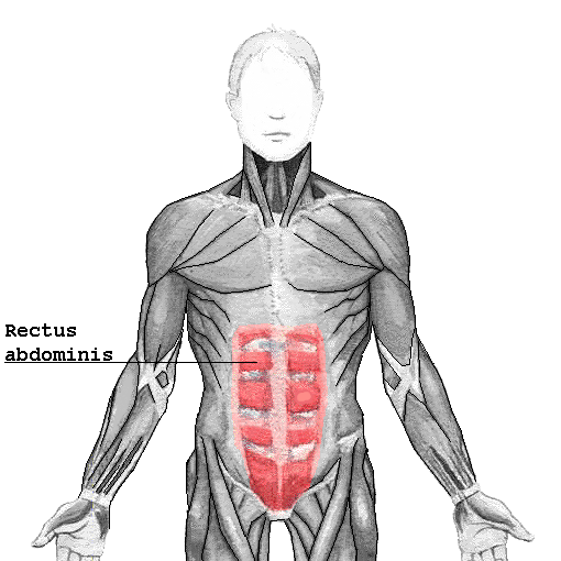
The human rectus abdominis muscle, part of the human abdomen
The human abdomen (from the Latin word meaning "belly") is the part of the body between the pelvis and the thorax. Anatomically, the abdomen stretches from the thorax at the thoracic diaphragm to the pelvis at the pelvic brim. The pelvic brim stretches from the lumbosacral angle (the intervertebral disk between L5 and S1) to the pubic symphysis and is the edge of the pelvic inlet. The space above this inlet and under the thoracic diaphragm is termed the abdominal cavity. The boundary of the abdominal cavity is the abdominal wall in the front and the peritoneal surface at the rear.
Functionally, the human abdomen is where most of the alimentary tract is placed and so most of the absorption and digestion of food occurs here. The alimentary tract in the abdomen consists of the lower oesophagus, the stomach, the duodenum, the jejunum, ileum, the cecum and the appendix, the ascending, transverse and descending colons, the sigmoid colon and the rectum. Other vital organs inside the abdomen include the liver, the kidneys, the pancreas and the spleen.
The abdominal wall is split into the posterior (back), lateral (sides) and anterior (front) walls.
Muscles of the abdominal wall

Henry Gray (1825–1861). Anatomy of the Human Body.
| Muscle | Origin and insertion |
| The obliquus externus (external oblique) muscle is the outermost muscle covering the side of the abdomen. It is broad, flat, and irregularly quadrilateral. | It originates on the lower eight ribs, and then curves down and forward towards its insertion on the outer anterior crest of the ilium and (via the sheath of the rectus abdominis muscle) the midline linea alba. |
| The obliquus internus (internal oblique) muscle is triangularly shaped and is smaller and thinner than the external oblique muscle that overlies it. | It originates from Poupart's ligament/inguinal ligament and the inner anterior crest of the ilium. The lower two-thirds of it insert, in common with fibers of the external oblique and the underlying transversus abdominis, into the linea alba. The upper third inserts into the lower six ribs. |
| The transversus abdominis muscle is flat and triangular, with its fibers running horizontally. It lies between the internal oblique and the underlying transversalis fascia. | It originates from Poupart's ligament, the inner lip of the ilium, the lumbar fascia and the inner surface of the cartilages of the six lower ribs. It inserts into the linea alba behind the rectus abdominis. |
| The rectus abdominis muscles are long and flat. The muscle is crossed by three tendinous intersections called the linae transversae. The rectus abdominis is enclosed in a thick sheath formed, as described above, by fibers from each of the three muscles of the lateral abdominal wall. | They originate at the pubic bone, run up the abdomen on either side of the linea alba, and insert into the cartilages of the fifth, sixth, and seventh ribs. |
| The pyramidalis muscle is small and triangular. It is located in the lower abdomen in front of the rectus abdominis. | It originates at the pubic bone and is inserted into the linea alba half way up to the umbilicus (belly button). |
Abdominal organs

The relations of the viscera and large vessels of the abdomen.
The abdomen contains most of the tubelike organs of the digestive tract, as well as several solid organs. Hollow abdominal organs include the stomach, the small intestine, and the colon with its attached appendix. Organs such as the liver, its attached gallbladder, and the pancreas function in close association with the digestive tract and communicate with it via ducts. The spleen, kidneys, and adrenal glands also lie within the abdomen, along with many blood vessels including the aorta and inferior vena cava. Anatomists may consider the urinary bladder, uterus, fallopian tubes, and ovaries as either abdominal organs or as pelvic organs. Finally, the abdomen contains an extensive membrane called the peritoneum. A fold of peritoneum may completely cover certain organs, whereas it may cover only one side of organs that usually lie closer to the abdominal wall. Anatomists call the latter type of organs retroperitoneal.
Surface landmarks of the abdomen
In the mid-line a slight furrow extends from the ensiform cartilage/xiphoid process above to the symphysis pubis below, representing the linea alba in the abdominal wall. At about its midpoint sits the umbilicus or navel. On each side of it the broad recti muscles stand out in muscular people. The outline of these muscles is interrupted by three or more transverse depressions indicating the lineae transversae. There is usually one about the ensiform cartilage, one at the umbilicus, and one between. It is the combination of the linea alba and the linea transversae which form the abdominal "six-pack" sought after by many people. A body fat of around 10% or below with a bodyweight that is not underweight is required to see them.
The upper lateral limit of the abdomen is the subcostal margin formed by the cartilages of the false ribs (8, 9, 10) joining one another. The lower lateral limit is the anterior crest of the ilium and Poupart's ligament, which runs from the anterior superior spine of the ilium to the spine of the pubis. These lower limits are marked by visible grooves. Just above the pubic spines on either side are the external abdominal rings, which are openings in the muscular wall of the abdomen through which the spermatic cord emerges in the male, and through which an inguinal hernia may rupture.
One method by which the location of the abdominal contents can be appreciated is to draw three horizontal and two vertical lines.
Horizontal lines
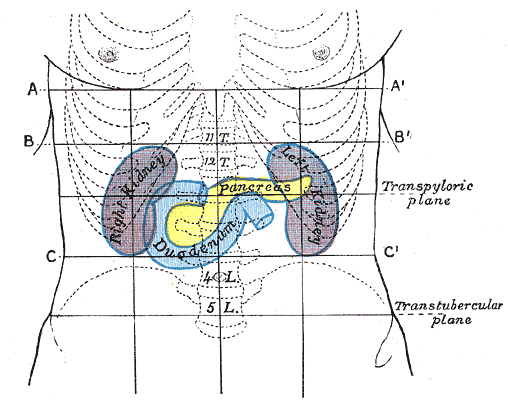
Front of abdomen, showing surface markings for duodenum, pancreas, and kidneys.
- The highest of the former is the transpyloric line of C. Addison, which is situated half-way between the suprasternal notch and the top of the symphysis pubis, and often cuts the pyloric opening of the stomach an inch to the right of the mid-line. The hilum of each kidney is a little below it, while its left end approximately touches the lower limit of the spleen. It corresponds to the first lumbar vertebra behind.
- The second line is the subcostal, drawn from the lowest point of the subcostal arch (tenth rib). It corresponds to the upper part of the third lumbar vertebra, and it is an inch or so above the umbilicus. It indicates roughly the transverse colon, the lower ends of the kidneys, and the upper limit of the transverse (3rd) part of the duodenum.
- The third line is called the intertubercular, and runs across between the two rough tubercles, which can be felt on the outer lip of the crest of the ilium about two and a half inches (60 mm) from the anterior superior spine. This line corresponds to the body of the fifth lumbar vertebra, and passes through or just above the ileo-caecal valve, where the small intestine joins the large.
Vertical lines
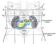
The two vertical or mid-Poupart lines are drawn from the point midway between the anterior superior spine and the pubic symphysis on each side, vertically upward to the costal margin.
- The right one is the most valuable, as the ileo-caecal valve is situated where it cuts the intertubercular line. The orifice of the vermiform appendix lies an inch lower, at McBurney's point. In its upper part, the vertical line meets the transpyloric line at the lower margin of the ribs, usually the ninth, and here the gallbladder is situated.
- The left mid-Poupart line corresponds in its upper three-quarters to the inner edge of the descending colon.
The right subcostal margin corresponds to the lower limit of the liver, while the right nipple is about half an inch above the upper limit of this viscus.
Regions of the abdomen

Surface lines of the front of the thorax and abdomen.
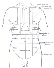
These three horizontal and two vertical lines divide the abdomen into nine "regions." (Note that "hypo" means "below" and "epi" means "above", while "chond" means "cartilage" (in this case, the cartilage of the rib) and "gast" means stomach. The reversal of "left" and "right" is intentional, because the anatomical designations reflect the position on the patient.)
| right hypochondriac/hypochondrium | epigastric/epigastrium | left hypochondriac/hypochondrium |
| right lumbar/flank/lateral | umbilical | left lumbar/flank/lateral |
| right inguinal/iliac | hypogastric/pubic | left inguinal/iliac |
Another way of dividing the abdomen is by using quadrants:
| upper right quadrant (RUQ) | upper left quadrant |
| lower right quadrant | lower left quadrant |
Animal abdomen
In vertebrates such as mammals the abdomen (belly) constitutes the part of the body between the thorax (chest) and pelvis. The region enclosed by the abdomen is termed the abdominal cavity. In arthropods it is the most distal section of the body which lies behind the thorax or cephalothorax [1][2].
Vertebrates
In vertebrates, the abdomen is a large cavity enclosed by the abdominal muscles, ventrally and laterally, and by the vertebral column dorsally. Lower ribs can also enclose ventral and lateral walls. The abdominal cavity is continuous with the pelvic cavity. It is separated from the thoracic cavity by the diaphragm. Structures such as the aorta, inferior vena cava and esophagus pass through the diaphragm. Both the abdominal and pelvic cavities are lined by a serous membrane known as the parietal peritoneum. This membrane is continuous with the visceral peritoneum lining the organs[3]. The abdomen in vertebrates contains a number of organs belonging, for instance, to the digestive tract and urinary system.
Abdominal organs
- Digestive tract: Stomach, small intestine, large intestine with cecum and appendix
- Accessory organs of the digestive tract: Liver, gallbladder and pancreas
- Urinary system: Kidneys and ureters
- Other organs: Spleen
Abdominal organs can be highly specialized in some animals. For example the stomach of ruminants (a suborder of mammals) is divided into four chambers - rumen, reticulum, omasum and abomasum.[4]
Invertebrates
In the worker ant, the abdomen consists of the propodeum fused to the thorax and the metasoma, itself divided into the narrow petiole and bulbous gaster.
The invertebrate abdomen is built up of a series of concave upper plates known as tergites and convex lower plates known as sternites, the whole being held together by a tough yet stretchable membrane.
The abdomen contains the insect's digestive tract and reproductive organs, it consists of eleven segments in most orders of insects though the eleventh segment is absent in the adult of most higher orders. The number of these segments does vary from species to species with the number of segments visible reduced to only seven in the common honeybee. In the Collembola (Springtails) the abdomen has only six segments.
The abdomen is sometimes highly modified. In ants, the first segment of the abdomen is fused to the thorax and called the propodeum. The second segment forms the narrow petiole. Some ants have an additional postpetiole segment, and the remaining segments form the bulbous gaster.[1] The petiole and gaster (abdominal segments 2 and onward) are collectively called the metasoma.
Unlike other Arthropods, insects possess no legs on the abdomen in adult form, though the Protura do have rudimentary leg-like appendages on the first three abdominal segments, and Archaeognatha possess small, articulated "styli" which are sometimes considered to be rudimentary appendages. Many larval insects including the Lepidoptera and the Symphyta (Sawflies) have fleshy appendages called prolegs on their abdominal segments (as well as their more familiar thoracic legs), which allow them to grip onto the edges of plant leaves as they walk around.
See also
- Abdominal epilepsy
- Abdominal pain
- Abdominal reflex
- Alimentary canal
- List of muscles of the human body
- Waist
References
- ↑ Abdomen. (n.d.). Dictionary.com Unabridged (v 1.1). URL: http://dictionary.reference.com/browse/abdomen [Accessed: 22 Oct 2007]
- ↑ Abdomen. Dictionary.com. The American Heritage Dictionary of the English Language, 4th Edition. URL: http://dictionary.reference.com/browse/abdomen [Accessed: 22 October 2007].
- ↑ Peritoneum. The Veterinary Dictionary. Elsevier, 2007. http://www.answers.com/topic/peritoneum [Accessed: 22 October 2007)
- ↑ "Ruminant." The Veterinary Dictionary. Elsevier, 2007. http://www.answers.com/topic/ruminant [Accessed: 22 October 2007].
- Tortora, Gerard J., Anagnostakos, Nicholas P. (1984) Principles of Anatomy and Physiology, Harper & Row Publishers, New York ISBN 0-06-046656-1
- Gray, Henry, (1977) Anatomy, Descriptive and Surgical (Gray's Anatomy) Bounty Books
- Taber, Clarence Wilber, (1981) Taber's Cyclopedic medical dictionary 14 Edition, F.A Davis Company, Philadelphia ISBN 0-8036-8307-3
- KMLE Medical Dictionary definition of abdomen retrieved from KMLE Medical Dictionary on 2007-03-25
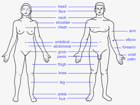 |
HEAD: Forehead – Eye – Ear – Nose – Mouth – Tongue – Teeth – Jaw – Face – Cheek – Chin TORSO: Shoulders – Spine – Chest – Breast – Ribcage – Abdomen – Belly button LIMBS: Arm – Elbow – Forearm – Wrist – Hand – Finger (Thumb - Index finger - Middle finger - Ring finger - Little finger) – Leg – Lap – Thigh – Knee – Calf – Heel – Ankle – Foot – Toe (Hallux) |
| This page uses Creative Commons Licensed content from Wikipedia (view authors). |